
Abstract
Ionizing radiations generate reactive oxygen species in irradiated tissue that induces several pathophysiological changes in the body. Radiotherapy induced toxicity is a major dose-limiting factor in anticancer treatments. Radioprotective agents are of significant importance in medical, industrial, environmental, military, and space applications. Radioprotective effect of polysaccharide protein complex (PPC-Pr) isolated from mushroom, Phellinus rimosus, was evaluated in Swiss albino mice. PPC-Pr (5 and 10 mg/kg bwt, i.p.) significantly increased leukocyte count, bone marrow cellularity, glutathione content, and activities of antioxidant enzymes such as catalase, superoxide dismutase, and glutathione peroxidase in blood as well as intestinal mucosa when compared with the irradiated control group. Histopathological observation of intestinal jejunal mucosa revealed the tissue protective effects of PPC-Pr. Further radioprotective activity of PPC-Pr was in a dose-dependent manner. The findings suggest potential radioprotective efficacy of PPC-Pr.
Introduction
Cancer remains a major public health problem worldwide, claiming over six million lives each year. More than half of all people with cancer are treated with radiotherapy today. It is used for curative purposes, preventing recurrence as well as for palliative purposes. In radiotherapy, ionizing irradiation is used to damage target cells or tissues. However, it causes severe side-effects and damage to normal tissues. Ionizing radiations cause macromolecular damage by the increased generation of reactive oxygen species (ROS) and reactive nitrogen species (RNS). ROS-mediated bimolecular reactions and their relationship with radiation sickness is the current subject of scientific investigations in radiotherapy. Radiation attenuates the endogenous antioxidant enzymes, which are considered as a first-line defense mechanism to maintain redox balance and normal biochemical processes. These alterations in the metabolic redox status of the body regulate activation or inhibition of various signal transduction pathways, transcription factors, and gene expression. 1
Exposure to artificial (man-made) radiation comes from various sources such as diagnostic radiotherapy and nuclear medicine apart from natural background radiation. In radiotherapy, total body irradiation is used for systemic treatment of lymphoma and leukemia and as a method of immunosuppression before bone marrow transplantation. Further, many human beings are exposed to total body irradiations, such as survivors of Hiroshima and Nagasaki and military personnel exposed to fall out from nuclear tests. Whole-body irradiation causes hematological, gastrointestinal, and neurovascular syndromes in the body. However, at doses used in clinical radiotherapy, hematopoietic and gastrointestinal syndromes are more pronounced. 2
The hematopoietic syndrome occurs at doses in the range of 2–8 Gy in humans (3–10 Gy in rodents). Whole-body irradiation causes loss of proliferative capacity of stem cells in renewal tissues such as bone marrow. In these cells, radiation upregulates the genes such as Fas, Bax, and caspase-3, which can facilitate apoptosis, and downregulates genes such as Bcl2, which prevents apoptosis. It leads to degeneration of precursor cells in the bone marrow and ultimately severe depletion of blood elements. 2 Various side-effects including leucopenia, thrombocytopenia, and loss of electrolyte and fluid balance have been observed in patients undergoing radiotherapy. Further radiotherapy causes immunosuppression in the host, significantly increasing patients' risk for infection.
The intestine is another important dose-limiting organ during radiation therapy of tumors in the pelvis or abdomen. Radiation responses in intestine are manifested by changes in cellular function and alterations in morphology. 3,4 Severity of intestinal radiation toxicity directly depends on cell death in intestinal crypts. Radiation treatment damages the mucosal lining of gastrointestinal tract, which result in diarrhoea and vomiting, ultimately leading to loss of electrolyte and fluid balance. The endogenous antioxidant defenses are inadequate to reduce the radiation-induced damages. So administration of immunomodulating drugs in combination with conventional radiotherapy strengthens patient's tolerance to treatment. A large number of drugs of natural and synthetic origin have been screened for evaluation of their radioprotective potential. Chemicals such as WR-2721, cysteamine, cysteine, mercaptopropionyl glycine, etc., have been evaluated in this context. 5 However, owing to the toxicity associated with the therapeutic doses, the search for more effective and less toxic agents remains continued.
Mushrooms represent a major and as-yet largely untapped source of new powerful pharmaceutical products. Influence of mushroom metabolites on immune function and overall hormonal system appears to be major beneficial effects of higher fungi. 6,7 Mushroom-derived polysaccharides had been reported to boost immune cell production and ameliorate chemotherapy symptoms. 8 Phellinus species are mostly tropical mushrooms and 18 species are known from Kerala. Phellinus rimosus is a parasitic host-specific polypore macrofungus often found growing on jackfruit trees (Atrocarpus heterophyllatus) trunks. 9 Earlier investigations showed that ethyl acetate and methanol extracts of P. rimosus possessed antioxidant, antitumor, and hepatoprotective activities. 10 –12 Recent investigations have also demonstrated the profound antioxidant, anti-inflammatory, and antiarthritic activities of polysaccharide protein complex (PPC-Pr) isolated from the aqueous extract of P. rimosus. 13,14
Phytochemical investigations revealed that total polysaccharide and protein content in the isolated PPC-Pr complex was 52% and 33.8%, respectively. Further, it had been reported that P. rimosus fruiting body possesses oxides of Na, K, Ca, Mg, Al, Mn, Ti, and Fe in varying proportions. 15 When using an antioxidant or radioprotective compound, its inherent toxicity should be evaluated. For this purpose, toxicity of PPC-Pr was evaluated using male Swiss albino mice. Acute toxicity studies showed that PPC-Pr produced no mortality up to 100 mg/kg bwt (i.p.). Further, subacute toxicity studies with PPC-Pr showed no hematological toxicity. Blood parameters, liver function, and kidney function tests as well as histopathology also showed no significant change in the treated groups (unpublished observation). The aim of the present study was to evaluate the radioprotective efficacy of PPC-Pr.
Materials and Methods
Chemicals
Glutathione (GSH), 5,5′dithio-dinitro bisbenzoic acid (DTNB), nitroblue tetrazolium, thiobarbituric acid (TBA), riboflavin, and sodium azide were obtained from SRL, Mumbai, India, and hydrogen peroxide (H2O2) from Merck India Ltd., Mumbai, India. Amifostine (Cytofos) was purchased from Sun pharmaceutical India Ltd., Gujarat, India. All other chemicals and reagents used were of analytical grade.
Animals
Male Swiss albino mice, 8–10 weeks old and weighing 20–25 g, were purchased from Small Animal Breeding Station of Kerala Agriculture University, Mannuthy, Thrissur, and were kept under environmentally controlled conditions (12 hours of light–dark cycle, 26°C–28°C temperature, and relative humidity of 60%–70%) with free access to standard food (Sai Durga Feeds, Bangalore, India) and water ad libitum. All the animals were acclimatized for 1 week before starting the experiment. Experiments were conducted according to the rules and regulations of the Institutional Animal Ethics Committee, Amala Cancer Research Centre, Amala Nagar, Thrissur, Kerala, India, which followed the guidelines of the Committee for the Purpose of Control and Supervision of Experiments on Animals, Govt. of India.
Isolation of PPC-Pr
Fruiting bodies of P. rimosus were collected from the outskirts of Thrissur, Kerala, India. The voucher specimen was deposited in the Herbarium of Centre for Advanced Studies in Botany, University of Madras, Chennai, India (HERB MUBL 3171). Fruiting bodies of P. rimosus were dried at 45°C–50°C for 48 hours and powdered. The powdered material (500 g) was defatted with petroleum ether using a Soxhlet apparatus for 8–10 hours. The defatted material was extracted with double-distilled water at 95°C for 8–9 hours. PPC-Pr was isolated from the aqueous extract by the method described by Meera et al. 13 Briefly, polysaccharides in the aqueous extract were precipitated with ethanol and lyophilized to obtain PPC-Pr complex. The yield of preparation was 0.5% of total weight of dry powder. The carbohydrate content of the PPC-Pr was determined by the phenol–sulphuric acid method using glucose as the standard 16 and protein content was determined using Folin's phenol reagent 17 with bovine serum albumin as the standard.
Irradiation
The animals were treated with a single dose of radiation of 400 rads (4 Gy). The source of radiation was a 60Co-Theratron Phoenix Teletherapy Unit (Atomic Energy Ltd., Ottawa, Canada) at Amala Cancer Hospital, Thrissur, Kerala, India. The animals were exposed to whole-body irradiation at a rate of 1.41 Gy/min.
Experimental design
The animals were divided into 5 groups and drugs were administered as follows: • Group 1—Normal 0 Gy • Group 2—Irradiated control 4 Gy • Group 3—Amifostine (300 mg/kg bwt, i.p.) administered 1 hour before irradiation of 4 Gy (positive control) • Group 4—PPC-Pr (5 mg/kg bwt, i.p.) administered daily for 5 consecutive days and irradiated with 4 Gy, 1 hour after the last dose • Group 5—PPC-Pr (10 mg/kg bwt, i.p.) administered daily for 5 consecutive days and irradiated with 4 Gy, 1 hour after the last dose
Blood was collected from caudal vein into heparinized tubes at 1st, 5th, 7th, and 14th day after irradiation and the following parameters were checked: (a) total WBC count (hemocytometer method) and (b) hemoglobin (cyanometh hemoglobin solution). Animals were sacrificed at the1st, 5th, 7th, and 14th day after irradiation by decapitation. Bone marrow cells were collected from the femur. Intestine was quickly excised, washed with saline, and blotted with a piece of filter paper. Intestinal jejunal mucosa was scraped and 10% homogenate was prepared using phosphate buffer (50 mM/L, pH 7.0). A portion of jejunal tissue was fixed in 10% formaldehyde, embedded in paraffin, and sectioned. The sections were stained with hematoxylin–eosin (H&E) and observed under light microscope for histopathological observations.
Determination of bone marrow cellularity and total WBC count
For the determination of bone marrow cellularity, femurs of each animal were dissected out and were flushed into phosphate-buffered saline (pH 7.4) containing 2% fetal calf serum. The cells were washed, bone marrow viability was determined with a hemocytometer, and expressed as total cells ( × 106 per femur). 18 The results were expressed as number of live bone marrow cells × 106 per femur. Total WBC count was done using hemocytometer method. Briefly, 20 μL of heparinized blood was mixed well with 380 μL of diluting fluid. 19 The diluting fluid contains acetic acid to lyse RBCs and crystal violet for staining the cell. The cells were allowed to settle at the bottom of the chamber for 2 minutes. The number of cells in the large four-corner squares were counted (N) under a microscope with a 10 × objective. Total leukocyte count/mm3 was expressed as (N * Dilution factor * Depth factor)/Area counted, which is equivalent to N * 50.
Determination of antioxidant enzyme status
Antioxidant enzyme assay was carried out in both heparinized blood and homogenate of jejunum mucosa spectrophotometrically in a double-beam spectrophotometer (Systronics India Ltd., Hyderabad, India). Reduced glutathione content (GSH) was assayed by the method of Moron et al. 20 based on the reaction with DTNB to produce a yellow-colored complex. The concentration of GSH was determined from the standard graph of GSH. Glutathione peroxidase (GPx) was assayed by the method of Hafeman et al. 21 based on the decrease in GSH content after incubating the sample in the presence of H2O2 and NaN3. One unit of GPx activity was defined as decease in log GSH activity by 0.001 per minute with respect to nonenzymatic reaction. Activity of the enzyme superoxide dismutase (SOD) was assayed from the ability of the sample to scavenge superoxide anion generated from the photoillumination of riboflavin according to the method of McCord and Fridovich. 22 Catalase (CAT) activity in blood was estimated by the method of Aebi 23 by measuring the rate of decomposition of hydrogen peroxide (H2O2) at 240 nm and expressed as K/mg Hb. Activity of CAT in tissue was estimated by the method of Beer and Siezer 24 and calculated using the molar extinction coefficient of H2O2 (43.6 M−1 Cm−1) and expressed in mmoles of H2O2 decomposed/min/mg protein (U/mg protein). Hemoglobin was estimated by the cyanmethemoglobin solution using Drabkin's method. 25 Protein content in tissue was determined using Folin's phenol reagent 17 and compared with bovine serum albumin standard.
Statistical analysis
All experimental data were expressed as mean ± SD. The data were analyzed by one-way analysis of variance (ANOVA) followed by the Bonferroni's multiple comparison test (using the Graph Pad Instat Software Package). A p-value of <0.05 was considered significant.
Results
Effect on bone marrow cellularity and total WBC count
Gamma irradiation (4 Gy) induced a significant decrease in both total WBC count and bone marrow cellularity (Fig. 1). Total WBC count was found to be decreased significantly from 1st day onward after irradiation in the control group compared with normal (p < 0.001). PPC-Pr at doses of 5 and 10 mg/kg bwt showed 3.7- and 4.2-fold increase in total WBC count than the control on 7th day (p < 0.001), wherein maximum radiation toxicity was observed.
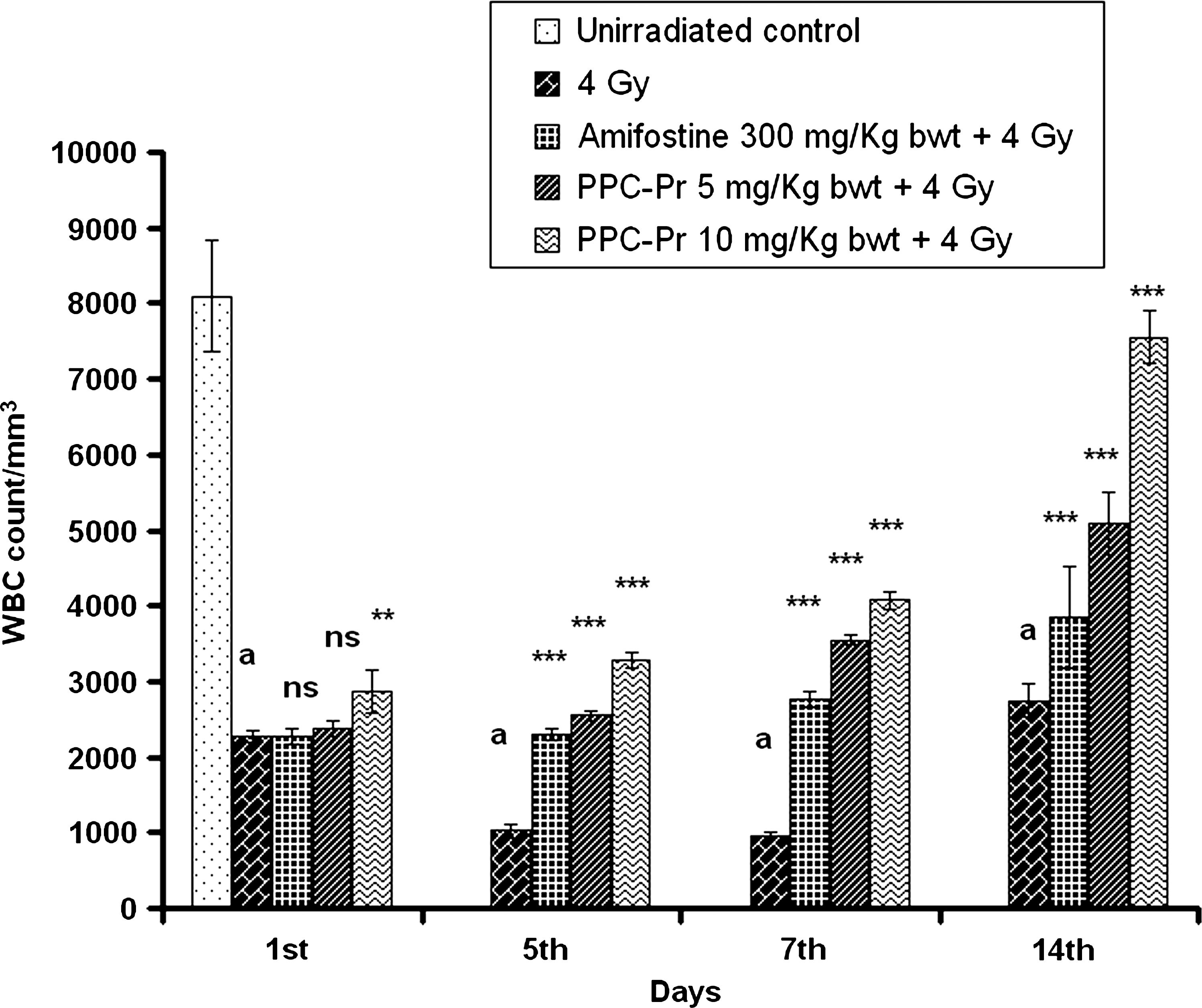
Effect of administration of polysaccharide protein complex (PPC-Pr) on total white blood cell (WBC) count of 4 Gy γ-irradiated animals. Values are mean ± SD; n = 6. **p < 0.01, ***p < 0.001, and ns indicates p > 0.05, not significant when compared with the irradiated control group. a denotes p < 0.001 compared with normal.
The protection offered was further supported by the improved bone marrow cellularity found in the treated group than that of the control (Fig. 2). PPC-Pr at both doses (5 and 10 mg/kg bwt) as well as amifostine showed significant improvement in bone marrow cellularity from 5th day onward when compared with the control (p < 0.001). On 14th day, both bone marrow cellularity and total leukocyte count were significantly restored in the treated group when compared with the irradiated control (p < 0.001). PPC-Pr at 10 mg showed 1.1 times increase in bone marrow cellularity than 5 mg on 14th day. Total leukocyte count in 10 mg treated group was 1.5 times higher than 5 mg treated group on 14th day after irradiation (p < 0.001).
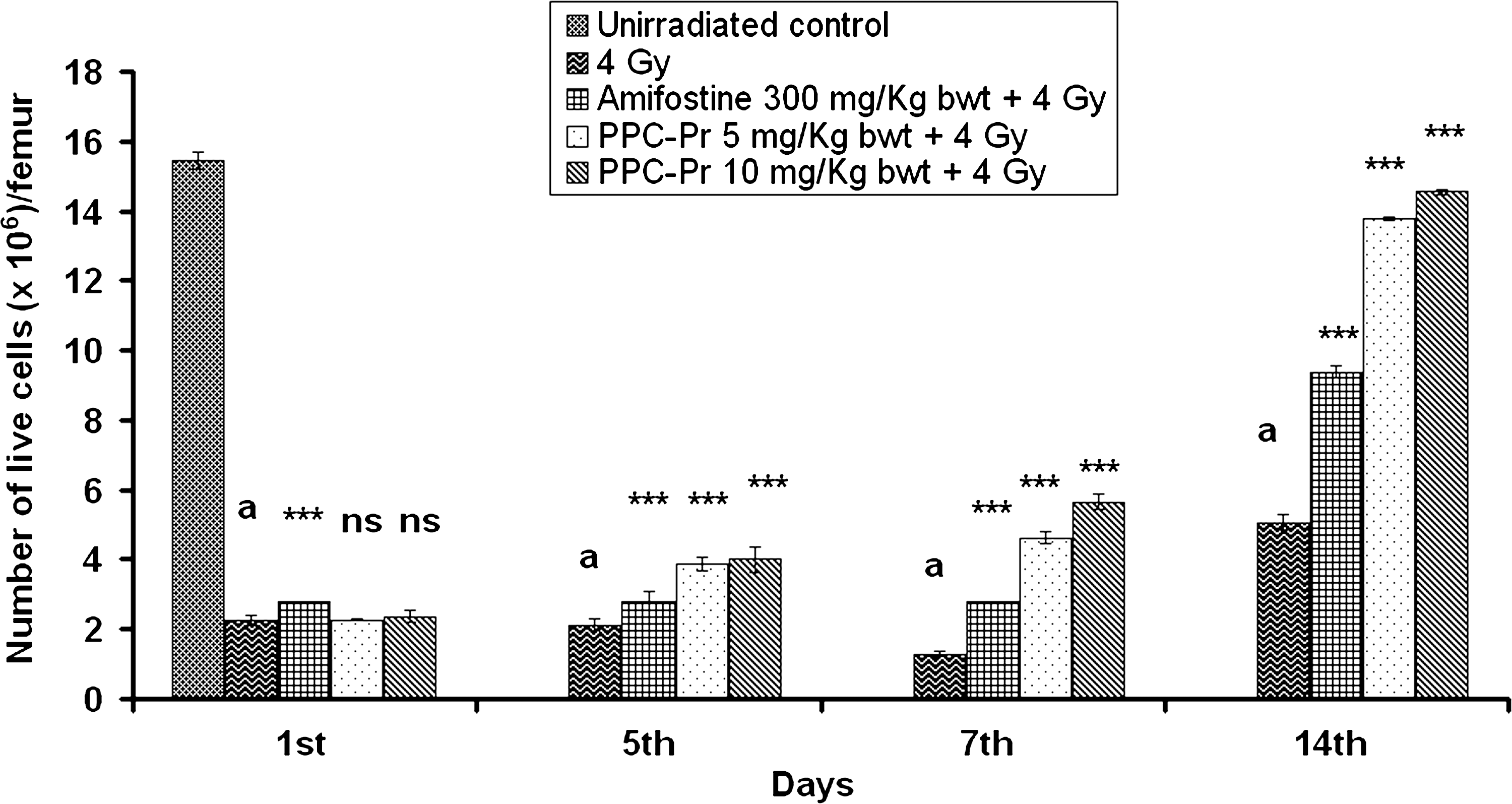
Effect of administration of PPC-Pr on bone marrow cellularity of 4 Gy γ-irradiated animals. Values are mean ± SD; n = 6. ***p < 0.001 and ns indicates p > 0.05, not significant when compared with the irradiated control group. a denotes p < 0.001 compared with the normal.
Effect on blood antioxidant status
The level of blood GSH, a major cellular antioxidant, was brought down significantly after irradiation up to 14th day with respect to that of normal (p < 0.001) (Fig. 3). However, treatment with PPC-Pr was able to ameliorate the effects of radiation to a great extent. GSH level in the PPC-Pr–treated group (5 and 10 mg/kg bwt) was found to be increased 1.6- and 1.9-fold higher than the control on 7th day (p < 0.001). PPC-Pr at 10 mg showed 1.3 and 1.2 times increase in GSH activity than 5 mg on 7th and 14th day, respectively. Activity of GPx in blood of control animals was found to be decreased significantly after whole-body irradiation when compared with the normal (p < 0.001) (Table 1). Administration of PPC-Pr prevented initial fall in GPx activity. Increase in GPX activity on 7th day in the treated group (5 and 10 mg/kg bwt) was found to be 2.9 and 3.8 times higher than control (p < 0.001). PPC-Pr at 10 mg showed 1.3 and 1.1 times increase in GPx activity than 5 mg on 7th and 14th days, respectively. Further, PPC-Pr at 10 mg showed significant increase in blood GPx activity in all the days tested when compared with the 5 mg treated group (p < 0.001).
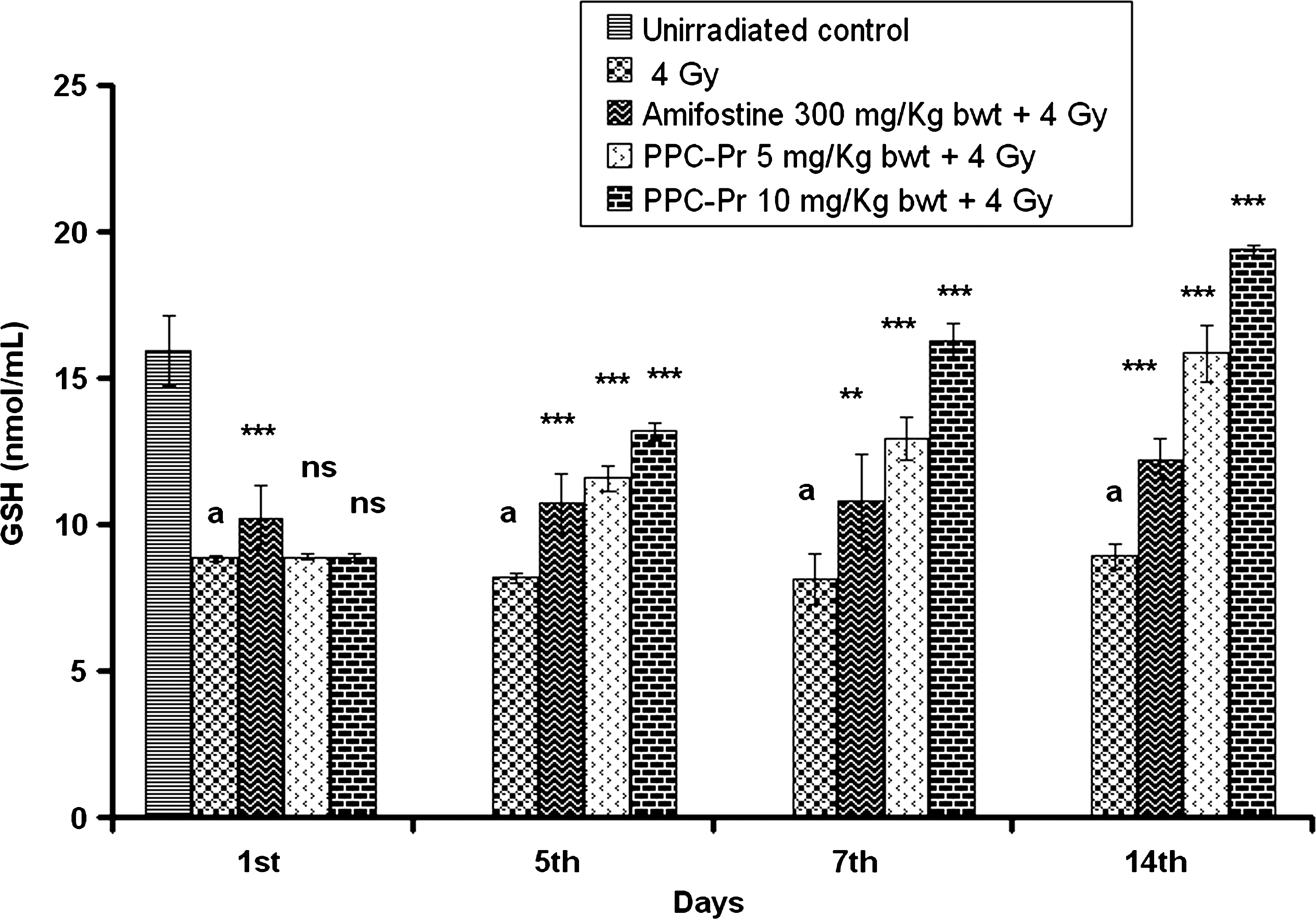
Effect of administration of PPC-Pr on blood GSH levels of 4 Gy γ-irradiated animals. Values are mean ± SD; n = 6. **p < 0.01, ***p < 0.001, and ns indicates p > 0.05, not significant when compared with the irradiated control group. a denotes p < 0.001 compared with the normal.
Values are mean ± SD; n = 6. *** p < 0.001 compared with the irradiated control and nsindicates p > 0.05, not significant when compared with the irradiated control group (Bonferroni test). a denotes p < 0.001 compared with the normal (Bonferroni test).
PPC-Pr, polysaccharide protein complex.
SD, standard deviation.
Gamma irradiation induced a significant decrease in the activity of SOD and CAT (Tables 2 and 3). Preadministration of PPC-Pr resulted in significant elevation in the activities of these enzymes. PPC-Pr (5 and 10 mg/kg bwt) showed 1.7 and 2.2 times increase in SOD activity than control (p < 0.001) on 7th day. Blood SOD activity in the 10 mg treated group was found to be 1.3 times higher than the 5 mg treated group on 7th day (p < 0.001). CAT activity was also decreased significantly by irradiation in the control group compared with the normal (p < 0.001). However, activity was restored significantly by both PPC-Pr and amifostine treatments. PPC-Pr at 10 mg showed 1.1 and 1.2 times increase in CAT activity than 5 mg on 7th and 14th days, respectively.
Values are mean ± SD; n = 6. *** p < 0.001 compared with the irradiated control and nsindicates p > 0.05, not significant when compared with the irradiated control group (Bonferroni test). a denotes p < 0.001 compared with the normal (Bonferroni test).
SD, standard deviation.
Values are mean ± SD; n = 6. *** p < 0.001 compared with the irradiated control and nsindicates p > 0.05, not significant when compared with the irradiated control group (Bonferroni test). a denotes p < 0.001 compared with the normal (Bonferroni test).
SD, standard deviation.
Effect on antioxidant enzyme status in the intestinal mucosa
GSH and GPx activities in intestinal mucosa were significantly decreased by whole-body irradiation in the control group when compared with the normal (p < 0.001) (Fig. 4 and Table 4). GSH and GPx activities in the PPC-Pr–treated group (10 mg/kg bwt) were found to be 1.7 and 4.3 times higher than the control on 7th day. Mucosal GSH activity in the 10 mg treated group was found to be 1.3 times higher than the 5 mg treated group on 14th day (p < 0.001).

Effect of administration of PPC-Pr on mucosal GSH levels of 4 Gy γ-irradiatezd animals. Values are mean ± SD; n = 6. ***p < 0.001 and ns indicates p > 0.05, not significant when compared with the irradiated control group. a denotes p < 0.001 compared with the normal.
Values are mean ± SD; n = 6. *** p < 0.001 compared with the irradiated control and nsindicates p > 0.05, not significant when compared with the irradiated control group (Bonferroni test). a denotes p < 0.001 compared with the normal (Bonferroni test).
SD, standard deviation.
SOD activity was restored significantly by PPC-Pr treatment compared with the control. SOD activity in the PPC-Pr–treated group (10 mg/kg bwt) was found to be 2.4 times higher than the control on 7th day (p < 0.001) (Table 5). Gamma irradiation induced significant decrease in the activity of mucosal CAT enzyme (Table 6). PPC-Pr administration showed significant increase in CAT activity from 5th day onward in the treated groups. Further, PPC-Pr at 10 mg showed significant increase in mucosal SOD and CAT activity in all the days tested when compared with the 5 mg treated group (p < 0.001). On 14th day, significant restoration of mucosal antioxidant enzyme activity was observed in the treated group than control. Amifostine treatment also significantly increased mucosal antioxidant status when compared with the control group.
Values are mean ± SD; n = 6. *** p < 0.001 compared with the irradiated control and nsindicates p > 0.05, not significant when compared with the irradiated control group (Bonferroni test). a denotes p < 0.001 compared with the normal (Bonferroni test).
SD, standard deviation.
Values are mean ± SD; n = 6. *** p < 0.001 compared with the irradiated control and nsindicates p > 0.05, not significant when compared with the irradiated control group (Bonferroni test). a denotes p < 0.001 compared with the normal (Bonferroni test).
SD, standard deviation.
Effect of radiation-induced histopathological changes
Microscopic examination of tissue slices revealed that γ irradiation led to prominent damage of small intestine and villi atrophy as well as mucosal erosion in the tissue, whereas in animals administrated with both doses of PPC-Pr and amifostine, these changes were less pronounced (Fig. 5).
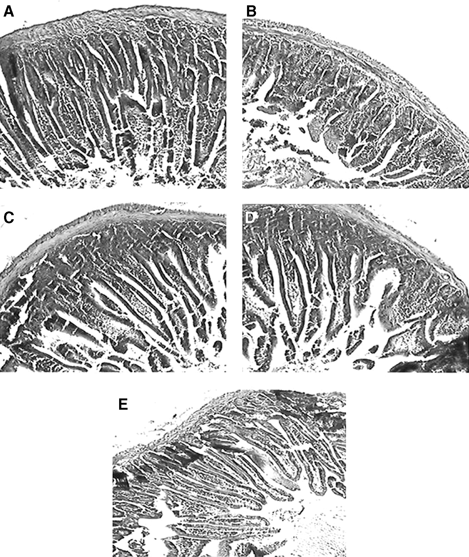
Histopathological sections of intestinal jejunal mucosa at 7 days after 4 Gy γ irradiation.
Discussion
Whole-body irradiation in mice causes oxidative tissue damage in various organs. However, acute responses to radiation primarily occur in tissues with rapid cell renewal, where cell division is required to maintain the function of the organ. Rapidly dividing cells of gastrointestinal tract and hematopoietic system are more prone to radiation-induced damages. One of the major syndromes of hematopoietic system by total-body exposure of ionizing radiation is bone marrow aplasia. Protection of PPC-Pr on bone marrow cellularity and total WBC count was more pronounced on 7th day after irradiation, where maximum radiation-induced toxicity was observed with respect to control. Further, on 14th day, PPC-Pr at both concentration exhibited dose-dependent improvement in the recovery of bone marrow count and total WBC count. The polysaccharides possess immunomodulating property, 26 so its action remains for long time. Thus, they significantly stimulate proliferation of stem cells in the bone marrow. Similarly, protein-bound polysaccharide complex isolated from mushroom Lentinus lepideus (PG 101) had been reported to recover radiation-induced bone marrow suppression very efficiently. 27 The dose and selection of amifostine as a positive control was selected from a comparative study that reported tissue protective effects of fullerenol and amifostine in irradiated rats. 28 Amifostine had been reported to protect against acute radiation syndrome and reduce gastrointestinal death of lethally irradiated mice. 29 Further, amifostine (300 mg/kg bwt) was used as a reference in radioprotection studies. 30
Exposure to ionizing radiations such as γ rays result in hydroxyl radical (•OH) production. Hydroxyl radical damages cellular DNA, protein, and lipids. Further, •OH generates peroxyl radical (RO2•) and alkoxyl radical (RO•) from organic compounds. Organisms contain innate antioxidant enzymes to remove free radicals and ROS. Major antioxidant enzymes include SOD, CAT, GPx, and low-molecular-weight antioxidant moiety GSH, which scavenges ROS and RNS. SOD is the only enzyme that disrupts superoxide radicals and is present in all cells with high amounts in erythrocytes. Superoxide radical formed by radiolysis of water generates peroxynitrite and H2O2. Peroxynitrite in turn generates a wide range of noxious species under physiological conditions. 31 Elevated SOD activity in PPC-Pr–pretreated mice can prevent the formation of toxic peroxynitrite radicals by attacking the superoxide radicals.
The primary role of CAT is to scavenge H2O2 generated by SOD in removal of superoxide anions and to convert it to water. Elevated CAT activity from the pretreatment of PPC-Pr reduces H2O2 and thus prevents •OH formation. PPC-Pr increased CAT activity in a dose-dependent manner when compared with control. The stimulatory effect of related species Phellinus linteus (P. linteus) on CAT activity had already been reported. 32 GPx converts H2O2 to H2O by coupling its reduction with oxidation of GSH. They can also catalyze GSH-dependent reduction of fatty acid hydroperoxides and catalyze the conversion of peroxides into alcohol. 33 PPC-Pr pretreatment significantly elevated GPx activity in blood and intestinal mucosa when compared with the control.
Gamma irradiation denatures proteins and causes conformational change in the structure, which renders them inactive. This could be correlated to the declined antioxidant status in the irradiated mice with respect to the normal. GSH Prevents protein-SH groups from oxidizing and crosslinking. Pretreatment with PPC-Pr significantly increased GSH content in irradiated mice, which helps to maintain the normal physiological process. Both blood and mucosal GSH levels increased significantly in the treated group when compared with the control. The association of polysaccharides with proteins is known to potentiate free radical scavenging activity. 34 Therefore, the improved antioxidant status in blood and intestinal mucosa can be ascribed to the free radical scavenging property of the PPC-Pr complex. PPC-Pr complex from P. rimosus had been already reported to increase in vivo antioxidant activity in arthritis-induced animals. 14 The radioprotective effects of PPC-Pr are further supported by histopathological examinations of intestinal mucosa of irradiated animals. Reduction in villus height and mucosal erosion were observed in irradiated control. Overall improvement is evident from the gross anatomy of the sections in the treated group. Recruitment of cells to compensate for damaged cell contributes toward the recovery of mucosal tissue.
PPC-Pr from a closely related species P. linteus had been already shown to stimulate immune system and enhance the production of interleukins. 35 Proteoglycans from P. linteus had been reported to stimulate host defense immune system by boosting both humoral and cellular immune responses. 36 Mushroom products such as Krestin, Lentinan, and Shizophyllan has been used as anticancer drugs. Majority of them are β-glucan polysaccharides or polysaccharide peptide complexes. Further, protein-bound polysaccharide isolated from P. linteus had an antiproliferative effect for SW480 human colon cancer cells. 37 Polysaccharides isolated from medicinal mushrooms act as immunopotentiator and enhance immune status in the body by a variety of mechanisms including production of immune mediators such as cytokines. 38 Hence, they are excellent agents against radiation-induced immunosuppression.
Conclusions
Polysaccharides are biological response modifiers and cause no harm and additional stress on the body. This mushroom polysaccharide appears to be well tolerated and compatible with radiation therapy. The results of the present study conclude that PPC-Pr isolated from the aqueous extracts of the mushroom Phellinus rimosus imparted significant protection against radiation-induced hematological toxicities as evident from increased bone marrow cellularity as well as WBC count in the treated groups. They also restored antioxidant status in a dose-dependent manner in radiosensitive tissues such as blood and intestinal mucosa. Further histopathological observation of mucosal morphology revealed improved radioprotective potential of PPC-Pr. However, the exact biochemical and molecular mechanism that mediates the radioprotective effect of PPC-Pr is still not clearly understood and requires further investigations.
Footnotes
Acknowledgments
The authors sincerely thank Dr. T.A. Ajith, Associate Professor, Amala Institute of Medical Sciences, Thrissur, Kerala, India, for his valuable suggestions.
Disclosure Statement
The authors declare that there are no conflicts of interest.