
Abstract
Bombesin (BBN) is a peptide showing high affinity for the gastrin-releasing peptide receptor. Tumors such as prostate, small cell lung cancer, breast, gastric, and colon cancer are known to over express receptors to BBN and gastrin-releasing peptide (GRP). The goal of this study was to evaluate a new 67Ga radiolabeled BBN analog based on the bifunctional chelating ligand DOTA (1, 4, 7, 10-tetraazacyclododecane-1, 4, 7, 10-tetraacetic acid), which could be used as a tool for diagnosis of GRP receptor-positive tumors. DOTA-GABA-BBN (7–14) NH2 was synthesized using a standard Fmoc strategy. Labeling with 67Ga was performed at 95°C for 30 minutes in ammonium acetate buffer (pH = 4.8). Radiochemical analysis involved ITLC and HPLC methods. The stability of radiopeptide was examined in the presence of human serum at 37°C up to 24 hours. The receptor-bound internalization and externalization rates were studied in GRP receptor expressing PC-3 cells. Biodistribution of radiopeptide was studied in nude mice bearing PC-3 tumor. Labeling yield of >90% was obtained corresponding to a specific activity of ≈2.6 MBq/nmol. Peptide conjugate showed good stability in the presence of human serum. The radioligand showed a good and specific internalization into PC-3 cells (16.13% ± 0.71% at 4 hours). After 4 hours, a considerable amount of activity (52.42% ± 1.86%) was externalized. In animal biodistribution studies, a receptor-specific uptake of radioactivity was observed in GRP-receptor-positive organs. After 4 hours, the uptake in mouse tumor and pancreas was 1.30% ± 0.18% ID/g (percentage of injected dose per gram of tissue) and 1.21% ± 0.13% ID/g, respectively. These data show that [67Ga]-DOTA-GABA-BBN (7–14) NH2 is a specific radioligand for GRP receptor positive tumors and is a suitable candidate for clinical studies.
Introduction
Radiolabeled peptides are of increasing interest in nuclear imaging. To extend the range of radiopeptide imaging, wide spectra of peptides have been proposed to target specific tumor cell types in vitro and in vivo. A lot of malignant human cancer cells overexpress different peptide hormone receptors on their cell surface. These receptors have become important and useful as targets for molecular imaging and targeted therapy of tumors using receptor eager small peptides. Several peptide receptors such as somatostatin, neurotensin, and bombesin (BBN) receptors have attracted considerable interest in recent years. BBN is a 14-aminoacid peptide isolated from frog skin. The mammalian counterparts of the frog peptide are neuromedin B (NMB) and gastrin-releasing peptide (GRP). The receptors for these ligands to be cloned were designated BB1 and BB2 and were originally referred to as the NMB and GRP preferring BBN receptors. Over-expression of receptors for both NMB and GRP has been reported to be found on the cell surfaces of several malignant tissues, particularly in the cases of lung cancer, colon cancer, prostate cancer, and breast cancer. 1 –6
In auto radiographic studies, Reubi and Markwalder found the GRP receptor to be expressed in high density on invasive prostate carcinomas and proliferative intraepithelial prostate lesions, whereas normal prostate tissues were GRP receptor negative. 7,8 These findings suggest that the GRP receptor can be used as a molecular basis for diagnosing and treating prostate tumors.
Up to now, many types of radiolabeled BBN analogs have been designed to target GRP receptor expressing tumors. 9 –19 For example, 99mTc, 111In, and 67Ga labeled BBN analogs have been developed for SPECT and 64Cu and 68Ga labeled analogs for positron emission tomography imaging. 7,9,12,20 –2690Y and 177Lu labeled analogs have been described as promising tools for targeted radiotherapy of these tumors. 7,8
We have recently developed and evaluated the radiolabeled peptide [99mTc/HYNIC0, D-Tyr6, D-Trp8] BBN (6–14) NH2, which internalized rapidly into GRP receptor positive tumor cells. 27,28 To extend our previous study, we developed our work to synthesize a DOTA coupled BBN analog with improved affinity to GRP receptors and increased uptake in GRP receptor expressing tumors.
Here we present data on the synthesis and labeling of DOTA-GABA-BBN (7–14) NH2 with a gamma and auger electron emitter 67Ga. In addition, we studied stability in human serum, receptor bound internalization, efflux in PC-3 cells, and in vivo tumor uptake and tissue biodistribution of radiolabeled compounds.
Materials and Methods
Rink amide MBHA (4-Methylbenzhydrylamine) resin and all of the Fmoc-protected amino acids were commercially available from NovaBiochem. The prochelator 1,4,7,10-tetraazacyclododecane-1,4,7-tris(aceticacid-t-butyl ester)-10-acetic acid [DOTA-tris(tBu ester)] was obtained from Macrocyclics. Other reagents were purchased from Fluka and used without further purification.
The reactive side chains of the amino acids were masked with one of the following groups: Trp, t-butoxycarbonyl; His, trityl; Gln, trityl. The cell culture medium was Dulbecco's modified Eagle's medium (DMEM) supplemented with 10% fetal bovine serum (FBS), amino acids, vitamins, and penicillin/streptomycin from Gibco. The production of 67Ga was performed at a 30 MeV cyclotron (Cyclone-30, IBA) from cyclotron division (AEOI). Analytical reverse phase high performance liquid chromatography (RP-HPLC) was performed on a JASCO 880-PU intelligent pump HPLC system equipped with a multiwavelength detector and a flow-through Raytest-Gabi γ-detector. CC 250/4.6 Nucleosil 120-5 C-18 column from Teknokroma was used for analytical HPLC, and a VP 250/10 Nucleosil 100-5 C-18 column was used for semipreparative HPLC. The gradient systems consisted of 0.1% trifluoroacetic acid/water (Solvent A) and acetonitrile (Solvent B). For analytical HPLC, Gradient I was used: 0 minute 95% A, 5 minutes 95% A, 25 minutes 0% A, 27 minutes 0% A, flow = 1 mL/min, γ = 280 nm; for semipreparative HPLC Gradient II: 0 minute 80% A, 2 minutes 80% A, 17 minutes 50% A, 19 minutes 0% A, 21 minutes 0% A, flow = 2 mL/min, γ = 280 nm. Mass spectrum was recorded on an Agilent 1100/Bruker Daltonic (Ion trap) VL instrument (LC/MS). Quantitative gamma counting was performed on an ORTEC Model 4001 M γ-system well counter.
Synthesis
The peptide was synthesized by standard Fmoc solid phase synthesis on Rink Amide MBHA resin with substitution, 0.69 mmol/g. Coupling of each amino acid was performed in the presence of 3 mol excess of Fmoc–amino acid, 3 mol excess of N-hydroxybenzotriazole (HOBt), 3 mol excess of diisopropylcarbodiimide (DIC), and 5 mol excess of diisopropylamine (DIPEA) in dimethylformamide (DMF). Completeness of coupling reactions was monitored by the Kaiser test, and the Fmoc groups were removed by adding 20% piperidine in DMF. Coupling of DOTA to peptide was performed in the presence of 1.2 mol excess of DOTA-(tBu)3, 2.5 mol excess of (2-(7-Aza-1H-benzotriazole-1-yl)-1, 1, 3, 3-tetramethyluronium hexafluorophosphate) (HATU), and 5 mol excess of diisopropyletylamine (DIPEA) in Dimethylformamide (DMF). The peptide DOTA conjugate was removed from the resin, and amino acid side chains were also deprotected by treatment with a cocktail of trifluoroaceticacid (TFA), triisopropylsilane, and water (95:2.5:2.5). After removing the organic solvents in vacuum, the crude product was precipitated with Cold Petroleum Ether and Diisopropyl Ether (50:50). The crude peptide DOTA conjugate was dissolved in water/methanol and purified by semipreparative RP-HPLC; then, the purified product was characterized by LC/MS and analytical HPLC.
Labeling of DOTA-GABA-BBN (7–14) NH2 with 67Ga
A stock solution of DOTA-GABA-BBN (7–14) NH2 (concentration 1 mmol/L) was prepared by dissolving the peptide in distilled water. Fifteen (15) microliters of the stock solution (20 μg of peptide) was added to an eppendorf tube containing 0.2 mL ammonium acetate buffer (pH 4.8, 0.5 mol/L). Then, 37 MBq 67Ga in 0.02 mL/0.1 mol/L HCl was added to a reaction solution, and the mixture was kept for 30 minutes at 95°C. After cooling down to room temperature, the preparation was checked for bound and free 67Ga.
Characterization of 67Ga-labeled DOTA-GABA-BBN (7–14) NH2
67Ga-labeled DOTA-GABA-BBN (7–14) NH2 was characterized by Paper chromatography and HPLC techniques. Paper chromatography was performed using Whatman No.1 and methanol/0.01 mol/L acetate buffer (pH 6.2) at a ratio of 55:45 as a mobile phase to check for bound and free 67Ga. HPLC was used to ensure that the labeled molecule was present as a single peak and to determine the complexation yield. For Analytical HPLC a C-18, reversed phase column with gradient system 1 was used with 0.1% trifluoroacetic acid/water (Solvent A) and acetonitrile (Solvent B) as the mobile phase. Partition coefficients for each conjugate were also evaluated in octanol/H2O. The partition ratios of the labels were calculated by dividing the counts in the organic phase with those in the aqueous phase per volume unit.
Serum stability
To 1 mL of freshly prepared human serum (pH = 7.2), 1.4 nmol [67Ga]-DOTA-GABA-BBN (7–14) NH2 was added and the mixture was incubated in a 37°C environment. At different time points, 100 μL aliquot was removed, and the activity was counted and then treated with 100 μL of alcohol. Sample was centrifuged for 15 minutes at 3000 rpm to precipitate serum proteins. The sediment was counted, and the activity in the pellet was compared with the total activity of aliquots to give the percentage of radiopeptide transferred to serum proteins. Supernatant was analyzed with HPLC Gradient I to determine the stability of labeled compound.
Cell culture
The PC-3 cells were cultured in DMEM supplemented with 10% FBS, 2 mM glutamine, and penicillin-streptomycin. Cells were maintained in a humidified 5% CO2/air atmosphere at 37°C. For all cell experiments, the cells were seeded at a density of 1 million cells per well in 6-well plates and incubated overnight with internalization medium (DMEM with 1% FBS).
Internalization and nonspecific membrane binding
The medium was removed from the 6-well plates containing PC-3 cells with the density of 1 million cells per well, and the cells were washed once with 2 mL of internalization medium (DMEM with 1% FBS). Further, 1.5 mL internalization medium was added to each well, and the plates were incubated at 37°C for about 1 hour. Afterward, about 6.5 KBq (2.5 pmol total peptide mass per well) was added to the medium, and the cells were incubated at 37°C for various time periods. To determine nonspecific membrane binding and internalization, we incubated the cells with the radioligand in the presence of 150 μL, 1 μmol/L BBN. The cellular uptake was stopped at the appropriate time periods (30 minutes, 1 hour, 2 hours, and 4 hours) by removing the medium from the cells and washing twice with 1 mL of ice-cold phosphate-buffered saline (PBS). An acid wash for 10 minutes with a glycine buffer (pH = 2.8) on ice was also performed twice. This step was to distinguish between the membrane-bound (acid releasable) and the internalized (acid resistant) radioligand. Finally, the cells were treated with 1 N NaOH. The culture medium and the receptor-bound and internalized fractions for both with and without cold peptide were measured radiometrically in a gamma counter.
Externalization
For externalization studies, the PC-3 cells (106 per well) were incubated with radioligand. After 2 hours internalization at 37°C and 5% CO2, the medium was removed, and the cells were washed twice with 1 mL ice cold PBS. Acid wash for a period of 5 minutes twice with a glycine buffer of pH 2.8 was done to remove the receptor-bound ligand. The cells were then incubated again at 37°C with fresh internalization medium. After different time points (15 minutes, 30 minutes, 1 hour, 2 hours, and 4 hours), the external mediums were removed for quantification of radioactivity in a gamma counter. The cells were solubilized in 1 N NaOH and removed, and the internalized radioactivity was quantified in a gamma counter. The externalized fraction was expressed as percentage of the total internalized amount per 1 million cells.
Biodistribution
Animal experiments were performed in compliance with the regulations of our institution and with generally accepted guidelines governing such work (see
Statistical methods
The calculations of means and standard deviations for internalization and biodistribution were performed on Microsoft Excel. Student's t-test was used to determine statistical significance. Differences at the 95% confidence interval (p < 0.05) were considered significant.
Results
Synthesis and radiolabeling
DOTA-GABA-BBN (7–14) NH2 (Fig. 1) was synthesized in an overall yield of 55%, and its purity obtained by HPLC was 97.3% (Table 1). The labeling yield of [67Ga]-DOTA-GABA-BBN (7–14) NH2 was 91% at a specific activity of 2.6 MBq/nmol. The HPLC elution times (Gradient I) were 3.67 minutes for 67GaCl3 and 14.54 minutes for [67Ga]-DOTA-GABA-BBN (7–14) NH2 (Fig. 2). The log p-value of the complex was found to be −0.86, thus reflecting its low lipophilicity.
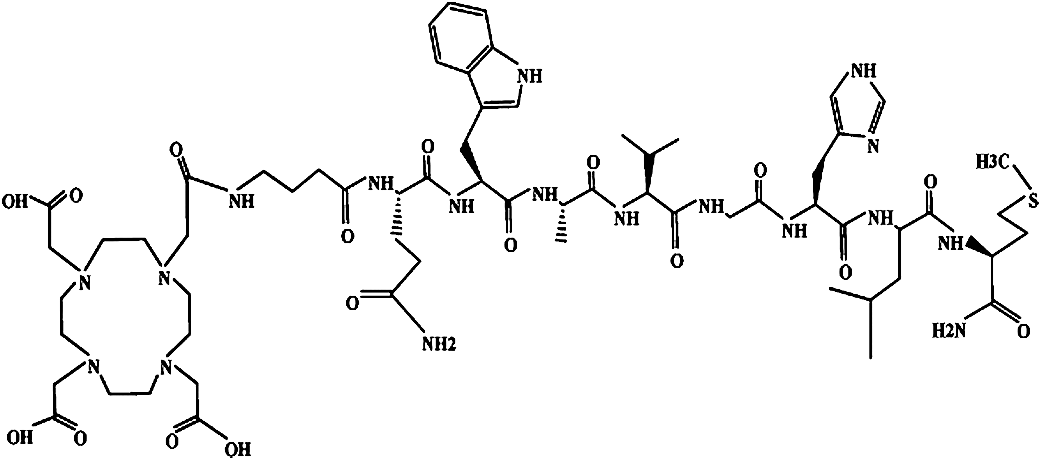
Structure of DOTA-GABA-BBN (7–14) NH2, which could be labeled with 67Ga. BBN, bombesin.
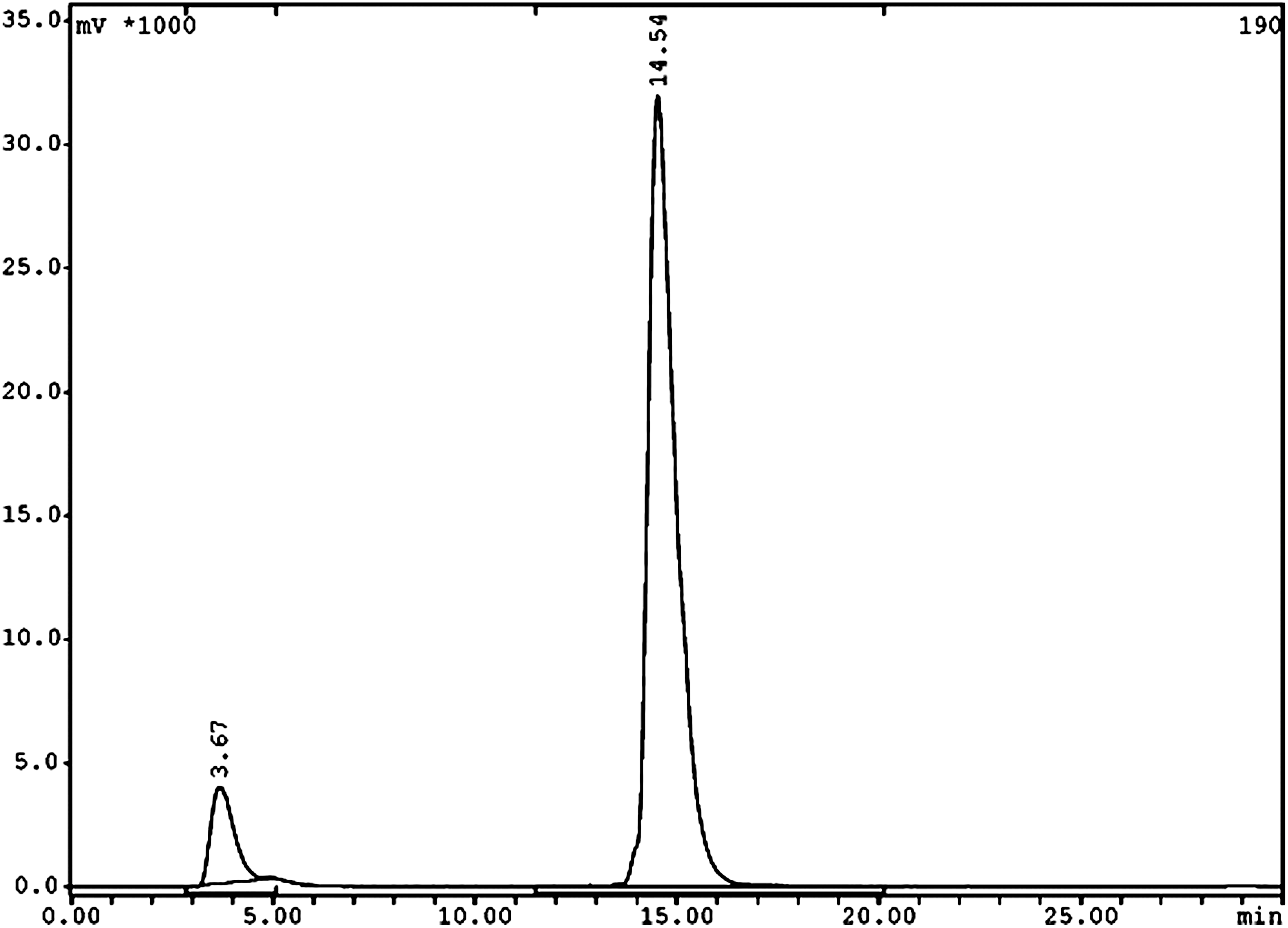
RP-HPLC profile of the [67Ga]-DOTA-GABA-BBN (7–14)NH2. 67GaCl3 and labeled peptide have retention times of 3.67 and 14.54 minutes, respectively.
In vitro internalization and stability
Figure 3 shows the result in respect of the time-dependent and specific internalization of the radioligand. During 60 minutes, the radioligand showed 5.48% ± 0.95% specific cell uptake, which increased to 16.13% ± 0.71% up to 4 hours (p < 0.05). In all experiments, the internalization was strongly reduced in the presence of excess cold (p < 0.05). In fact, nonspecific internalization was 0.56% ± 0.21% after 4 hours, and the surface-bound peptide (acid removable) was 1.1% ± 0.52% of the added activity after 4 hours.
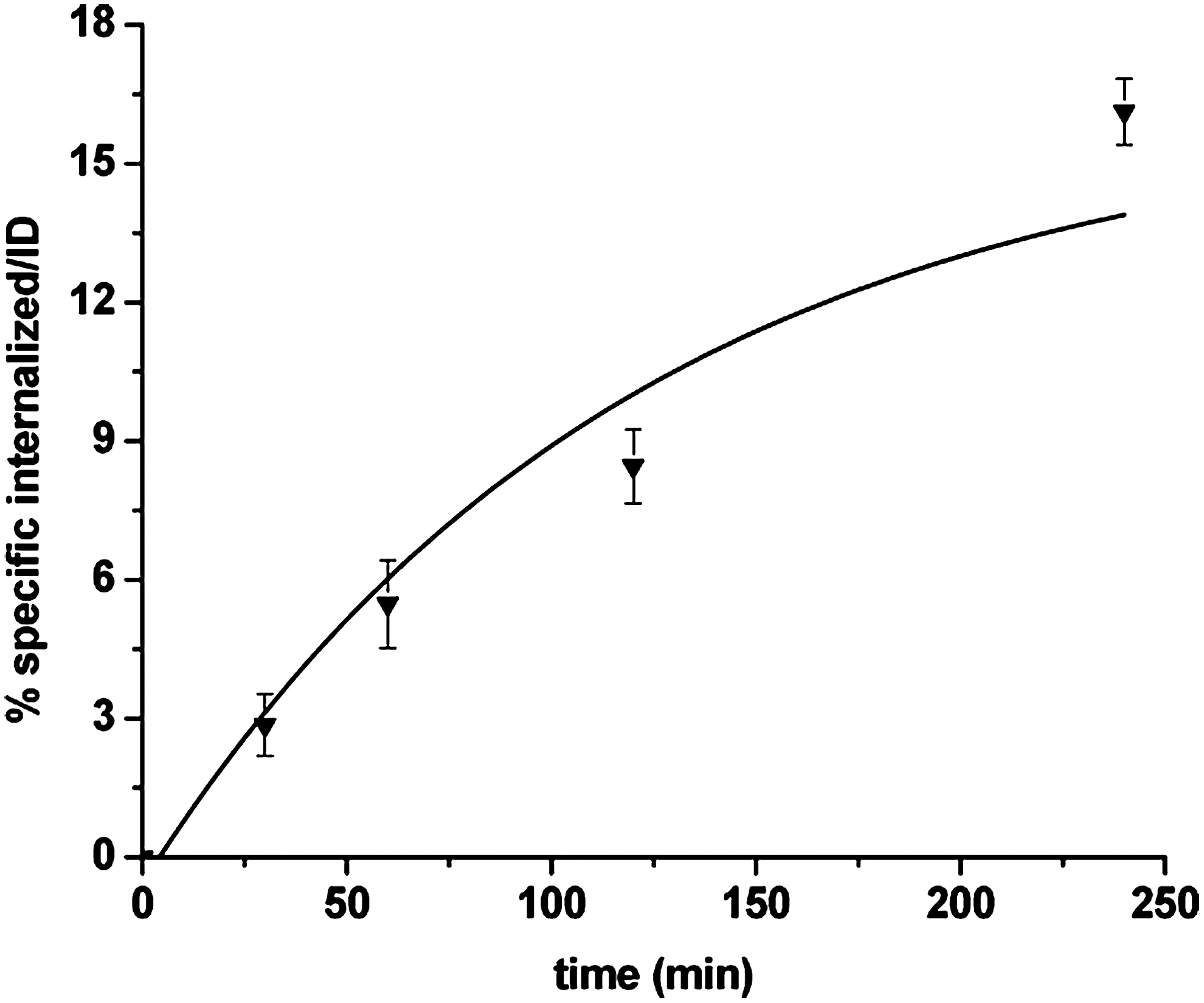
Internalization rate of [67Ga]-DOTA-GABA-BBN (7–14) NH2 into PC-3 cells. Data are from three independent experiments with triplicates in each experiment and are expressed as specific internalization.
In stability study, the protein fraction obtained in sedimentation form contained 9.2% ± 1.5% of radioactivity. Up to 16 hours incubation in human serum, the radiochemical purity remained 90% ± 2.3%.
Externalization
After 15 minutes, for a 2-hour internalized radioligand, 14.77% ± 1.41% of activity was externalized (85.23% ± 1.41% remained), which increased to 52.42% ± 1.86% at 4 hours (p < 0.05). With more time, the percentage of externalization reached a plateau (Fig. 4).
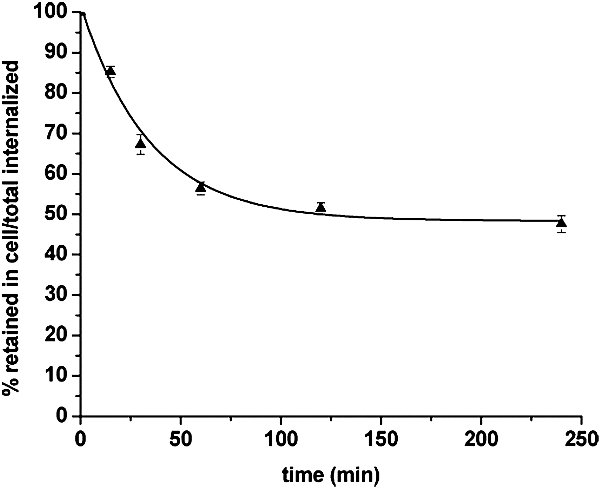
Externalization over time for [67Ga]-DOTA-GABA-BBN (7–14) NH2 in PC-3 cells. Data result from two independent experiments with triplicates in each experiment and are expressed as a percentage of total internalized amount.
Animal biodistribution
[67Ga]-DOTA-GABA-BBN (7–14) NH2 displayed rapid blood clearance with 0.1% ± 0.02% ID/g at 4 hours (Table 2). Fast clearance from the GRP receptor-negative tissues except the kidneys was found as well. Labeled peptide showed high uptake values in the PC-3 tumor and in the GRP receptor-positive organs. By blocking the receptor, the uptake in tumor and pancreas diminished, confirming the specificity of radioconjugate. The percentages of reduction uptake were 76% (1.3% ID/g vs. 0.3% ID/g at 4 hours [p < 0.05]) and 91% (1.21% ID/g vs. 0.1% ID/g at 4 hours [p < 0.05]), respectively (Fig. 5). On the other hand, the uptake reduction in nontargeted tissues due to blocking dose was not significant.

Biodistribution of [67Ga]-DOTA-GABA-BBN (7–14) NH2 in PC-3 tumor-bearing mice 4 hours after injection. Nonblocked (4h) and blocked (4h block).
Data are presented as % injected dose per gram organ ± SD, n = 3.
Discussion
The successful application of radiolabeled somatostatin analogs in nuclear medicine for diagnostics and therapy of neuroendocrine tumors has stimulated the research in receptor targeting of additional tumor types. 22 GRP receptors were shown to be overexpressed on a variety of human tumors such as breast and prostate cancer. Based on this fact and the experience with other peptides such as somatostatin and ubiquicidin, 29,30 we concluded that targeting the GRP receptor with an optimized analog of BBN is very important for scintigraphy of prostate and breast tumors.
According to the published data, the direct coupling of the chelate to the peptide results in a significant loss of binding affinity to the GRP-receptor; the introduction of a spacer between the peptide and the chelate seems to be necessary for retaining the biological properties of the nonchelated peptide. Hoffman and his coworker synthesized a series of DOTA-X-BBN (7–14) NH2 analogs containing no spacer (0) and 3, 5, 8, and 11 carbon spacers. Their results demonstrated that analogs containing spacers with 3, 5, and 8 carbons bind to PC-3 cells with high specifity and affinity, whereas for spacers with 0 and 11 carbons the binding affinity 100-fold decreased. Previous studies carried out by the same group and based on [105Rh]-S4-X-BBN (7–14) NH2 and [99mTc]-N3S-X-BBN (7–14) NH2 show that increasing the hydrophobicity of the linker group to excessive levels will reduce the receptor binding affinity. 31 –33 In another study, the results for binding of different spacer groups such as hydrocarbon, ether, and aromatic moieties suggest that the aromatic linking group has more tumor retention than hydrocarbon or ether linking groups but the former showed higher hepatobiliary retention. In studies by Hanwen et al., they used a 4 carbon spacer (GABA) between DOTA and peptide and reported that metallated DOTA-GABA-[D-Tyr 6 , βAla 11 , Thi 13 , Nle 14 ] BN (6–14) (BZH2) was shown to bind to GRP- and NMB-receptors with high affinity. Based on this knowledge and our previous experience with BBN analog [99mTc]-HYNIC0, D-Tyr6, D-Trp8] BBN (6–14) NH2, 28,29 in the present study, we introduced a 4 carbon spacer into the prototype DOTA-X-BZ (7–14) NH2, which could be labeled with hard Lewis acid type metallic radionuclides such as 111In, 90Y, 177Lu, 67Ga, and 68Ga.
DOTA (1,4,7,10-tetraazacyclododecane-1,4,7,10-tetraacetic acid) was chosen as a chelator, as it chelates a large number of radiometals with high in vitro and in vivo stability. 34 DOTA chelator enables labeling with 111In and 67Ga for SPECT, 68Ga for positron emission tomography imaging, and 177Lu and 90Y as a beta emitter for radiotherapy of gastrin releasing peptide receptor positive tumors. In addition to the above-mentioned features, an increased GRP receptor affinity and tumor uptake with replacement of DOTA instead of DTPA as a chelator in [pro1, tyr4] BBN analog has also been reported. 35
The biological activity of [67Ga]-DOTA-GABA-BBN (7–14) NH2 was determined through internalization and efflux studies in PC-3 cells. GRP receptors belong to G-protein–coupled receptors groups, which, after agonistic binding, go through endocytosis and internalization of the complex. 12,21,22,29 High rate of internalization was observed in our compound (16.13% ± 0.71% up to 4 hours), which was not unexpected, as BBN (7–14) NH2 sequence offers agonistic property to compounds. In addition to efflux curve of [67Ga]-DOTA-GABA-BBN (7–14), NH2 in PC-3 cells after 2 hours of internalization showed an acceptable intercellular trapping. Previous studies with [111In]-DOTA-8-Aoc-BBN (7–14) NH2 12,36 and [111In]-DTPA or [111In]-DOTA-GABA-[D, Tyr 6 , β-Ala11, Thi13, Nle14] BN (6–14) [BZH1 and BZH2] 7 also demonstrated internalization and receptor mediated trapping of labeled compounds but due to differences in sampling method and times, direct comparison of them was not possible. 12 In contrast to our pervious works, this new analog showed higher rate of internalization compared with [99mTc/tricine/HYNIC0, D-Tyr6, D-Trp8] BN (6–14) NH2 after 4 hours in PC-3 cells (16.13% ± 0.71% vs. 10.7% ± 1.2%). 28 It could be due to the replacement of a three carbon chain spacer (GABA) instead of D-Tyr6. By placing the bifunctional chelating agent farther from the receptor binding region in BBN peptide, the negative effect of the chelator on receptor binding has been reduced. It has been shown that the positive charge in sequence of the peptide tends to interact faster and in a strong way with proteins; moreover, it is not targeted to a specific receptor. 37 The cell internalization was receptor specific as was demonstrated with uptake results in gastrin releasing peptide receptor blocked cell experiments, which indicate the balance of charge for the complex.
In suitability of a radiopeptide for diagnostic or specially targeted radiotherapy, an important aspect is stability in human serum, which allows a high concentration of intact radiopeptide for binding with receptors. Our BBN analog showed metabolic stability in human serum up to 16 hours after labeling and incubation. Results from Hanwen et al. 7 show relatively low metabolic stability for [111In]-BZH1 and [111In]-BZH2. They found two degradation sites in their peptides sequences, one between β-Ala11 and His12 and another between Gln7 and Trp8. Also, the study by M. de Visser et al. 35 has shown that changes in the BBN amino acid sequence can have a marked effect on the peptides stability. They found that substitution of native amino acids in BBN sequences can enhance receptor affinity but not the serum stability. As the previous studies by different groups show, the attribution of various factors could affect the in vivo stability of peptide, which requires more studies in the future.
In biodistribution studies, clearance from the blood circulation was fast with <0.13% ID/g remaining in the blood at 4 hours, and the whole body clearance proceeded via the urinary system. Clearance from GRP receptor negative tissues was also rapid except from the kidneys. Accumulation of radiopeptide in BBN receptor positive tissues such as the pancreas, the stomach, the intestines, and the xenografted tumor was observed. Tumor to the pancreas ratio and tumor to the kidney ratio were higher than reported for [111In]-DOTA-8-AOC-BBN (7–14) NH2 36 (1.07 and 0.65 vs. 0.05 and 0.51 respectively), which could be due to the balance in lipophilicity for our compound. In comparison with our pervious study, 28 uptake of radioactivity in the pancreas increased as the lipophilicity of the conjugate increased with the replacement of a four carbon chain spacer instead of D-Tyr6 (1.55% ± 0.17%ID/g vs. 1.04% ± 0.11%ID/g at 1 hour). The uptake in the tumor, pancreas, adrenals, and intestine was specific and receptor mediated, as shown by the coinjection of cold peptide, indicating that these organs are also GRP receptor positive.
The choice of radionuclide also has an important role, whereas the change in the M3+ radiomethal used for DOTA labeling could alter the biodistribution of a DOTA conjugated peptide. 38 –41 Jochen Schuhmacher and coworker found about 20% reduced uptakes and retention of 177Lu-BZH3 in the tumor in comparison with [67Ga]-BZH3. 22,38 Also, it has been demonstrated that gallium 67Ga and [68Ga]-DOTA-octapeptides have distinctly better preclinical pharmacological performances than 111In-labelled peptides, especially on SSTR2-expressing cells and the corresponding animal models. 41 Accumulation in the tumor and pancreas along with demonstrating good pharmacokinetic properties such as low tendency to accumulate in the liver and intestine and also high kidney excretion due to its moderate lipophilicity are the major advantages of our radioconjugate. The ability of DOTA-GABA-BBN (7–14) NH2 for labeling with β emitter radionuclides such as 90Y and 177Lu and its important role to formulate a useful therapeutic GRP receptor targeting radiopharmaceutical is another advantage.
Conclusions
In this study, we have shown the synthesis and radio labeling of DOTA-GABA-BBN (7–14) NH2. 67Ga labeled peptide prepared with high yield at an acceptable specific activity of 2.6 MBq/nmol, which may be used as SPECT imaging agent. The radiolabeled conjugate was able to internalize in GRP receptor positive cancer cells. The prepared conjugate showed accumulation in tumor and pancreas as positive GRP receptors targeted tissues followed by excretion via the kidney. These promising characteristics make our new designed labeled peptide conjugate a very suitable candidate for diagnosis of GRP receptor positive tumors in nuclear medicine.
Footnotes
Acknowledgments
This work was funded by Nuclear Sciences and Technology Research Institute. The authors wish to thank Dr. Rajabi, Dr. Roshanzamir, Mr. Shafiei, Mr. Mirfallah, Mr. Mazidi, and Mr. Talebi of the radioisotope department (AEOI) for providing 67GaCl3 and assistance in quality control tests.
Disclosure Statement
The authors declare no conflict of interest. Animal rights: The institutional and international guide for the care and use of laboratory animals was followed. See