
Abstract
The purpose of this study was to investigate the feasibility and optimization of protocols using superparamagnetic iron oxide (SPIO) nanoparticles to label human prostate cancer cell lines PC3 in vitro. The PC3 cells were labeled with different concentrations (28–252 μg Fe/mL) of SPIO and increasing incubation time (6–24 hours), in the presence or absence of a transfection agent poly-
Introduction
The field of molecular imaging has recently seen rapid advances in the development of novel contrast agents and the implementation of insightful approaches to monitor biological processes noninvasively.
1
In particular, superparamagnetic iron oxide (SPIO) nanoparticles have demonstrated their utility as an important tool for enhancing magnetic resonance (MR) contrast, allowing researchers to monitor not only anatomical changes but also physiological and molecular changes.
2
–4
Although macrophages exhibit a high rate of spontaneous iron oxide uptake, for nonphagocytic cells, spontaneous SPIO particles uptake is generally low. Various approaches for magnetic nonphagocytic cell labeling have involved the synthesis and modification of SPIO particles through linking to lectins,
5
HIV transactivator transcription peptide,
6
or monoclonal antibodies.
7
These labeling strategies do increase the labeling efficiency of SPIO nanoparticles. However, they have potential disadvantages in terms of technical complexity, species specificity, and biosafety. Recently, a convenient method to improve SPIO internalization has been developed using polycationic transfection agents, which are explored as a vehicle for intracellular iron oxide delivery in the cells by endocytosis and pinocytosis through electrostatic interactions.
8,9
Among those transfection agents, high-molecular-weight poly-
Materials and Methods
Cell culture
Human prostate-derived cell line PC3 was obtained from the American Type Culture Collection (ATCC, Rockville, MD). The PC3 cell line was androgen receptor-negative, androgen-insensitive human prostate cancer cell line, which can be grown as xenografts in nude mice. PC3 cells were cultured in F-12 medium (Invitrogen Life Technologies, Gaithersburg, MD) supplemented with 10% fetal bovine serum (Invitrogen Life Technologies) at 37°C in 95% air/5% CO2.
Superparamagnetic iron oxides
Ferucarbotran (SHU 555 A, Resovist®; Schering AG, Berlin, Germany) is a clinically established contrast agent for MRI. It consists of SPIO nanoparticles (average diameter, 65 nm; core size, 3–5 nm) coated with carboxydextran. One (1) milliliter of solution contains 540 mg ferucarbotran, corresponding to 0.5 mmol (28 mg) iron.
Transfection agents
PLL (catalog No. P1524; Sigma, St. Louis, MO) (molecular weight, >388,000; cell culture grade) was used as the transfection agent in the present study, and a stock solution was made at a concentration of 1 mg/mL in distilled water and kept frozen at −20°C until required. For experimental usage, PLL was diluted to 0.1 mg/mL as recommended.
Preparation of the Resovist–PLL complex
Resovist was put into a 3-cm round culture plate containing complete medium and then PLL was added to the solution. The volume ratio of Resovist to PLL was 1:7.5 and the corresponding mass ratio of Resovist (iron) to PLL was 1:0.03 μg/mL. Then, the solution containing Resovist and PLL was allowed to mix for 60 minutes at room temperature on a rotator shaker (QILINBEIER TS-100 orbital shaker; Kylin-bell Lab Instruments Co., Ltd., Jiangsu, China).
SPIO labeling of PC3 cells
PC3 cells were seeded in 24-well plates and placed in an incubator. When the cells reached 80%–90% confluence on the surface area of culture plate, the old medium was discarded, and the fresh complete medium containing Resovist–PLL complexes was added. The cells were then incubated with increasing concentrations of SPIO-PLL complexes (equivalent to 0–28/0.75–42/1.13–84/2.25–126/3.38–252/6.75 μg/mL) for different incubation periods (6–12–24 hours). Further, to evaluate the effect of PLL on labeling efficiency, some other cells were incubated with aforementioned different iron concentrations of SPIOs in the absence of PLL for 24 hours.
Determination of labeling efficiency
SPIO labeling efficiency was determined by manual counting of Prussian blue-stained and unstained cells. After incubation in SPIO-containing medium, cells were washed three times in phosphate-buffered saline (PBS) to remove any free SPIO, then were fixed with 4% paraformaldehyde for 20 minutes, washed with distilled water for two times, acted with Perls reaction liquid (4% potassium ferrocyanide/6% HCl, 50:50 [vol/vol]) for 20 minutes, then washed with deionized water for two times, restained by 0.5% eosin for 3 minutes, and observed under a light microscope (Olympus IX51; Olympus, Tokyo, Japan) at 200×magnification. The cells were considered Prussian blue positive if intracytoplasmic blue granules were detected. The percentage of labeled cells was determined from the average of 5–10 high-powered fields.
Cellular viability test
The viability of SPIO-labeled cells was evaluated using trypan blue dye exclusion test. After incubation with different concentrations of SPIO-PLL for 24 hours, the cells were collected and labeled with 0.4% of Trypan Blue solution (Sigma-Aldrich Co., St. Louis, MO), which is excluded by live cells but accumulates in dead cells. Enumeration of viable and dead cells was carried out using a light microscope with a hemocytometer at 200×magnification (Olympus IX51; Olympus).
MRI of labeled cells
SPIO-labeled cells were thoroughly washed with PBS to ensure complete removal of unbound SPIO. For MR detection, 1 mL of SPIO-labeled and unlabeled control cell suspensions with a cell density of 2×10⁵ cells/mL were added into 1.5-mL Ependoff pipes. The pipes were then put into a water tank for the in vitro MRI, which was performed using a 3 Tesla (T) MR unit (Signa HDxt; GE Healthcare, Milwaukee, WI) with eight-channel phased array brain coil. Transversal images were acquired using a fast spin echo (FSE) T2WI sequence with repetition time (TR)=4360 ms, echo time (TE)=201 ms, field of view (FOV)=80 mm, matrix size=512×224, slice thickness=2 mm, and number of excitation (NEX)=2.0. Eight contiguous slices were acquired to image the whole hypointense region attributable to the presence of labeled cells. Region of interest (ROI) analysis of signal intensities (SIs) was performed using a circular 30-mm2 ROI.
Statistical analysis
Statistical analysis was performed with SPSS 12.0 (SPSS, Chicago, IL). All the data were presented as means±standard deviations. Two-tailed Student's t-test and Mann–Whitney test were employed to compare viability of labeled and unlabeled cells and labeling efficiency. Pearson correlation coefficients were calculated to characterize (1) the relationship between labeling efficiency, SPIO concentration, and incubation time and (2) the relationship between MRI signal and number of SPIO-labeled cells. Statistical significance was accepted with a p-value of <0.05.
Results
Cell labeling and Prussian blue staining
The results of Prussian blue stains of PC3 cells labeled with increasing concentration of SPIO-PLL for different incubation times are presented in Table 1. The incorporation of SPIOs by PC3 cells was dependent on dose and time. There was a positive linear correlation between SPIO uptake, the incubation time, and the iron concentration in the culture medium (correlation coefficient ranging between 0.76 and 0.99; p<0.05) (Fig. 1). Blue cytoplasmatic inclusions with typical perinuclear localization were present only in cells incubated with the SPIO. At the lowest doses and shorter incubation times, few Prussian blue-labeled cells were recognized among the cell population. As the concentration of SPIO nanoparticles and the incubation time increased, the amount of intracellular iron increased, resulting in heavy labeling in almost all the cells. SPIO-PLL concentration in the range of 42–126 μg Fe/mL with 12–24 hours incubation time could effectively label 85%–100% of the PC3 cells. With the iron concentration as low as 28 μg Fe/mL, the labeling efficiency was only 19.4% after incubating for 12 hours. When prolonging the incubation time to 24 hours, the labeling efficiency could increase to 83.1%; however, the intracellular iron of these cells was relatively less than that of other effectively (>80%) SPIO-labeled cells. On the other hand, although the doses up to 252 μg Fe/mL with 12–24 hours incubation times could result in 100% labeling efficiency, the exorbitant iron concentration made large extracellular blue-stained particles, which were difficult to eliminate from the culture plate and the external surface of the cells with simple cell washes.

Photomicrographs show Prussian blue staining of PC3 cells treated with different incubation condition. No blue dots are observed in the untreated PC3 cells
p<0.01 versus the comparably treated cells incubated with PLL for 24 hours.
PLL, poly-
Table 1 also demonstrates the results of Prussian blue staining of PC3 incubated with SPIO alone or with SPIO-PLL complexes for 24 hours. The cellular iron uptake with PLL was significantly increased than that without PLL in each iron concentration (p<0.01). When the cells were incubated with SPIO alone, cellular labeling was low or not detectable on Prussian blue staining (Fig. 1). However, when PC3 cells were incubated in the simultaneous presence of SPIO and PLL, the increased numbers of detectable labeled cells and of cells displaying high density of blue cytoplasmic inclusions could be observed, which reflected higher levels of intracellular iron accumulation (Fig. 1).
Cell viability determined by the trypan blue dye method
Figure 2 demonstrates that the cell viability of SPIO-labeled PC3 with a concentration of 0, 28, 42, 84, 126, and 252 μg Fe/mL were 90.93%±1.39%, 90.83%±1.87%, 91.13%±1.04%, 90.61%±1.61%, 89.91%±1.68%, and 77.41%±2.81%, respectively. The cellular viability of PC3 cells labeled with SPIO at 252 μg Fe/mL was significantly lower than that of the unlabeled PC3 cells (p<0.01). However, no statistically significant decrease in viability was observed for the labeled PC3 cells with SPIO nanoparticles at a concentration of ≤126 μg Fe/mL, compared with unlabeled cells (p>0.05).
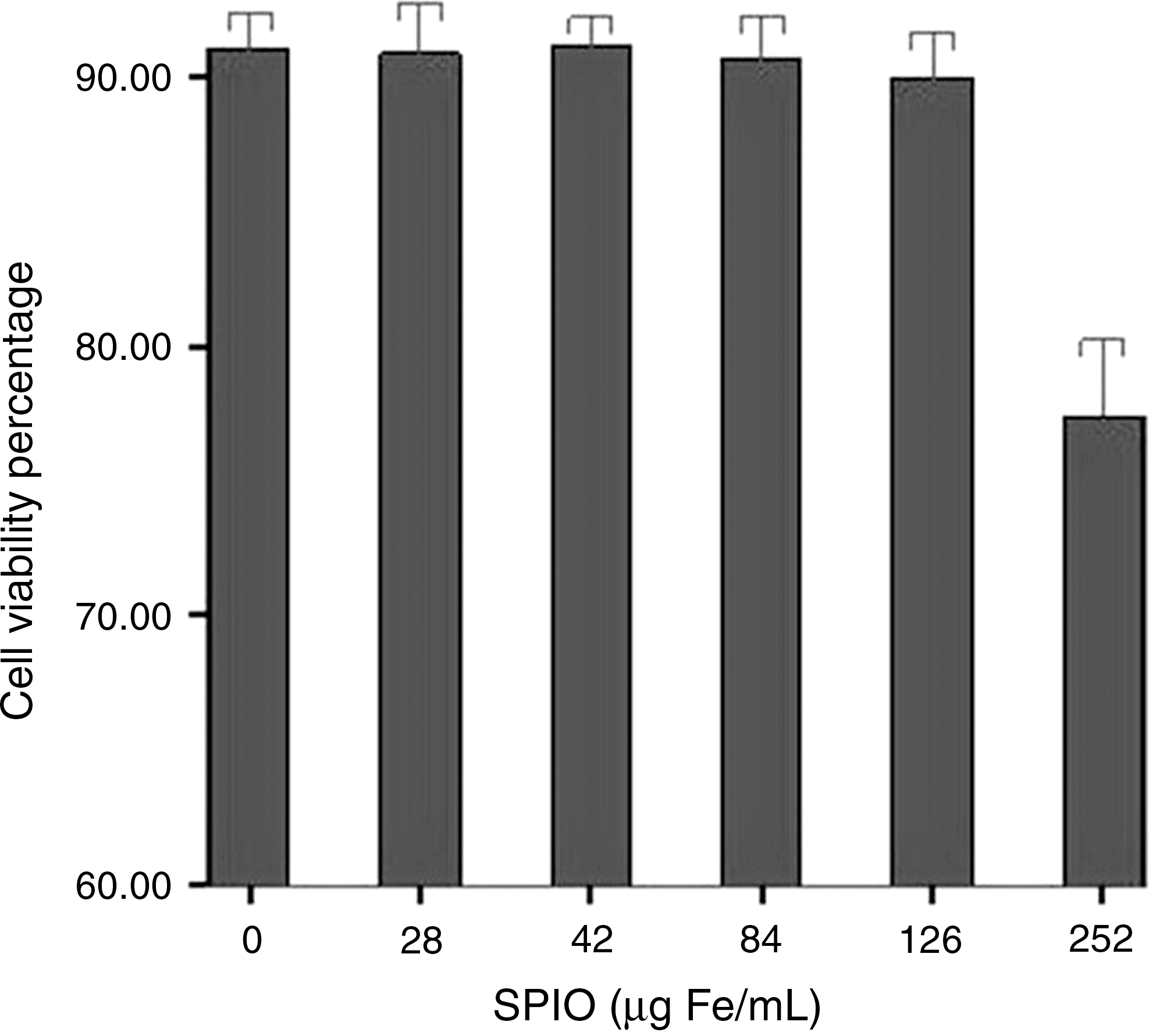
Results for the viability of SPIO-labeled cells with a concentration of 0, 28, 42, 84, 126, and 252 μg Fe/mL for 24 hours, obtained by the trypan blue dye exclusion method, expressed in percentages (bars represent mean value and error bars show standard deviation).
In vitro MRI of SPIO-labeled PC3 cells
FSE T2-weighted images of cross-sections through the 1.5-mL EP pipe revealed the presence of a hypointense signal (Fig. 3). The SI of the labeled cells with different incubation conditions is summarized in Table 2. Unlabeled cells were not detectable in MR images and its SI was 2750.3±220.1. The number of SPIO-PLL–labeled cells correlated well with the hypointense pixels in the MR images and the Pearson correlation coefficient was 0.82 (p<0.01). In most cases, MRI could efficiently detect the SPIO-labeled PC3 cells with a labeling efficiency of >80%. However, the SI of SPIO-labeled cells incubated with 28 μg Fe/mL showed no significant difference between that of the unlabeled cells, although its labeling efficiency was 83.1%. For SPIO-labeled PC3 cell incubation without PLL, the SI of cells treated with 252 μg Fe/mL was lower than that of unlabeled cells (p<0.05), although its labeling efficiency was less than 80%.
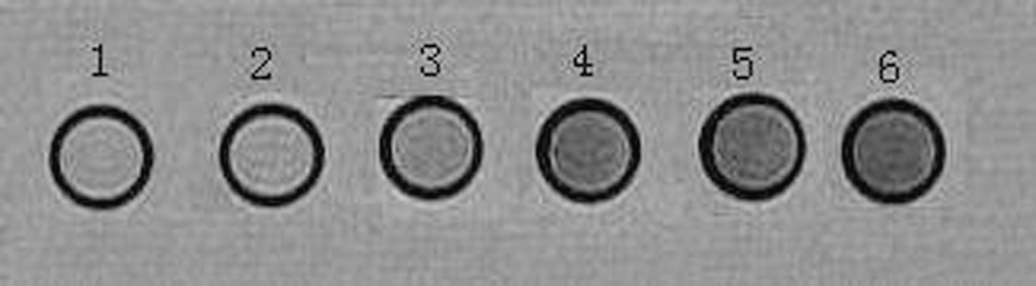
Magnetic resonance image of magnetically labeled PC3 cells. 1, Unlabeled cells; 2–6, labeled cells treated with 28, 42, 84, 126, and 252 μg Fe/mL for 24 hours, respectively.
p<0.05 versus signal intensity of unlabeled cells=2750.3±220.1.
Discussion
Molecular imaging can be broadly defined as the in vivo characterization and measurement of biologic processes at the cellular and molecular levels. The in vivo localization and tracking of specific types of cell is one of the main challenges of molecular imaging. Because of its high spatial resolution, MRI appears ideally suited for imaging the biodistribution of magnetically labeled cells. SPIO particles have been used to label these cells to provide future possibilities to monitor the cell migration and proliferation by MRI noninvasively. Researches about SPIO labeling of embryonic, mesenchymal, and hematopoietic stem cells were abundant in the literature, and the protocols to label these cells with SPIO had been systematically described. However, a detailed documentation of protocol to label human prostate cancer PC3 cell lines with SPIOs did not come forth. The appropriate concentration of SPIOs and incubation time for the labeling of PC3 cells remained dubious. Under such ground, this study was carried out to explore the efficiency of different SPIO concentrations and incubation time in in vitro labeling of PC3 cells as well as the influence on cellular viability.
The present study demonstrated that the incorporation of SPIO by PC3 cells in the presence of PLL was dependent on dose and time. There was a positive correlation between SPIO uptake and both iron concentration and the incubation time. The results were in line with previous studies on human precursor cells 12 and hematopoietic stem cells. 17 In the labeling protocol of the present study, a final concentration of SPIO nanoparticles at a dose of 42–126 μg/mL with 12–24 hours incubation times could result in efficient (>80%) cell labeling, without impairment of cell viability. The number of Prussian blue-stained cells increased with increasing concentration of iron oxide in the incubation medium at a fixed incubation time. Likewise, prolonging the incubation time did improve the intracellular iron uptake at the same concentration of SPIO particles. For the cells incubated with an iron concentration as low as 28 μg/mL, its labeling efficiency was only 19.4% even after extending the incubation time to 12 hours. However, with prolongation of the incubation time to 24 hours, the percentage of magnetic-labeled cells could also reach 80% or above, and relatively fewer iron oxide nanopaticle uptake was observed in these cells. When the cells were cultured with SPIOs at a dose of 252 μg/mL, the labeling efficiency could reach 100% after incubating for 12 hours. Under the microscope, a lot of aggregates of SPIO-PLL particles were observed both on the external surface of cells and on the bottom of culture plate and were difficult to clean with PBS washes. Further, the morphology of some cells was changed. The trypan blue dye exclusion test showed that the viability of those cells was significantly lower than unlabeled cells. Emerit et al. 18 had pointed out that the increase of iron in cells might result in an increased production of reactive oxygen species because of iron-mediated free radical formation resulting in damage to DNA in the nucleus. Neri et al. 12 observed important cell death and impairment in neurosphere-forming ability in the heavily labeled human neural precursor cells soon after the first postlabeling subculturing passage, which confirmed the theory that the intracellular free iron overload eventually results in functional impairment and cell death. 19 In addition, PLL, as a polymer with great molecular weight, could result in a loss of cell viability with certain range of dosage. Arbab et al. 11 reported that PLL was toxic to cells at a concentration above 7.5 μg/mL. However, when PLL was complexed with SPIOs, there was a clear shift in the maximal tolerated concentration by the cells before significant cell death. Arbab et al. 16 demonstrated in another research that a ratio of ferumoxides to PLL between 1:0.03 and 1:0.05 μg/mL results in no free PLL in the medium, thereby not affecting cell viability. But, an increase in cell death could be observed when PLL was added in 10-fold or more excess to the amount needed to complex with ferumoxides (ratio of iron to PLL >1:0.3 μg/mL). Based on the previous studies as well as the highest concentration of PLL (6.75 μg/mL) and the fixed ratio of iron to PLL (1:0.03 μg/mL) applied in the present study, the increase in cell death was presumably due to iron overload of the cells and not due to free uncomplexed PLL in the medium.
It has been previously reported that PLL might improve the efficiency of SPIO intracellular uptake. The labeling efficiency of SPIO alone was low, because the carboxydextran coating for Resovist is an anionic material, that is, the surface charge of the cell membrane is negative and thus anionic nanoparticles have repulsive interaction with the proteins of the cell membrane. On the contrary, the PLL is positively charged. Complexing of PLL and SPIO occurs through electrostatic interactions, then allowing for highly efficient incorporation of iron oxide nanoparticles into endosomes of magnetic cell labeling. Song et al. 13 observed that with using PLL, the iron content in the ferumoxide-labeled, MION-47–labeled, and CLIN-NH2–labeled cells increased to 27-fold, 202-fold, and 7-fold uptakes, respectively, compared with the comparably treated cells without PLL. In this series, the transfection agent PLL significantly increased the iron load of cells. The incubation of iron nanoparticles with PLL resulted in not only an increase in the number of labeled cells but also more efficient accumulation and detection of SPIO particles in the cytoplasm, with no increase in toxicity, which led to the conclusion that PLL could play an important role in labeling efficiency. Therefore, the present study's results seem to reconfirm the previous reports that SPIO-PLL complexes can be easily taken into cells. 12,13,16,17
SPIOs, such as Resovist, are a class of MRI contrast agents used for in and ex vivo cellular MRI. The SPIOs generate strong T2-negative contrast in MRI and, therefore, increase the 1/T2 of tissue and decrease SI. 1,4 FSE T2WI sequence is commonly used for both SPIO-labeled cellular MRI 9 and clinical SPIO-enhanced MRI. 20 In the present study, the SPIO-labeled PC3 cells also could be efficiently demonstrated by FSE T2WI, and their SI was changed with the iron concentration and the incubation time, which resulted in different labeling efficiency. In most cases, MRI allowed to achieve excellent detection of SPIO-labeled PC3 cells at a labeling efficiency more than 80%. However, the SPIO-labeled cells incubated in a 28 μg Fe/mL-containing medium for 24 hours with a labeling rate of 83.1% failed to be detected on MRI, which was supposed to be related to the observation through the light microscope that the iron within each cell in this incubation condition was relatively less when compared with that in other cells detectable on MRI. The result indicated that the intensity of MRI signal correlated not only with the numbers of labeled cells but also with the intracellular accumulation of nano-iron particles. The result also demonstrated that the SPIO-labeled cells incubated with 252 μg Fe/mL plus or minus PLL all allowed to achieve excellent detection on MRI, even with one case of them having a labeling efficiency of 59.7% only. Considering the fact that many blue-stained aggregates could be observed on the culture plate and the external surface of the cells, it was supposed that the low SI on MRI was generated as a result of these extracellular complexes, which impeded the accuracy of quantitative MRI analysis. Pawelczyk et al. 21 also observed from their research that the cells incubated with excessive iron concentration would lead to large FE-Pro complexes that would attach to the outer cellular surface and remain extracellular, thereby causing interference when monitoring the migration of labeled cells by MRI.
Conclusions
Human prostate cancer PC3 cells could be easily and safely labeled with SPIO-PLL complex. A final concentration of Resovist of 42–126 μg/mL and 12–24 hours incubation times could be sufficient to label cells. The range of iron concentration resulted in no toxicity to the cells and dramatically reduced SI on MRI. A simple and efficient protocol for labeling PC3 cells with SPIO-PLL complexes was developed to be used for monitoring the growth and dissemination of cancer cells in vivo by MRI, which would represent a potentially powerful technology in the development of effective prostate cancer therapies.
Footnotes
Acknowledgments
Financial support from the Science and Technology Commission of Shanghai Municipality Foundation (Nos. 10JC1411400, 09411963800 and 10411952000) and Shanghai University Foundation (jdy 10120) is gratefully acknowledged.
Disclosure Statement
The authors declare that no conflicts of interest exist.