
Abstract
A facile, viable, “green” two-step, inexpensive technique was developed for the preparation of 32P patch for the treatment of skin cancer. This technique consists of impregnation of H3 32PO4 solution into an inert solid carrier followed by immobilization into a nonleachable matrix by lamination. The morphology of the impregnated paper was evaluated by scanning electron microscope and energy-dispersive spectral analyses. Radioactive patches containing up to ∼37 MBq/cm2 of 32P could be prepared. Distribution of 32P on sources was uniform and release of 32P from the sealed source in water and saline was found to be well within the permissible levels of 185 Bq. Custom-shaped 32P-patches after quality assurance were supplied to AIIMS, New Delhi, for clinical evaluation. 32P-impregnated paper protected by a laminated film holds promise for treatment of superficial cancers.
Introduction
Skin cancer is a serious concern among the fair-skinned population, a majority of which are basal cell carcinomas. More than 95% of basal cell carcinomas, the most common form of skin cancer, occur in patients more than 40 years of age. 1 Curative modalities for skin cancer are local destruction, radiotherapy, and surgery. Each therapeutic modality has its own advantages and disadvantages. Although surgery is usually preferred, recurrence rates are high and it is not always possible to be practiced. Radiotherapy using external beam therapy is often used for treatment owing to the advantage of treating a visible tumor. However, external radiotherapy has certain disadvantages, such as the necessity for expensive radiation therapy units and the adverse effects of penetration into underlying bone and soft tissues. To overcome some of the disadvantages of external beam radiotherapy such as high cost of therapeutic units, radiation protection of patients and paramedical staff, etc., mould brachytherapy using high-energy β− emitters has been explored as an alternative treatment modality for achieving adequate dose delivery to the affected area without causing undesired radiation exposure or injury to the neighboring normal tissues. Compared with external radiotherapy, radionuclide therapy is simpler, less traumatic, and less expensive. The first radionuclide patch for treating skin cancer was produced by bombarding a 165Ho-incorporated patch in a nuclear reactor by Korean scientists. 2 Extending this theme, preparation of skin patches incorporating β−-emitting isotopes such as 90Y and 188Re for treatment of superficial tumors have been reported by the present study group and other research groups. 2 –7 The availability of radionuclides, their physical characteristics, and their logistic advantages are of importance when considering the possibility of a treatment employing them. The limitation of using the aforementioned radionuclides is their limited availability and short half-lives, which are not long enough for wide deployment.
32P has emerged as an important radionuclide for such therapy owing to its physicochemical properties and commercial availability on demand. It is a pure β− emitter with a maximum β− energy of ∼1.7 MeV and the advantage of adequately long half-life of 14.2 days suitable for wide use, and hence, it is a better choice for use in the proposed application. In recent years, specially designed patches containing 32P have been developed for contact brachytherapy of skin lesions. 8 –11 In a previous work, the authors of the present study have reported the preliminary results on the preparation of 32P-based skin patch to test the efficacy of 32P in controlling tumor growth. 12 In this communication, a detailed investigation on the preparation, properties, and quality of 32P-based skin patch has been evaluated.
Materials and Methods
Radioactive 32P as orthophosphoric acid was obtained from the Radiochemicals Section of our Division. For the preparation of 32P patches, different kinds of adsorbent papers were procured from reputed local suppliers. Nontoxic, laminating films were obtained from Hi
Scanning electron microscope (SEM) Model-Vega MV-2300T/40, supplied by M/s TESCAN, was used for SEM analysis. Optical density (OD) measurements of exposed films were carried out using an OPTEL transmission densitometer (Model No. 125). A radiochromic densitometer (3E NDT, LLC. Model-37-443) was also used for OD measurements of exposed GAF-chromic films. Both radiochromic densitometer and GAF-chromic films were procured from M/s Nuclear Associates (a division of M/s Victoreen, Inc.). A GM counter procured from M/s PLA Electro Appliance Pvt. Ltd. was used for radioactivity measurements. A portable laminating unit obtained from M/s Avanti Pvt. Ltd. was used to laminate the 32P-adsorbed paper.
Adsorption of 32P on paper strips
32P as H3 32PO4 solution was adsorbed on Whatman-541 filter paper strips of predetermined sizes. Initial tracer experiments were performed to adsorb the radioactivity on unconditioned as well as on the treated paper strips (preconditioned with phosphoric acid of varying molarities). Circular paper strips of known dimensions (∼25–50 mm) were taken and known volumes of radiochemical containing up to ∼3.7 MBq of 32P was dispensed. Different means of contacting the paper strips with the radioactive substance were tried. The method of applying the 32P in the form of H3 32PO4 to the Whatman-541 filter paper was carried out using a semiautomatic micropipette. To enhance the receptivity of the paper strips for H3 32PO4 solution, a known volume of radiochemical containing appropriate amount of radioactivity was dispensed at the center of the strip. An empirical relationship was established between the volume of the dispensed liquid and the surface area of the strip. Care was taken to mount the strip in such a way that no loss of liquid takes place during spread of dispensed liquid over the paper strip. The strip was subsequently allowed to dry at different temperatures by placing the activity-impregnated filter paper under an infrared (IR) lamp inside a well-ventilated glove box. The source to lamp distance was suitably maintained to obtain the desired drying temperature. In addition, various experiments were carried out, in which the paper adsorbed with known weight of 32P solution was dissolved in a suitable medium. Subsequently, after required dilution, the radioactivity associated with an aliquot of resulting solution was counted in a liquid scintillation counter. To prepare sources with higher levels of radioactivity whenever needed, the radioactive solution was concentrated by controlled evaporation in a specially designed quartz container under IR lamp at a temperature of 60°C–70°C. Precalculated volume of 32P in desired concentration (ranging from 7.0 to 10.5 GBq/mL) was used to obtain a source with specific strength of ∼37 MBq/cm2 on the intended date of application.
Source lamination
It is desirable to have a source that is as thin as possible for minimum loss of radiation intensity, with good flexibility and adequate structural integrity, which will prevent cracking of the patch or leakage of activity. For this purpose, lamination was carried out with thermoplastic urethane (TPU) sheets. 32P-impregnated adsorbent papers were immobilized within TPU sheets (varying thicknesses ranging from 25 to 100 μm) with the help of a laminating unit. The optimum thickness of film was used in preparation of final set of sources. Attenuation of radiation by the encapsulating sheets was calculated using the standard value of β− absorption coefficient (i.e., 9.22 cm2/g) for 32P and subsequent determination of intensity of β− radiations was carried out after traversing the distance equivalent to thickness of encapsulating sheet using the knowledge of fundamental attenuation principles.
Surface texture characterization of inactive patches
Prototype sources incorporated with inactive phosphorus were prepared under optimized conditions and were used for energy-dispersive X-ray spectrum (EDS) analysis using SEM. The distribution of elements on the source core was mapped to assess the uniformity of various elements on the source core. Scanning electron micrographs were also obtained to examine the morphology of phosphorus-incorporated paper strips.
Quality evaluation
Determination of source strength
Source strength was indirectly determined by estimating the weight of 32P solution adsorbed on the strip. Experiments were carried out to assess the percentage of activity adhered on the strip, and finally, the average of such values was used for assessment of the radioactive strength of sources. Using the value of radioactive concentration of 32P solution used, the amount of radioactivity transferred onto the paper strip was calculated to estimate the strength of the source prepared.
Uniformity of 32P in the patches
Uniformity of 32P in the sources of different shapes (∼37 MBq/cm2) was checked by autoradiography, by exposing industrial X-ray films with 32P-patches for a predecided duration. Additionally, autoradiographs were also taken with radiosensitive GAF-chromic films by exposing suitably cut portions of the films to 32P patches for a period of 5 hours. At the end of these experiments, OD was measured at various points of exposed films and the variation difference in the values of OD was taken as a measure of variation of distribution of 32P in the source.
Determination of surface contamination
The radioactive patches were tested for absence of loosely held activity (surface contamination) by swiping the sources using cotton wool immersed in alcohol and measuring the radioactivity associated with swipes with the help of a GM counter of known efficiency.
Leachability of 32P from the patches
Leachability of the immobilized 32P patches containing up to ∼37–39 MBq/cm2 of 32P was assessed by determining the release of radioactivity on immersing them in 100 mL of distilled water at room temperature over a period of 48 hours as per the method prescribed by the Atomic Energy Regulatory Board (AERB), India. 13 At the end of these tests, a measured aliquot of test solution was transferred over an aluminium planchette and the radioactivity associated with it was determined with the help of a GM counter of known efficiency. The total activity associated with 100 mL of the test solution was calculated. A similar test was carried out in saline solution for 48 hours at a temperature of 37°C. 13
Results
The present investigation outlines the approach of making 32P skin patches and evaluation of their suitability for therapeutic applications.
Preparation of 32P patch
The insoluble matrix chosen for retaining radioactivity should be porous, inert, and capable of adsorbing liquid. The type of adsorbent used for preparation of source has a marked effect on the reliability of the results obtained. Whatman-541 filter paper was chosen owing to its ability to retain 32P activity from solution phase on account of adequate porosity and sufficient pliability. The initial attempts to impregnate 32P using H3 32PO4 solution on the untreated Whatman-541 filter paper failed because of the nonuniform distribution of deposited activity on the substrate, as indicated by preliminary studies.
It was also established that the use of paper strips preconditioned with 0.1–0.2 M phosphoric acid resulted in more homogeneous distribution of phosphorus on source core in comparison to the cores prepared with unconditioned paper strips. Beyond this concentration, a yellowish tinge was noticed on the surface of strips, which could be indicative of saturation with phosphate species resulting in poor absorption and nonhomogeneity of radioactivity on the source core. In the present study, the empirical relationship established for spread of radioactive liquid on paper strips holds good under practical conditions and ∼5.2 μL of liquid was needed to prepare a source having an area of ∼1 cm2. The concentration of radioactivity plays a role in the spread of radioactivity on paper strips, and whenever concentrated solutions were used, this effect was accounted for. Although the sources could be dried at different temperatures ranging from room temperature to 60°C, drying them in air at ambient temperature was found to be most effective. Attempts to dry the active paper at a temperature more than 40°C resulted in blackening of source strip at few points, which subsequently disintegrated in the course of time. The sources containing ∼37 MBq/cm2 on the intended date of use could be prepared by adjusting the initial radioactive concentration of 32P solution to an appropriate value, and excellent batch-to-batch reproducibility was observed. In the experiments carried out to estimate the radioactivity of dissolved paper, the estimated activity was found to be within±1.2% of the expected values. Additionally, the surface radiation doses of 32P-adsorbed strips adsorbed with same weight of 32P solution was measured and the radiation doses of patches were measured with the help of a radiation dosimeter. The variation in measured radiation doses of sources adsorbed with same weight of 32P solution was found to be within±1.6%. Both these results are well within the uncertainties of measurements, which are assumed to be within±5%.
On account of nonlinearity, β− dose measurements could not be used for estimating the source activity. Hence, weight difference method was used for estimating the amount of activity dispensed on paper strips.
The thickness of TPU laminating sheet was optimized and a thickness of 40 μm was found to be most suitable for sealing the sources. Sources sealed with such films were adequately flexible with acceptable β− radiation attenuation within 3.4%±0.5%, which can be accounted for in the dosimetric calculations. Beyond this thickness, the resulting sealed sources were not flexible enough to cover the areas contoured on the body surface.
Phantom dosimetry studies of the 32P patches were carried out using a specially designed phantom made of polymethyl methacrylate as a substitute of tissue and of aluminium as a substitute of bone. The results of such investigation have been reported. 14 The reported study revealed that the dose descends rapidly toward the tissue and the bone. The dose rate at 4 mm in tissue is nearly the same as that at 2 mm in bone. Also, there was a 79% fall in dose rate at 1 mm and 93% fall at 2 mm with respect to the contact dose of 272.8 mGy/MBq/hour.
SEM analysis
The scanning electron micrograph of inactive phosphorus-impregnated paper obtained under 2500 magnification factor is shown in Figure 1, which indicates that H3PO4 is possibly well accommodated in the porous network of the matrix.

Scanning electron micrograph of inactive patch.
EDS analysis
Further, the EDS of phosphorus-loaded film, as shown in Figure 2, revealed that the incorporation of phosphorus was uniform within the matrix. EDS analysis of inactive sources prepared under optimized conditions revealed that the variation in amount of phosphorus present at different points of the core was within±7.1%. The distribution of various elements on source core is depicted in Table 1.
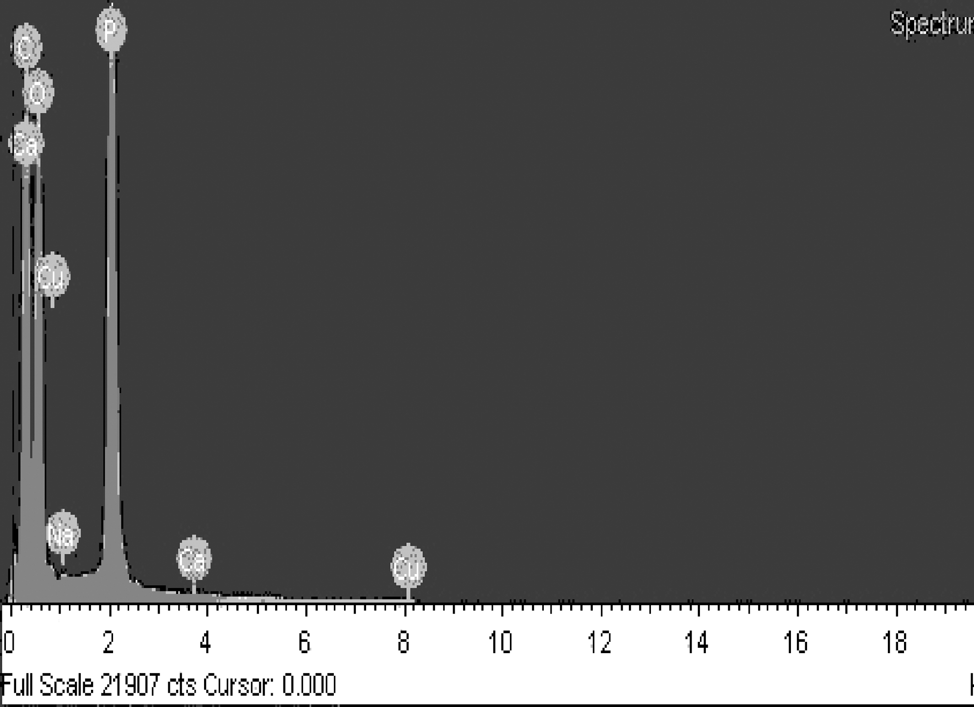
Energy-dispersive X-ray spectra of inactive patch.
SD, standard deviation.
Evaluation of distribution of 32P (autoradiography)
Uniformity of activity in 32P patches was confirmed by determination of OD variations at different points of X-ray films. Industrial X-ray films could be effectively used for taking the autoradiographs of 32P patches and an exposure time of ∼30–45 seconds was found to be optimum for the exposure of film with the source having ∼37 MBq/cm2 of 32P. The autoradiographs of the radioactive sources prepared in custom shapes are shown in Figure 3. The OD of these exposed films at different positions was measured as per the procedure described under the Materials and Methods section. The results are depicted in Table 2. It was observed that the OD variations were within±4.7%, indicating reasonably good homogeneity of 32P on the source. Autoradiographs taken with radiosensitive GAF-chromic films after exposing them for a period of 5 hours also showed good homogeneity of 32P with variations of OD over the exposed film within±2.9%.

Autoradiographs of custom-shaped
Quality assessment
The release of radioactivity from encapsulated source assembly was very low and barely any counts were recorded in the GM counting assembly over a typical counting time of 1 minute. Hence, the test samples were counted over a period of 10 minutes each to assess the extent of release of activity. The a priori knowledge of the counter efficiency was used to arrive at the activity values in Bq from the observed counts. It was observed that there was negligible release of radioactivity, when the sources were immersed in water at room temperature over a period of 48 hours, as well as in saline solutions at 50°C and 37°C, respectively. Surface contamination on the encapsulated patches was also found to be much less than the prescribed permissible level of 185 Bq. The results of leachability, leakage, and surface contamination tests are depicted in Table 3.
BDL, below detection limit.
Sources could be produced in desired shapes in a reproducible manner with desired radioactive strength and could comply with the quality norms in accordance with AERB, India. 32P patches developed by the present study group have also proven their efficacy in bioevaluation studies in C57BL6 mice bearing melanoma. 12
To evaluate the efficacy of patches, 8 patients aged between 45 and 74 years with histopathologically proven unifocal basal cell carcinoma on face were treated with custom-made 32P patch of different shapes and sizes matching the dimension of the lesions at the All India Institute of Medical Sciences, New Delhi, India. 15 After treatment, histopathological examination of biopsies from both center and margins of the lesions showed a negative result for any residual malignant disease in all the patients. Routine hematological and biochemical examinations did not reveal any toxicity. 15 There was minimal scarring of the tumor site and the cosmetic results were excellent.
Discussion
Mould brachytherapy using β−-emitting radionuclide-incorporated patches has been reported as one of the promising alternative therapeutic modalities for topical treatment of skin cancer in areas that are difficult to excise, especially on the face, including eyelids, nose, and lips. This modality received wide attention and appears to be poised for rapid growth. This mode of treatment has numerous advantages as it does not need expensive therapeutic units unlike external beam radiotherapy, and the procedure is simple for preparation as well as application and is noninvasive. The potential benefits of using 32P for such brachytherapy applications are as follows: It has a half-life of 14.2 days, which is adequately long to prepare skin patches at a centralized radiopharmaceutical laboratory and for supply to different destinations without much depletion in the radioactivity. This is of great advantage considering the need for applying the radiation source for brachytherapy more than once at each lesion, to achieve desired radiation dose in fractionated doses over a period of time. In case of 32P, the same patch can be reused over 1–2 weeks time, adjusting the time of exposure to achieve the desired dose, whereas this is practically impossible with radionuclides such as 166Ho, 188Re, and 90Y, which have comparatively shorter half lives than 32P. Its decay mode and emission of β− particles of 1.7 MeV, which have a maximum range in tissue of 8 mm without any γ emission, facilitate in avoiding the radiation dose to the underlying bone and healthy tissue.
Impregnation of H3 32PO4 solution into an inert solid carrier appears to be a feasible process to achieve uniform activity distribution and this simple, fast, effective technique yielded patches of satisfactory quality quickly and satisfactorily. Optimization of the amount of 32P activity to be impregnated in the matrix was desired and can be determined for convenient control of the activity content of the source and uniform spatial distribution, as the impregnation process is easily tuned by the manner of adding the H3 32PO4 solution and the amount of the 32P impregnated is adjusted by the radioactive concentration of the H3 32PO4 solution. This procedure does not generate radioactive waste, is free from the use of external reagents, has excellent batch-to-batch reproducibility, and could be considered a clean process.
Whatman-541 filter paper was chosen as the matrix for impregnating 32P activity from solution phase owing to its commercial availability and ability to absorb and retain H3 32PO4 solution in a predictable manner. The 32P-labeled filter paper was laminated using TPU sheet in order to secure the 32P activity in place and to ensure the integrity of the film during application. TPU film was used owing to its high toughness, mechanical strength, biocompatibility, and resistance against atmospheric moisture and bacteria. It is perfectly suitable for the intended application. The thickness of the barrier TPU film was kept 40 μm, as this was adequately thin to be contoured around skin lesions. The laminated layer protects the impregnated 32P layer from mechanical abrasion, which could result in damage or release of radioactivity from the source.
Production of 32P patches was simple and immobilization of the radioactive source was achieved after lamination. Prior to application, they were subjected to quality assurance tests such as source strength check, surface contamination test, leachability assessment, and uniformity of 32P distribution, to ensure the safety for employing them in superficial lesions. The protocols described in this communication are suitable for a centralized radiopharmacy, where instant quality control of radioactive patch could be performed prior its use. As the patches are sealed in thin plastic sheets, they can be reused after cleaning and sterilization. The present study recommends the reuse of 32P patches over a period of 2 weeks, as there was absolutely no radiation damage observed when the sources were tested for leachability, leakage, and surface contamination after 2 weeks. As the source core is adherently sealed between 40-μm-thick TPU sheets, the leakage of radioactivity in all these tests was almost nil over the period of observation. After about 20 days, the thickening of sources was observed because of prolonged radiation exposure, making them less flexible and hard for clinical applications at delicate sites, and hence, the reuse of patches was recommended only up to a period of 2 weeks.
After their useful life, the patches are not recommended to be recycled. The decayed sources were segregated, kept in a suitably shielded container, and stored in a specially designated area. They were allowed to decay for a period of about 5–6 months and were ultimately disposed off in accordance with the national radioactive waste management strategy.
The 32P patch prepared by the reported procedure is a simple and viable option for distribution from a centralized radiopharmacy. Most of the earlier reported source preparation procedures are tedious. Moreover, in many of them, the source integrity and uniformity of distribution of radioactivity are not maintained, especially if the sources are made from radioactive particulates. Salgueiro et al. have reported the procedure for preparation of 32P sources for treatment of skin diseases.10 However, their procedure needs multistage processing and makes the source preparation time consuming. They have also shown significant radioactivity leakage from their patches over a period of time. In addition, the sources prepared by them are of ∼1 mm thickness, which may attenuate the β− radiations emitted from 32P incorporated in rubber and may reduce the radiation flux. In contrast, in the present study, the source preparation is very simple and the sources can be prepared within a span of only 30–45 minutes with minimum radiation exposure to the working personnel. Also, very thin sources (∼0.2 mm) can be fabricated with extremely good reproducibility and minimum attenuation of radiation. In the present study, the method used also provides an added advantage of making custom-designed sources matching with the geometry of the cancerous lesion. The 32P-labeled patch has been clinically applied for treating skin cancer with excellent results in a small patient population. The therapeutic efficacy for different skin diseases requires additional studies in a large number of patients.
Conclusions
An effective, simple approach for the preparation of 32P patch has been developed and characterized. The 32P patch can deliver homogeneous radiation to skin cancers. Mould brachytherapy using 32P patch may be considered as a promising alternative in the treatment of skin cancer. Sources of different sizes matching the shape of malignant lesion have been supplied to AIIMS, New Delhi, for clinical efficacy evaluation. In view of facile preparation methodology, ease of making custom-shaped sources, advantage of repeated use, and low cost, the newly developed 32P patches could be an acceptable source in the brachytherapy of superficial cancers in humans.
Footnotes
Acknowledgments
The authors thank Prof. (Dr.) V. Venugopal, Director, Radiochemistry and Isotope Group, BARC, for his continuous encouragement and support. The authors express their thanks to Mr. P.V. Joshi, Head, Radiochemicals Section of this Division, for providing 32P activity for this work.
Disclosure Statement
The authors have neither received any outside funding nor any grant from any external agencies in support of this research. The authors' research institution is fully funded by Government of India. Neither any of the authors nor their institution has any financial relationship with any commercial entity that has an interest in the subject matter or materials discussed in this manuscript. The authors do not have any conflict of interest.