
Abstract
The metabolic comparison of bleomycin (BLM) and bleomycin-glucuronide (BLMG) radiolabeled with 99mTc (99mTc-BLM and 99mTc-BLMG, respectively) has been investigated in this study. Quality control procedures were carried out using thin-layer radiochromatography and high-performance liquid chromatography. To compare the metabolic behavior of BLM and its glucuronide conjugate radiolabeled with 99mTc, scintigraphic, and biodistributional techniques were applied using male New Zealand rabbits and Albino Wistar rats. The results obtained have shown that these compounds were successfully radiolabeled with a labeling yield of about 100%. Maximum uptakes of 99mTc-BLM and 99mTc-BLMG metabolized as N-glucuronide were observed within 2 hours in the liver, the bladder, and the spinal cord for 99mTc-BLM and the lung, the liver, the kidney, the large intestine, and the spinal cord for 99mTc-BLMG, respectively. Scintigraphy and biodistributional studies performed on the experimental animals have shown that radiopharmaceutical potentials of these compounds are completely different. At the same time, uptake of the 99mTc-BLMG was found to be better than that of 99mTc-BLM.
Introduction
In nuclear medicine, novel tumor-specific radionuclide-labeled agents still have great interest. The antibiotic and cytostatic bleomycin (BLM) seems to have this property with respect to some types of tumors. BLM is produced by a strain of Streptomyces verticillus as mixture of about 19 sulfur containing glycopeptides, which only differ in the terminal amine moiety (molecular weight about 1500). 1 –5
Certain enzymes in tissues and body fluids may play a role through the detoxification process and may influence the composition and availability of steroid hormones, toxins, or carcinogens. 6 One of the most important detoxification processes occurs through glucuronidation conjugation. The enzyme β-glucuronidase, which hydrolyses glucuronide conjugates, reversing one of the main detoxification and excretion pathways, was found to vary in concentration in different cysts and tumor tissues over a 300-fold range. 7 This enzyme has already been proven to be useful in tumor-specific bioactivation of glucuronide prodrugs of anticancer agents. 7,8 Indeed, several glucuronide prodrugs have already been selectively activated by β-glucuronidase, either present in high concentration in necrotic tumor areas 9,10 or previously targeted to the tumor sites antibody-directed enzyme prodrug therapy (ADEPT), gene-directed enzyme prodrug therapy (GDEPT) 11 and consequently demonstrated superior efficacy in vivo compared with standard chemotherapy. 12 These results were attributed to the increased drug deposition and retention in the tumor connected with reduced anticancer agent concentration in normal tissues, considerably lowering the destruction of normal cells. Therefore, glucuronide prodrugs are activated by human enzyme β-glucuronidase in ADEPT and GDEPT. These drugs enhance tumor selectivity and reduce the systemic toxicity of anti-cancer agents. At present, only a few glucuronide metabolites are commercially available. Both traditional chemical 13,14 and enzymatic 15 –22 methods have been presented for the synthesis of glucuronide conjugates. Each approach has the advantages and disadvantages of it. In the matter of biosynthetic reactions, yields are dependent on the enzyme activity, and it may be difficult to obtain a sufficient amount of product at reasonable cost. While traditional chemical syntheses of glucuronides offer good yields, frequently side products are formed as well. Enzyme catalyzed reactions are usually more regio- and stereospecific compared with chemical syntheses. 16
The aim of the current study was to synthesize a novel glucuronide derivative of BLM (BLMG) as a new radiopharmaceutical, which is able to be labeled with 99mTc, and to compare its radiopharmaceutical potential with that of BLM using biodistribution and scintigraphic studies in rats and rabbits.
Materials and Methods
In this study, BLM was purchased from Sigma Chemical. BLMG was enzymatically synthesized using microsome preparates separated from rat liver. 23 Their preliminary biological activities in the rabbit metabolism were examined using gamma camera imaging technique, and biodistribution studies. Na99mTcO4 was supplied by the Department of Nuclear Medicine of Ege University, as 99Mo/99mTc generator eluent (Monrol, Turkey). HEPES [4-(2-hydroxyethyl)-1-piperazineethane-sulfonic acid, sodium salt], tris buffer, uridine diphosphate glucuronic acid (UDPGA), and Triton×100 were purchased from Sigma. All other chemicals were purchased from Merck Co. Thin-layer radiochromatography (TLRC) and high-performance liquid chromatography (HPLC) chromatograms were obtained using a Bioscan AR-2000 Imaging Scanner and HPLC (LC-10ATvp quaternary pump and SPD-10A/V UV detector and a syringe injector equipped with a 1-mL loop and 7-μm reversed-phase [RP]-C-18 column 250×21 mm I.D., Macherey-Nagel), respectively. Scintigrams were obtained using a double-headed gamma camera (Infinia, GE).
Synthesis of BLM-glucuronide
Preparation of microsomal fraction from rat livers
Two male Albino Wistar rats were sacrificed by cervical dislocation. Preparation of microsomal fractions from the rat livers was carried out according to the procedure previously described by Zihnioglu. 24 Briefly, the liver was excised and placed in cold (0°C–4°C) 250 mM sucrose/5 mM HEPES 99% [4-(2-hydroxyethyl)-1-piperazineethane-sulfonic acid, sodium salt]. It was then chopped using scissors and was blended and homogenized after adding 35 mL of 250 mM sucrose/5 mM HEPES, pH 7.4 in a teflon/glass homogenizer at 1700 g (1500 rpm) for 30 minutes.
Purification of UDP-glucuronyl transferase from rats
Homogenates were then centrifuged at 12,000 g (10,500 rpm) for 10 minutes, and the resulting supernatant was decanted through glass wool to trap fat particles and centrifuged at 105,000 g (31,500 rpm) for 1 hour. All further operations were performed at +4°C. Microsomal pellets were solved by resuspension in a volume (in 2 mL) of 0.2 M potassium phosphate, 2 mM mercaptaethanol, and 0.4% TritonX100 (pH 7) buffer, equal to twice the wet weight (in grams) of the tissue. The suspension was stirred on ice for 30 minutes and centrifuged for 1 hour at 105,000 g to remove insoluble material. The resulting supernatant was stored at −80°C until use.
Estimation of protein in microsomal samples
The protein content in microsomal samples was estimated using the Bradford method. This method is based on the observation of maximum absorption for a solution of Coomasie Blue G-250 at acid pH shifts from 465 nm to 595 nm when binding to protein occurs. Using this method, standard curves covering range of protein concentration was constructed as follow: 0.02, 0.05, 0.01, 0.12, 0.15, 0.20, and 0.25 mg/mL. Bradford reagent was consist of 40 mg Coomasie Brilliant Blue and 55 mL 88% (w/v) phosphoric acid dissolved in 50 mL ethanol that was diluted to 1 L and filtered off. Standard curves, using bovine serum albumin, plotted by linear regression analysis, were prepared and protein concentrations of appropriately diluted samples calculated from the relevant standard curve. The protein content was found to be approximately 8.22 mg/mL which was similar to the value reported by Bradford. 25
Glucuronidation reaction
Microsomal enzyme preparate (0.98 mg protein/119 μL) was added to 5 mL of 50 mM tris buffer (pH 8.0) containing 6 mM CaCl2, 10 mM UDPGA, and 1 mM dithiothreitol at a temperature of 37°C. The reaction mixture (total volume 5 mL) containing UDP-glucuronyl transferase (UDPGT) was stirred at 37°C in a water bath for 10 minutes. The contents were then sonicated in an ultrasonic bath for 30 seconds to disperse the microsomes and the reactions were started by the dropwise addition of 0.5 mg/0.5 μL BLM in H2O, with stirring. Slow stirring at 37°C was continued for 18 hours. The reaction was terminated after 18 hours by addition of 300 μL of acetonitrile and the precipitated protein removed by centrifugation at 5200 rpm for 10 minutes by using a microcentrifuge. The supernatant was then analyzed by RP HPLC (Shimadzu 10 AVp). HPLC analysis indicated that the glucuronidation yield was obtained about 100% and it was obtained one peak for BLMG.
Radiolabeling procedure
Radiolabeling of BLM with 99mTc
In the labeling of BLM with 99mTc, 10 μg (10 μL) of BLM was added to a vial. About 50 μg (50 μL) of SnCl2·2H2O was added into the vial as a reducing agent and pH of this mixture was adjusted to 6 by using 0.1 M of NH3 solution. Then, argon was given into the mixture for 5 minutes. At the end of this time, 67 MBq (1.8 mCi) of Na99mTcO4 was added into the vial and then, argon was given into the mixture for 2 minutes once again. Finally, the vial was incubated at room temperature for 30 minutes.
Radiolabeling of BLMG with 99mTc
In the labeling of BLMG with 99mTc, 50 μg (290 μL) of BLMG was added to a vial. Fifty (50) micrograms (50 μL) of SnCl2−2H2O was added into the vial as a reducing agent and pH of this mixture was adjusted to 7 by using 0.1 M of HCl. Then, argon was given into the mixture for 5 minutes. At the end of this time, 74 MBq (2 mCi) of Na99mTcO4 was added into the vial and then, argon was given to the mixture for 2 minutes once again. Finally, the vial was incubated at room temperature for 30 minutes.
HPLC studies
BLM and BLMG (Figs. 1 and 2) were investigated in HPLC. Table 1 shows chromatographic conditions used analytical experiments in HPLC. For analytical experiments a 5-μm RP-C18 column (250×4.6 mm I.D.; Macharey-Nagel) and a syringe injector equipped with a 20 μL loop was used. The flow rate was set 1 mL/minute. UV detection was achieved at 260 nm.

Chemical structures of BLM A2 and BLM B2 isomers. BLM, bleomycin.
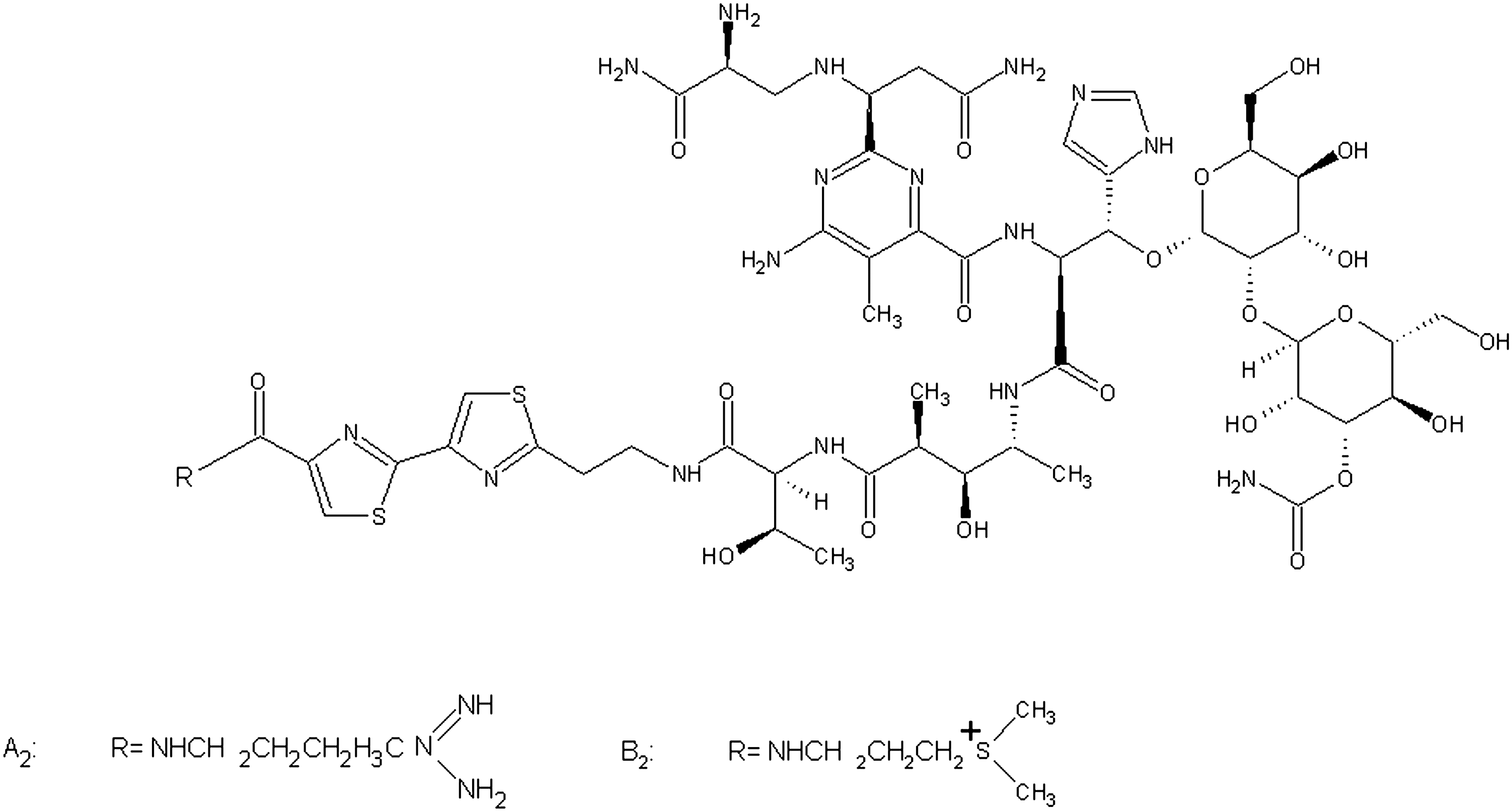
Chemical structure of BLMG. BLMG, bleomycin-glucuronide.
RP, reversed-phase C-18 column.
TLRC studies
For TLRC studies, TLC Aluminum sheets (Merck, 20×20 cm; code: 5552) were used. Acid-citrate-dextrose (ACD) (100%) and a mixture of 0.2 M potassium phosphate, 2 mM mercaptaethanol, and 0.4% TritonX100 (pH 7) buffer were used as the mobile phase for BLM and BLMG, respectively. The TLRC technique was used to determine the R f values of the radioiolabeled products. The R f values were determined and given in Tables 2. Radiochromatograms of labeled compounds were given in Figure 3.
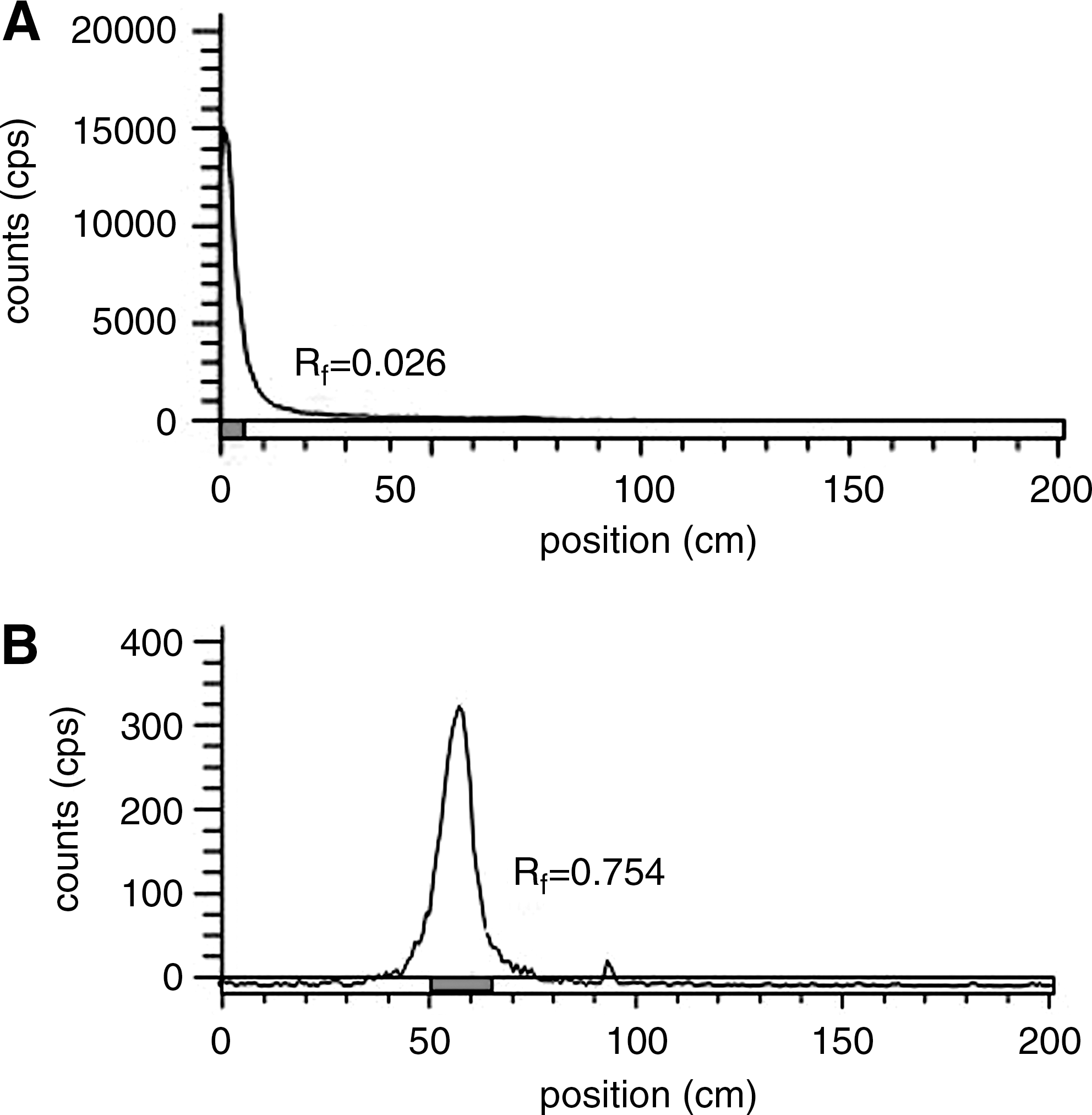
The radiochromatograms of 99mTc-BLM
BLM, bleomycin; BLMG, bleomycin-glucuronide.
Stability in human serum of radiolabeled compounds
In vitro stabilities of radiolabeled compounds in human serum were determined by incubating 100 μL (25 μg) of the labeled compounds with 300 μL of blood serum at 37°C. The aliquots were the analyzed in time intervals of 0, 30, 60,180, and 1440 minutes by TLRC technique after labeling.
Lipophilicity (partition coefficient)
The lipophilicity (logP) of the radiotracer was measured as follows: 100 μL of the radiolabeled compounds, 99mTc-BLM and 99mTc-BLMG, were added to a premixed suspension of 200 μL of octanol in 200 μL pH 7 buffer. The resulting solution was mixed for 15 minutes at room temperature and centrifuged for 30 minutes at 2500 rpm. Then, 0.1 mL aliquots of each phase were removed and counted by a Cd(Te) detector equipped with a RAD 501 single-channel analyzer. Experiments were conducted in triplicate.
Biodistribution studies in rats
Experiments with animals were approved by the Institutional Animal Review Committee of Celal Bayar University. The biodistribution data are expressed as percentage of injected radioactivity per gram of tissue (% ID/g) for selected organs as the mean value of 3 rats. The experiments were performed on 9 Albino male Wistar rats weighing approximately 180–200 g. The 99mTc-BLM and 99mTc-BLMG were sterilized by membrane filter and then injected into the tail vein of the animals [56 MBq (1.5 mCi)/4 μg of 99mTc-BLM per rat, 56 MBq (1.5 mCi)/6.6 μg of 99mTc-BLMG per rat]. Then, they were sacrificed at 30, 120, and 240 minutes postinjection under sodium pentabarbital anesthesia and the tissues of interest were removed. Blood was taken, and organs were excised. All tissues were weighed and counted for radioactivity with a Cd(Te) detector. The percent of radioactivity per gram of tissue weight (in% injected activity/g tissue,% ID/g) was determined.
Scintigraphic studies
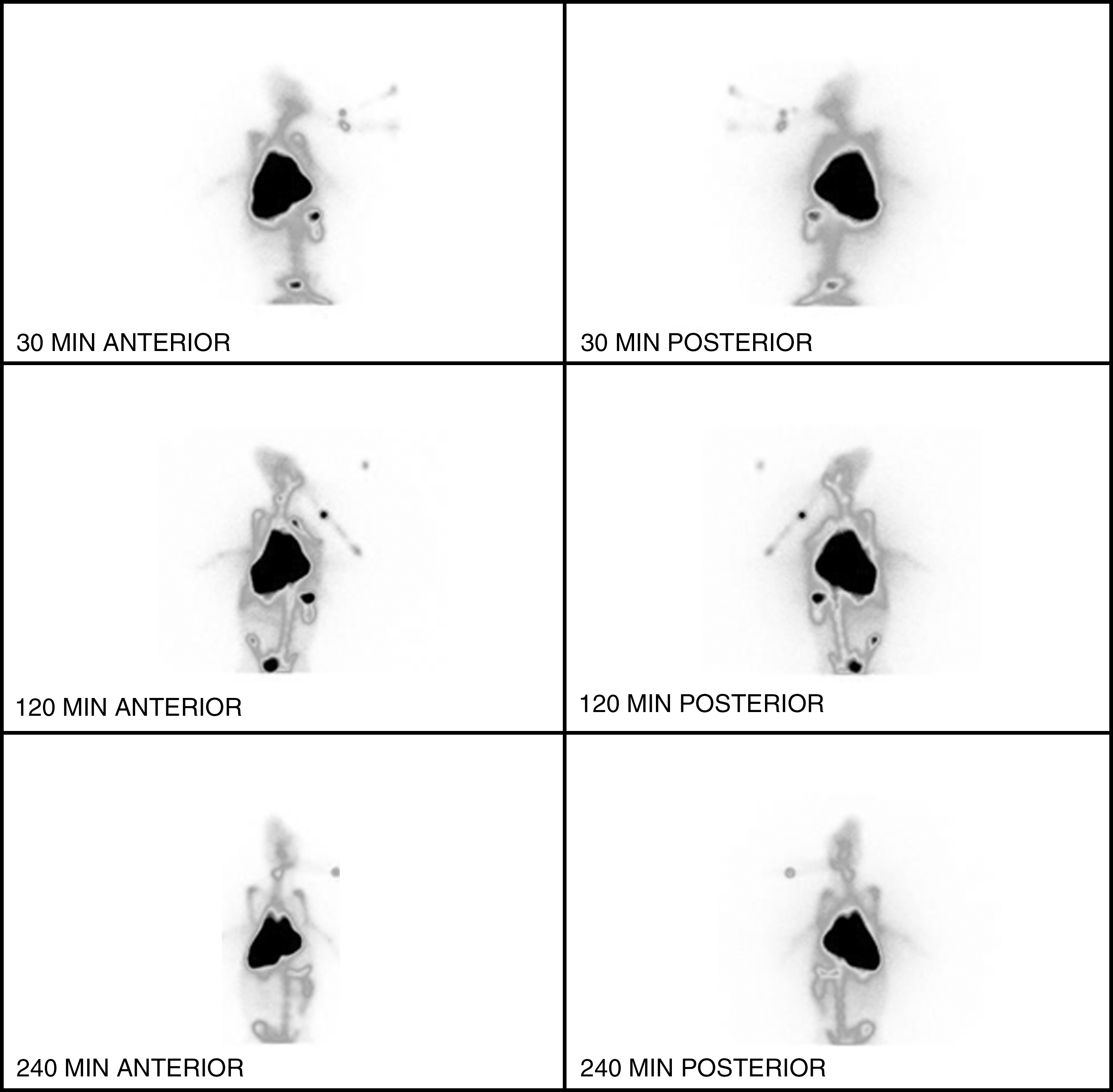
The imaging studies were performed on healthy adult male New Zelland rabbits using a gamma camera (Infinia, GE) at Department of Nuclear Medicine of Celal Bayar University. This work was approved by the Animal Ethics Committee of the Celal Bayar University (Manisa/Turkey) with approving number 2008/0015 (Date: 10/04/2008). The 99mTc-BLM and the 99mTc-BLMG were intravenously injected into 2 rabbits via the ear vein after anesthetizing by the mixture of xylazin and ketamine to determine the dynamic and static situations of 99mTc-BLM and 99mTc-BLMG in the metabolism. Dynamic and static scintigrams were obtained using a gamma camera (Infinia, GE), which was adjusted to detect γ radiations of 99mTc. Dynamic scintigrams were obtained over the first half hour with frames of 1 minute after the administration of the labeled compound. Static images were obtained from posterior projection after different time intervals up to about 4 hours after the administration of the radiolabeled compounds. Static and dynamic scintigrams of 99mTc-BLM and 99mTc-BLMG were given from Figures 4 to 7, respectively.

Static scintigram of 99mTc-BLM that was administered to a rabbit via the ear vein in 30 minutes. (The mixture of ksilazin and ketamin anesthesia was used in the scintigraphy studies. Static image was obtained from posterior projection after the administration of the 99mTc-BLM.)
Static scintigram of 99mTc-BLMG that was administered to a rabbit via the ear vein in 30 minutes. (The mixture of ksilazin and ketamin anesthesia was used in the scintigraphy studies. Static image was obtained from posterior projection after the administration of the 99mTc-BLMG.)

Dynamic scintigrams of 99mTc-BLM that was administered to a rabbit via the ear vein in 30 minutes (The mixture of ksilazin and ketamin anesthesia was used in the scintigraphy studies. Dynamic scintigrams were obtained over the first half hour with frames of 1 minute after the administration of the labeled compound.)

Dynamic scintigrams of 99mTc-BLMG that was administered to a rabbit via the ear vein in 30 minutes. (The mixture of ksilazin and ketamin anesthesia was used in the scintigraphy studies. Dynamic scintigrams were obtained over the first half hour with frames of 1 minute after the administration of the labeled compound.)
Statistical analysis
Differences in the mean values of measured activities were evaluated statistically by the SPSS 13 program (Univariate Variance Analyses and Pearson Correlation). Probability values <0.05 were considered significant. Pearson correlation was carried out between organs for 99mTc-BLM and 99mTc-BLMG.
Results
TLRC studies
The results of TLRC studies showed that ACD, 100%, and a mixture of 0.2 M potassium phosphate, 2 mM mercaptaethanol, and 0.4% TritonX100 (pH 7) buffer were the most suitable developing solvents for 99mTc-BLM and 99mTc-BLMG to establish their R f values given in Table 2. Radiochromatograms of 99mTc-BLM and 99mTc-BLMG were given in Figure 3. The best labeling yields were obtained at about 100% when the pH value was set to 6 and 7. The labeling yields of radiolabeled compounds were measured to be 99.40%±1.34% and 98.43%±1.32%, respectively.
HPLC studies
All HPLC chromatograms were obtained using UV detector at 260 nm. It was detected five peaks for BLM and only one peak for BLMG in the HPLC studies and also, the retention times of related compounds were different from each other as is seen in Figures 8 and 9. In the chromatographic studies, HPLC conditions were used given in Table 1.

HPLC chromatograms of BLM monitored at 260 nm: linear gradient 100% ammonium acetate buffer (10 mM) over 10 minutes. HPLC, high-performance liquid chromatography.

HPLC chromatograms of BLMG monitored at 260 nm: linear gradient 100% Ammonium acetate buffer (10 mM) over 10 minutes.
Lipophilicity (partition coefficient)
The n-octanol/water partition coefficients (lipophilicity) of 99mTc-BLM and 99mTc-BLMG were determined and the lipophilicities were found to be −0.73±0.03 and 0.42±0.03 (n=3), respectively. It was reported that octanol/water partition coefficient of BLM was −0.52 according to ACD/logP algorithm program. 26
Stability studies
Stability in human serum was investigated at time 0, 30, 60, 180, and 1440 minutes after radiolabeling. The results of the serum stability experiments demonstrated that approximately 100% of 99mTc-BLM and about 100% of 99mTc-BLMG existed as an intact complex in blood serum within 1440 and 180 minutes, respectively, as seen in Figure 10. Hence, stabilities of 99mTc-BLM and 99mTc-BLMG are sufficient for imaging procedures. For these reasons, both of them were directly injected into 1 rabbit without needing any separation or purification procedures.

Stabilities of 99mTc-BLM and 99mTc-BLMG in serum.
Scintigraphic studies
After administration of 99mTc-BLM and 99mTc-BLMG to rabbits, static and dynamic scintigrams were obtained and were given in Figures 4 to 7. Figures 4 and 5 show the static images corresponding to 30 minutes after the administrations of 99mTc-BLM and 99mTc-BLMG, respectively. Figures 6 and 7 show the dynamic images of these compounds. As is seen dynamic scintigrams of 99mTc-BLM, it is clearly observed that there is an important accumulation in the abdominal and the breast zone. 99mTc-BLM was significantly localized in the bladder within 11 minutes. Another result of dynamic images of related compound is the presence of high uptake in the intestine and the kidneys. In the static images of the same compounds, activities mentioned above is seem to be decreased at 120 minutes, but this activity does not seem to be completely cleared at the end of the 240 minutes. When scintigraphy and biodistributon data are analyzed together, accumulation in these zones can probably be caused by the liver, the lung, the stomach, and the spleen. Since BLM is a drug used to treatment of lung diseases, it can be clearly expressed that the accumulation in the lung is an inevitable situation. In addition to these, high uptake in the liver can be caused due to glucuronidation of BLM. As is observed on the scintigrams, no thyroid uptake was appeared. Since labeling yields of labeled compounds are about 100%, it is an expected result.
As is seen dynamic scintigrams of 99mTc-BLMG, it is clearly seen that the activity between the stomach and the breast region is more than that of 99mTc-BLM in the same regions. The most important curious difference between the two labeled compund was bone uptake in rabbit given BLMG. The results of scintigraphic and biodistribution of 99mTc-BLMG can be evaluated together. Accordingly, it can be said that accumulations at these sites are probably originated by the liver, the lungs, the kidneys, and the large intestine. It is known that glucuronides are exposed to deglucuronidation. Thus, it is estimated that the bladder uptakes seen static scintigrams of 99mTc-BLMG at 30, 120, and 240 minutes are probably caused by deglucuronidation of this compound.
Results of biodistribution studies
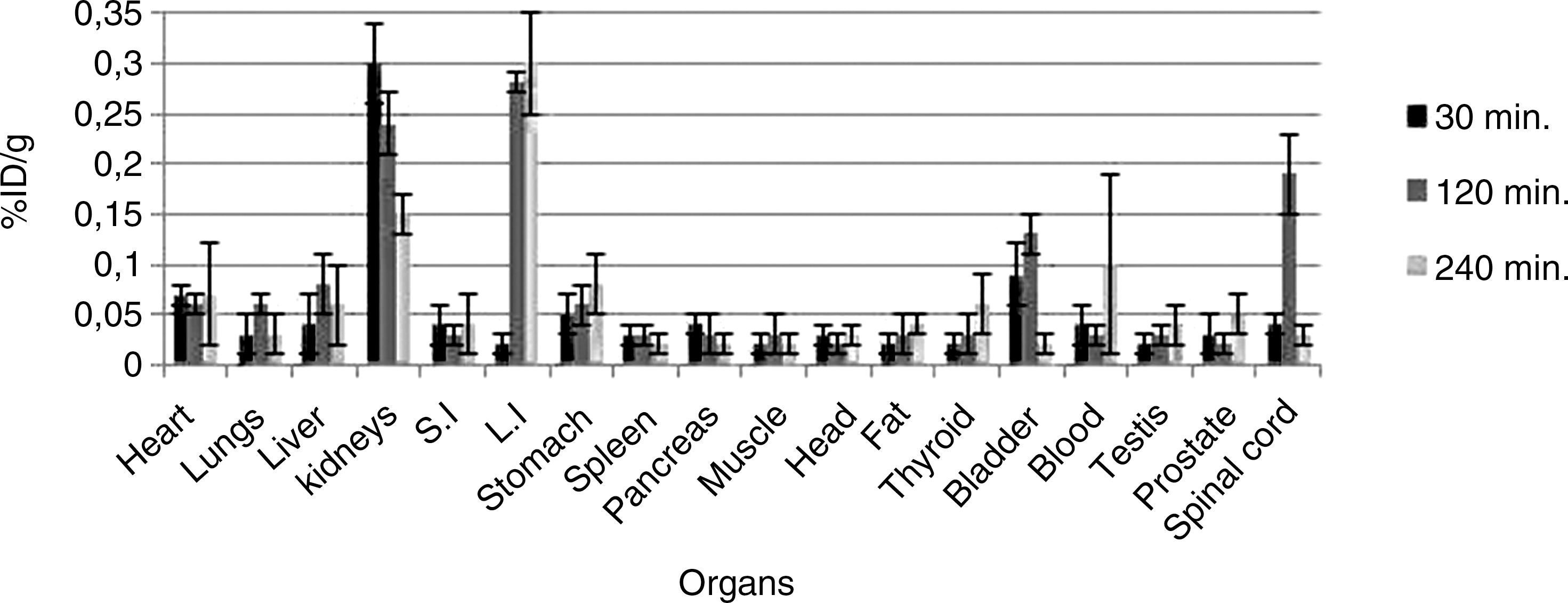
Biodistribution results (in % injected activity/g) of 99mTc-BLM and 99mTc-BLMG in rat tissues are given in Tables 3 and 4. Figures 11 and 12 represent the biodistribution results of 99mTc-BLM obtained 240 minutes after its administration to 1 rabbit. The high accumulation of 99mTc-BLM was observed within 240 minutes after the administration of 99mTc-BLM in main organs such as the liver, the spleen, and the spinal cord. Some amount of radioactivity was seen in the stomach at 120 minutes. Activity uptake in thyroid and, evenmore, in the stomach is a (well known) strong indication for the presence of 99mTc. Therefore, it is to be assumed that elimination of radioactivity is mainly due to the clearance of 99mTc beside the metabolism of radiolabeled BLM. Radioactivity in this organ was not cleared after 240 minutes. Obtained biodistribution data results agreed well with that of scintigraphic results. The%ID/g values of the radiolabeled BLM in the liver, the spleen, and the spinal cord were 0.58±0.43, 0.17±0.11, and 0.15±0.10 at 120 minutes, respectively. The uptakes in these organs decreased through time. It is also observed that 99mTc-BLM remained for a sufficiently long time in the metabolism (longer than 2 hours) and was not cleared rapidly.
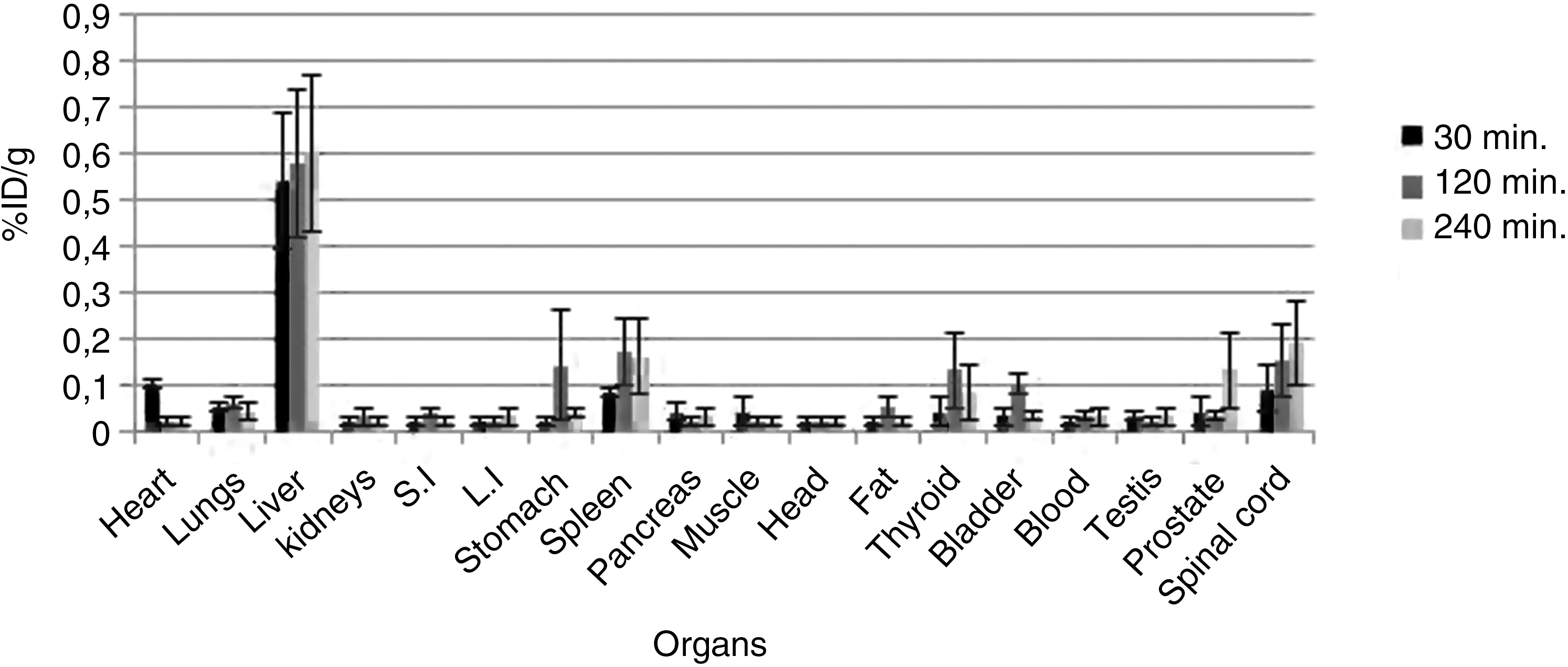
Activity (%ID/g) of 99mTc-BLM in major organs of the rats in the given time intervals (n=3); error bars mean standard deviation, %ID/g=percent of the injected dose per gram tissue. All the presented data are decay corrected. S.I., small intestine; L.I., large intestine.
Activity (%ID/g) of 99mTc-BLMG in major organs of the balb/c mice in the given time intervals (n=3); error bars mean standard deviation, %ID/g=percent of the injected dose per gram tissue. All the presented data are decay corrected.
SD, standard deviation, SI, small intestine; LI, large intestine.
Figure 12 represents the biodistribution results of 99mTc-BLMG obtained 240 minutes after its administration to 1 rabbit. The high accumulation of 99mTc-BLMG was observed within 240 minutes in main organs such as the kidney, the large intestine, and the spinal cord. Some amount of radioactivity was seen in the bladder in 120 minutes. Activity uptake in thyroid and, evenmore, in the stomach is considerably low. Radioactivity in this organ was not cleared after 240 minutes. The %ID/g values of the radiolabeled BLMG in the kidney, the large intestine and the spinal cord were 0.24±0.03, 0.28±0.01, and 0.19±0.04 at 120 minutes, respectively. The uptake in these organs decreased through time. It is also observed that 99mTc-BLMG remained for a sufficiently long time in the metabolism (longer than 2 hours) and was not cleared rapidly.
As is seen Figure 11, 99mTc-BLM has high radioactivity between 30 and 120 minutes after administration to rats; then, it rapidly decreases. This is probably due to rapid clearance of this compound from the metabolism.
In the biodistribution studies, the biological half-time of 99mTc-BLM and 99mTc-BLMG were measured to be 329 and 85 minutes by taking blood samples obtained from each rat group at 30, 120, and 240th minute.
Statistical results
According to statistical results, significantly positive correlation was shown in heart–large intestine (r=0.805, p<0.03), heart–blood (r=0.920, p<0.01), liver–spleen (r=0.867, p<0.02), kidney–stomach (r=0.978, p<0.01), spinal cord–small intestine (r=0.842, p<0.03), lung–large intestine (r=0.940, p<0.04), and bladder–testis (r=0.891, p<0.04) in rat's organs in the biodistribution studies. In addition to this, significantly positive correlation was shown in pancreas–kidney (r=0.805, p<0.04), large intestine–pancreas (r=0.920, p<0.05), prostate–muscle (r=0.867, p<0.02), spinal cord–large intestine (r=0.978, p<0.01), spinal cord–pancreas (r=0.842, p<0.04), and spinal cord–fat (r=0.940, p<0.01) in rat's organs.
Discussion
99mTc is eluted from the generator as pertechnetate. It is generally accepted that it has to be reduced to a lower valence state to form complexes. Many studies aimed at elucidating the chemical state of Tc in its complexes with radiopharmaceuticals have been reported, but the published data are somewhat contradictory. The yield of 99mTc incorporated into a radiopharmaceutical and the stability of the complex may depend on the valence state. The labeling of BLM and BLMG with 99mTc using SnCl2 as the reducer is a simple and rapid method. The reported preparation methods in this study differ for concentrations and pH of the reaction mixtures for BLM and BLMG.
To obtain the enzyme UDPGT, microsomal fractions of rat livers were separated and enzymatic synthesis of BLMG was performed using this enzyme. Enzymatic synthesis is a metabolic event conjugation of glucuronic acid to small molecules by means of UDPGA. Thus, drug molecules and other small lipophilic molecules convert into hydrophilic conjugates and excrete from the body. Enzyme UDPGTs catalyze the reaction. Thus, many endogenous and exogenous compounds, food molecules, hormones, and drugs may be glucuronidated with this way similar with in vivo. Enzymatic glucuronidation run according to SN2 reaction (second degree nucleophilic reaction) and at the end of the reaction glucuronic acid convert to beta anomer from alpha. The reason is the nucleophilic attack from behind of the substrate in the SN2 reactions. 27 Enzyme UDPGT-rich microsome preparates were extracted with a good yield and high purity from rat livers. BLMG can be deglucuronidated by the β-glucoronidase enzyme, which has an activity that is considerably high in certain kinds of cancer cell. Owing to this enzyme activity, BLMG can be considered as a potential anti-cancer drug.
It is known that logP has been calculated for the uncharged molecule theoretically. Theoretical lipophilicity of 99mTc-BLM could not be calculated due to the charge of this molecule. Experimental lipophilicity value of 99mTc-BLMG was obtained to be quite liphophilic than that of BLM and 99mTc-BLM. In the literature, it has been reported that compounds with high lipophilicity value will be able to pass to blood–brain barrier. Thus, 99mTc-BLMG can easily pass the blood–brain barrier.
It has been reported that the intestines have significantly β-glucuronidase enzyme activity by Avcıbaşı et al. 28 Peptides are absorbed by the intestines; then, they are removed to the liver. Thus, peptide is reversely absorbed and glucuronide conjugate is hydrolized again. This behavior called enterohepatic reverse circulation affects an important impact on metabolism and increases the half-life of the drug in organism. 29,30 It is seen that there is an important uptake in the large intestine at 120 minutes. Due to the liver–large intestine recirculation of glucuronide and glucoronide derivative compounds, uptake of 99mTc-BLMG in large intestine is thought to be caused by enterohepatic reverse circulation. Another remarkable point is that there is no accumulation in the stomach similar to the thyroid. Activity uptakes in this organ are 0.02±0.01, 0.02±0.01, and 0.03±0.01 at 30, 120, and 240 minutes, respectively. Due to labeling in a high yield, low uptake in the stomach is a sign of trace amount of radiochemical impurity and not occuring of pertecnetate.
BLM is a compound in the peptide form. Peptides are degraded by peptidases in the metabolism. Degradation is occured in the blood stream or the liver and/or the kidney excratory organs. In this respect, both 99mTc-BLM and 99mTc-BLMG are two different conjugates of the same form in basis. However, in case of the glucuronidation of BLM, physical and chemical properties of BLM change. Whenever these compounds are radiolabeled with 99mTc and investigated in vivo, it is appeared to have different properties. Considering in terms of biodistribution, the most important of these properties are specifity, uptake of the compound, clearance time, and clearance way from metabolism. As a result of this study, it was determined that 99mTc-BLM and 99mTc-BLMG were specific to different organs and each of them have different pharmacokinetic characteristics.
In a study carried out by Grove et al., to investigate the effects on pharmacokinetic properties of radiometal bounded to BLM, BLM was seperately radiolabeled with 57Co, 111In, and 67Ga. As a result of this, it was observed that 57Co-BLM has the highest tumor uptake and clearance. 31 In this respect, it can be said that 99mTc-BLM and 99mTc-BLMG formed after the conjugation of BLM and BLMG with 99mTc, have different clearances depend on the properties of 99mTc.
Jalilian et al. have reported that in consequence of the biodistribution study with 65Zn-BLM radiocomplex obtained after radiolabeling of BLM with [65Zn], radiolabeled compound showed a high activity uptake in the liver and the intestine between 24 and 48 hours after injection. It has been reported that the high uptake occured in mild liver tissues is probably caused by accumulation of metalloproteins in tissues. 32 Observation of high activity uptake of 99mTc-BLM after 4 hours in the liver is agreed with the reported idea above.
After biodistribution study carried out using 99mTc-BLM in Balb/C mice that were intravenously inoculated KHJJ tumor, it was observed high uptakes in the kidneys and a low uptakes in the liver, the muscle, and the brain. 33 In the biodistribution studies carried out using 99mTc-BLM, considerably high activity was observed in the liver, the spleen, and the spinal cord whereas fairly low activity was obtained in the kidney. On the contrary to this study, which expressed that BLM was renally excreted from metabolism, 99mTc-BLM displayed a hepatobiliary excretion. In addtion to this, at the same time intervals (30, 120, 240 minutes) it obviously can be said that 99mTc-BLMG was cleared more quickly than 99mTc-BLM from metabolism.
After the injection of radiolabeled compounds, blood samples were collected and radioactivity counts were measured at 30, 120, and 240 minutes. After measurements, biological half-times of 99mTc-BLM and 99mTc-BLMG were measured to be 329 and 85 minutes, respectively. As is seen, the biological half-time of 99mTc-BLMG is shorter than that of 99mTc-BLM. As a result of this, toxicity of 99mTc-BLMG is lower than 99mTc-BLM. Consequently, it can be used as a better chemotherapeutic agent than 99mTc-BLM. In this respect, in a study carried out by Crooke et al., it was determined that without any radiolabeling, biological half-time of BLM, which was directly given to patients for treatment was determined between 15 and 60 minutes. 34 In another study, Jalilian et al. have reported that biological half-time of 62Zn-BLM was suitable for imaging. Ryynänen has reported that biological half-time of 111In- BLM was detected to be 3.5±0.6 hours in the bladder. 35 In this study, since the half-time of 99mTc-BLM was measured as a close value to values given in the literatures above, this was an expected result. Because, all the investigated compounds were different BLM derivatives.
BLM was radiolabeled with three different radionuclides such as 97Ru, 67Cu, and 57Co and the efficiencies of radiolabeled BLM in tumor imaging were investigated and compared by Shao et al. 57Co>67Cu>97Ru sequence was obtained and it has been reported that 57Co has the highest ratio of target organ/nontarget organ, and also accumulation in the tumor was reported to be maximum. 36 In respect of this, the high ratio of target organ/nontarget organ increases accumulation of compound, so that it ensures to gain specifity in a certain organ. By taking advantage of the high ratio of target organ/nontarget organs of both 99mTc-BLM and 99mTc-BLMG, it can be said that 99mTc-BLMG will be able to be suitable for imaging and therapy of concerning organs in the organs such as the liver, the spinal cord, the spleen, the kidney, the large intestine, and the retinal cord as a proper radiopharmaceutic.
In conclusion, in this study, BLM and primarily enzymatically synthesized BLMG were radiolabeled with 99mTc and radiopharmaceutic potentials of compounds were investigated on experimental animals by using nuclear methods. In radiolabeling studies, direct labeling method was used. In all measurements, same labeling yields were obtained by using TLRC method. It was observed that parameters such as quantities of compounds, pH, temperature, reaction time, and amount of reductant were vitally important on obtaining the highest labeling yields. They were labeled by using different amounts of reducer and reagent in the different pHs (5–7) until the highest labeling yields were obtained. In the result of many different measurements, the optimum pHs of 99mTc-BLM and 99mTc-BLMG were obtained to be 5 and 7, respectively.
In the biodistrbution and scintigraphic studies carried out on experimental animals, it was anticipated that radiopharmaceutical potential of each compound was different and the highest uptakes of 99mTc-BLM and 99mTc-BLMG were observed in (the liver, the spinal cord, and the spleen), and (the kidney, the large intestine, and the spinal cord), respectively.
Satisfactory results were obtained for especially 99mTc-BLMG, which would be able to be used as a radiopharmaceutics in tumor imaging and therapy. In the next stage of the study with experiments that are going to be carried out using human cancer cells, uptake, toxicity, and apoptotic effects are going to be investigated for 99mTc-BLMG and the studies carried out using tumor bearing animals are going to contribute for obtaining advanced knowledge about this subject.
Footnotes
Acknowledgments
The authors thank for the financial supports from Celal Bayar University Research Fund (Contract no. 2008 FBE 017). The authors thank Dr. İlker Medine, M.Sc. student Gökçen Topal, and Ph.D. student Ya
Disclosure Statement
There is not any conflict of interest among the authors in this study.