
Abstract
Elemene (1-methyl-1-vinyl-2,4-diisopropenyl-cyclohexane) is a naturally occurring compound that can be isolated from the traditional Chinese medicinal herb Curcuma wenyujin. β-elemene, its active component, has recently been demonstrated to enhance the radiosensitivity of human cancer cell lines in vitro and of one animal tumor in vivo. The underlying mechanism, however, is still unclear. In this study, we demonstrated for the first time that β-elemene significantly improves the radiosensitivity of A549 lung adenocarcinoma xenograft in vivo as measured by tumor regrowth delay experiments. Our results showed that β-elemene, at 45 mg/kg, significantly inhibited radiation-induced expression of survivin and hypoxia-inducible factor (HIF)-1α proteins. Because HIF-1α is known to regulate survivin transcription and acts as upstream regulator of survivin, it is possible that β-elemene regulates the transcription of survivin through HIF-1α. Our study suggests that β-elemene is a promising drug to enhance tumor radioresponse, and survivin and HIF-1α are novel targets of β-elemene.
Introduction
Lung cancer is the most prevalent cancer in men and women and the leading cause of cancer-related death worldwide. 1 Despite significant advances in lung cancer treatments during the past several decades, the overall 5-year survival rate is poor. 1 One of the factors for treatment failures is radiation resistance. Therefore, finding novel drugs to overcome radioresistance may be a feasible way to improve effectiveness of radiotherapy and, thereby, overall survival.
Elemene (1-methyl-1-vinyl-2,4-diisopropenyl-cyclohexane), a naturally occurring compound that can be isolated from the traditional Chinese medicinal herb Curcuma wenyujin, is a mixture of β-, γ- and δ-elemene with β-elemene being the active component. The antitumor activity of elemene was first found by Chinese researchers in the 1980s; since 1994, this agent has been used in the clinic as one of the national class II novel antitumor drugs in China.
Many preclinical studies and clinical trials have demonstrated that elemene possesses several potential advantages over conventional chemotherapy agents, including broad-spectrum antitumor activity, favorable toxicity profile, and the ability to penetrate the blood-brain barrier. 2,3 Therefore, elemene has been used in the management of a variety of solid tumors, malignant pleural effusion, peritoneal effusion, and brain metastases. 4,5 β-elemene subsequently has been extracted from the elemene compound since 1996.
Recently, β-elemene was approved as a national first-class new agent, and phase II clinical trials are currently ongoing. Many laboratory studies have focused on the mechanisms underlying the anticancer effects of β-elemene. Several studies showed that this novel agent enhanced the radiosensitivity of human cancer cell lines in vitro. 6 –8 One study demonstrated improved radiation sensitivity of rabbit VX2 carcinoma by β-elemene in vivo. 9 No in vivo studies with human cancers, however, have been performed so far. While the in vitro studies have attributed the enhanced radiation response to increased apoptosis and cell G2/M arrest, 6,7,10,11 the underlying mechanisms are far from being clarified.
Survivin, a member of the family of inhibitors of apoptosis proteins (IAPs), has been found to be barely detectable in most normal adult tissues while being expressed at high levels in most human cancers, including non–small-cell lung cancer. 12 Survivin is regarded as a key regulator of cell division 13,14 and is expressed in a cell cycle-dependent manner. It is thought that survivin levels peak in the G2/M phase and that it can regulate microtubule dynamics by interacting with microtubules of the mitotic spindles. 13 –15 Moreover, current evidence suggests that survivin is compartmentalized in mitochondria and plays an antiapoptosis role. 16
Hypoxia-inducible factor-1α (HIF-1α), an oxygen-sensitive HIF-1 subunit, can transcriptionally activate many genes that are critical for cellular function under hypoxic conditions. 17,18 HIF-1α has been found to directly bind to the survivin promoter and to positively regulate the transcriptional activity of surviving. 19 In fact, survivin expression has been found to be significantly decreased by down-regulation of HIF-1α. 20,21
Human cancers with high expression levels of survivin or HIF-1α exhibit increased resistance to radiotherapy and are associated with higher risk of local recurrence and poor overall survival. 22 –26 Therefore, inhibition of survivin or HIF-1α may be a promising way to enhance tumor radiosensitivity. In this study, we investigated the antitumor activity of β-elemene at different dose levels on xenografts derived from A549 lung adenocarcinoma cell line in BALB/c nu/nu nude mice. Underlying mechanisms have been explored as well.
Materials and Methods
Chemicals and cell culture
β-elemene was purchased from Jingang Pharmaceutical Co. (Dalian, China). The human lung adenocarcinoma cell line A549 was purchased from the Cell Center of the Chinese Academy of Medical Sciences. Cells were grown in RPMI 1640 medium (Gibco-BRL/Invitrogen, Carlsbad, CA), supplemented with 10% fetal bovine serum (TBD Bio, Tianjin, China) at 37°C in a humidified atmosphere with 5% CO2. Polyclonal antisurvivin, HIF-1α antibodies and HRP-conjugated secondary immunoglobulin G (IgG) antibodies were purchased from Santa Cruz Biotechnology Inc. (Santa Cruz, CA)
Animals and tumor models
Female athymic BALB/c nu/nu mice aged 6 to 8 weeks were purchased from the Animal Experiment Center of Dalian Medical University and maintained in specific pathogen-free condition. The facilities and the protocol for these experiments were consistent with the regulations of animal use for biomedical experiments issued by the Ministry of Science and Technology of China and approved by the Animal Care Committee of Dalian Medical University. A549 cells were subcutaneously injected into the right hind leg (1×10⁷ cells per animal). The tumor sizes were measured every 2 days using Vernier calipers. At 4 to 5 weeks, tumor volumes reached the required size (0.8–1.0 cm3). Tumor volumes were calculated using the following formula: 0.5×largest diameter×smallest diameter 2 as previously reported. 27
Xenograft treatment with β-elemene and/or radiation
Nude mice with tumors of 0.8 to 1.0 cm3 were randomized to eight groups (five mice per group), including a control group and seven treatment groups: β-elemene alone, radiation alone, and β-elemene combined with radiation. β-elemene doses were 25 mg/kg, 45 mg/kg, and 100 mg/kg. Radiation treatments were performed 1 hour after β-elemene was injected intraperitoneally. For radiation treatments, mice were placed in a specially designed polyvinylchloride box, and the right hind legs bearing xenograft tumors were pulled out of the box. The mice were immobilized, and tumors were placed in the center of a 4 cm×3 cm radiation field, with the rest of the body remaining outside of the radiation field. Radiation was delivered by 6 MeV electron beams from a linear accelerator (Varian) at a single dose of 5 Gy.
Assessment of xenograft response to treatment
The average tumor inhibition rate (ATIR) after monotherapy was calculated every day until tumors in the control group doubled in size. The formula for ATIR is (1 – volume of treatment group/volume of control group)×100%. The degree of tumor growth delay was expressed either as: (a) the absolute growth delay (defined as the time for tumors in the radiation group to grow from baseline to twice the size, minus the time for the tumors in the untreated control group to reach the same size); or (b) normalized growth delay (defined as the time for tumors in the cotreatment group to grow from baseline to twice the size, minus the time for the tumors in the group receiving a single treatment to reach the same size). The activity of β-elemene to enhance tumor radioresponse was evaluated by EF, the Enhancement Factor, which was calculated using the following formula: EF=normalized growth delay/absolute growth delay. When EF was greater than 1, the drug at this dose level was considered to enhance tumor response to radiation. 28 The optimal radiosensitizing dose was selected for later experiments based on EF calculations.
Western blot analysis
Mice were sacrificed, and the tumors (five from each group) were excised and cut into small pieces. The tumor pieces were mixed in radioimmunoprecipitation assay buffer, homogenized by Polytron. Protein concentrations from tumor lysates were determined by Bio-Rad assays. Equal amounts of proteins were separated on sodium dodecyl sulfate-10% polyacrylamide gel electrophoresis, transferred to polyvinylidene difluoride membrane, and probed with the indicated primary antibodies followed by secondary HRP-conjugated secondary IgG antibodies. The protein bands were detected by an image analyzer (Labworks Software, UVP, Upland, CA).
Reverse transcription-polymerase chain reaction (PCR) analysis
Total RNA from tumor tissues (five from each group) was isolated using Trizol reagent (Invitrogen, Carlsbad, CA) according to the manufacturer's instructions. The reverse transcription-PCR analysis was performed using a RT-PCR kit (Takara, Otsu, Japan). The specific primer sequences were as follows: for survivin (5’ AGCCAGACTTGGCCCAGTGTTTC3’ and 5’ GCACTTTCTCCGCAGTTTCCTCA 3’) and β-actin (5’ CTGGGACGACATGGAGAAAA3’ and 5’ AAGGAAGGCTGGAAGAGTGC 3’); for HIF-1α (5′ ATGAAGTGTACCCTAACTAGCCG 3′ and 5′ GCTTGAGTTTCAACCCAGACATA 3′) and β-actin (5′ ATCATGTTTGAGACCTTCAACA3′ and 5′ CATCTCTTGCTCGAAGTCCA3′). The PCR protocol was as follows: Initial denaturation at 94°C for 2 minutes, followed by 35 cycles at 94°C for 30 seconds, annealing at 59°C (survivin) or 54°C (HIF-1α) for 30 seconds, and extension at 72°C for 1 minute. The final extension was performed by an incubation step at 72°C for 7 minutes. The PCR products were subjected to electrophoresis in agarose gel and visualized with ethidium bromide. The bands were analyzed with Quantity One software (Bio-Rad).
Hematoxylin and eosin staining and immunohistochemistry
The fresh tumor tissues were fixed in 10% formalin, embedded in paraffin, and cut into 4-μm thick sections. The sections were then stained with hematoxylin and eosin. Survivin and HIF-1α immunohistochemistry were performed as follows: Sections were stained with primary rabbit antihuman survivin and antihuman HIF-1α antibodies. After incubation with the respective HRP-conjugated goat antirabbit IgG, sections were visualized with the peroxidase substrate kit, followed by counterstaining with hematoxylin. We analyzed the results by IPP (Image-Pro Plus 6.0) software, using the method introduced by Wang and associates. 29
Terminal deoxynucleotidyl transferase dUTP nick end labeling (TUNEL) assay
The presence of apoptotic cells within the tumor sections was evaluated by TUNEL staining (Roche) according to the protocol. Percent apoptosis was determined by counting the number of apoptotic cells and dividing that by the total number of cells by using a light microscope.
Statistical analysis
The results of statistical analysis were presented as mean±standard deviation. The differences between the groups were tested by performing analysis of variance, and the levels of probability were noted.
Results
Antitumor activity of β-elemene alone
After treatment, tumor regression and regrowth were monitored until tumor volumes approximately doubled in size. Tumor volumes in each group were not statistically different on day 0. During the observation period, there was no significant β-elemene toxicity in any of the treatment groups. β-elemene, when used alone, showed antitumor activity in a dose dependent manner, as measured by ATIR
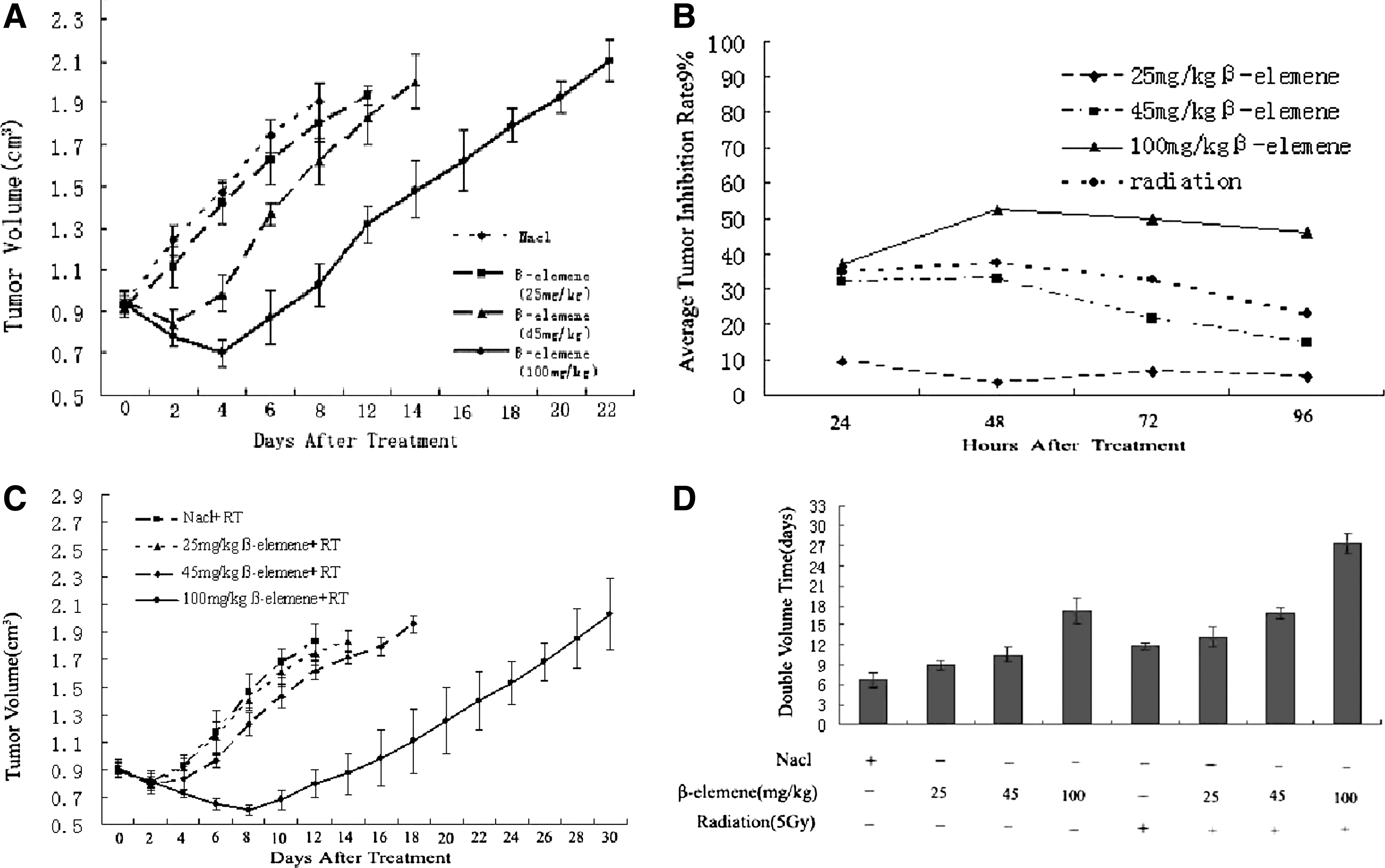
Effect of treatment on the growth of xenografts in athymic BALB/c nu/nu mice aged 6 to 8 weeks.
β-elemene enhanced radioresponse in xenografts
To determine whether β-elemene could improve radioresponse and to obtain an optimal dose to combine β-elemene with radiation, mice bearing 0.8 to 1.0-cm3 tumors were treated with 25 mg/kg, 45 mg/kg, or 100 mg/kg dose of β-elemene followed by radiation (5 Gy). There was significantly longer tumor regrowth delay when xenografts were treated with both β-elemene and radiation when β-elemene was given at or higher than the 45 mg/kg level
β-elemene decreases radiation-induced survivin mRNA/protein expression
Survivin expression levels were investigated by measuring mRNA and protein levels in tumor samples in the control, β-elemene alone (45 mg/kg), radiation (5 Gy) alone, and cotreatment groups
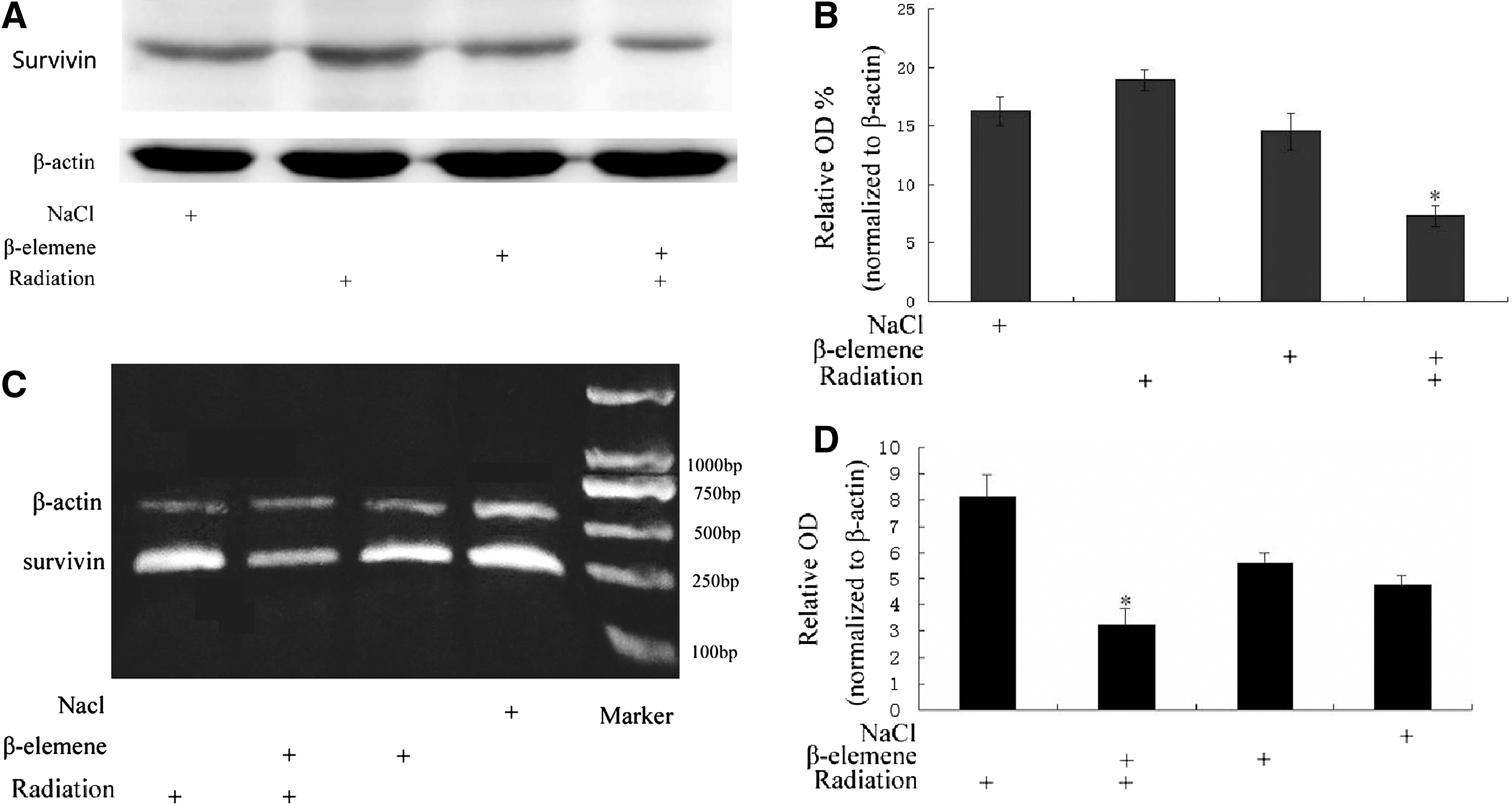
Survivin protein/mRNA expression in xenografts analyzed by Western blot
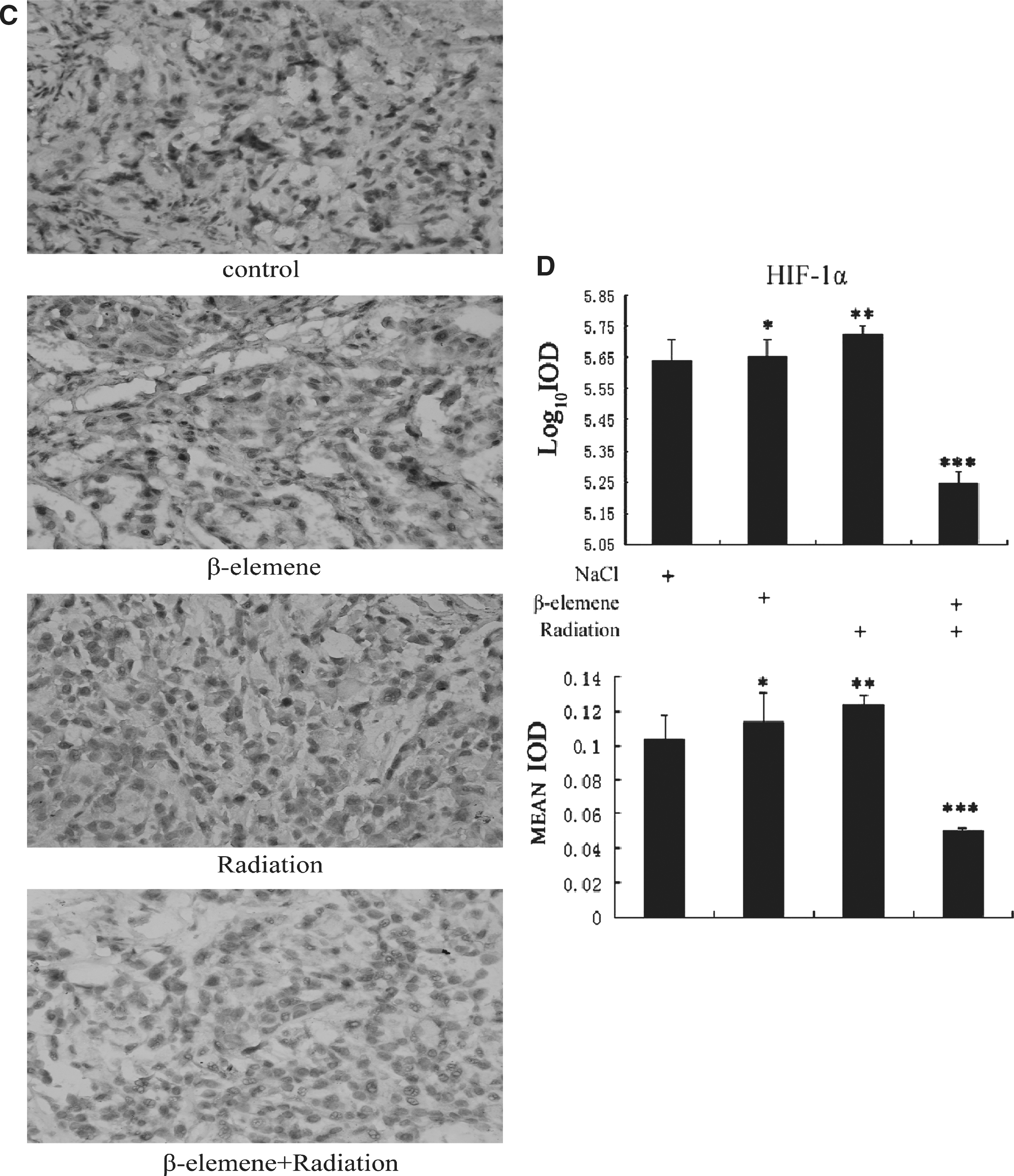
β-elemene decreases radiation-induced HIF-1α mRNA/protein expression
HIF-1α mRNA/protein expression in xenografts was also investigated for the four treatment groups
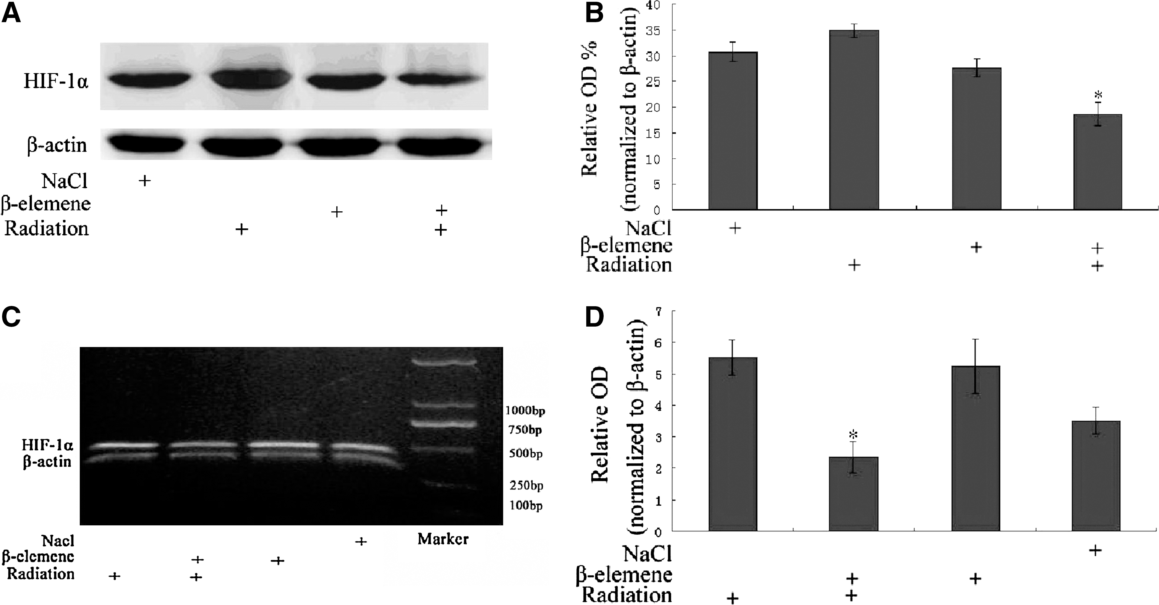
HIF-1α protein/mRNA expression in xenografts analyzed by Western blot
Survivin/HIF-1α staining by immunohistochemistry
We further investigated survivin and HIF-1α expression in xenografts by immunohistochemistry. The staining results are shown in
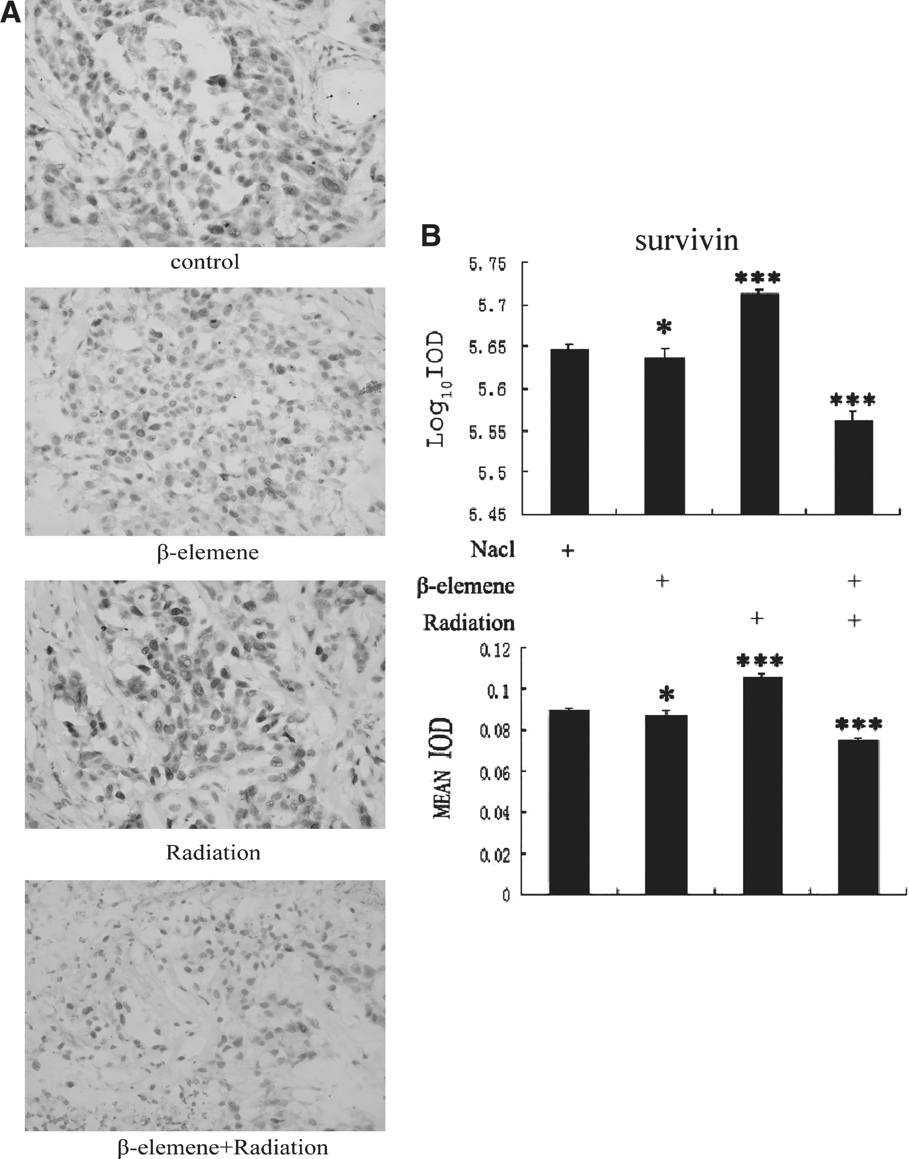
The immunohistochemistry staining of survivin and HIF-1α in xenografts among four treatment groups. The expression of survivin and HIF-1α was analyzed by Image-Pro Plus 6.0 software.
Increased apoptosis may be responsible for enhanced radiation response with β-elemene
Apoptosis in xenografts was determined by TUNEL assay. Apoptotic and live cells were counted under a light microscope, and percent apoptosis was recorded as the percent of apoptotic cells
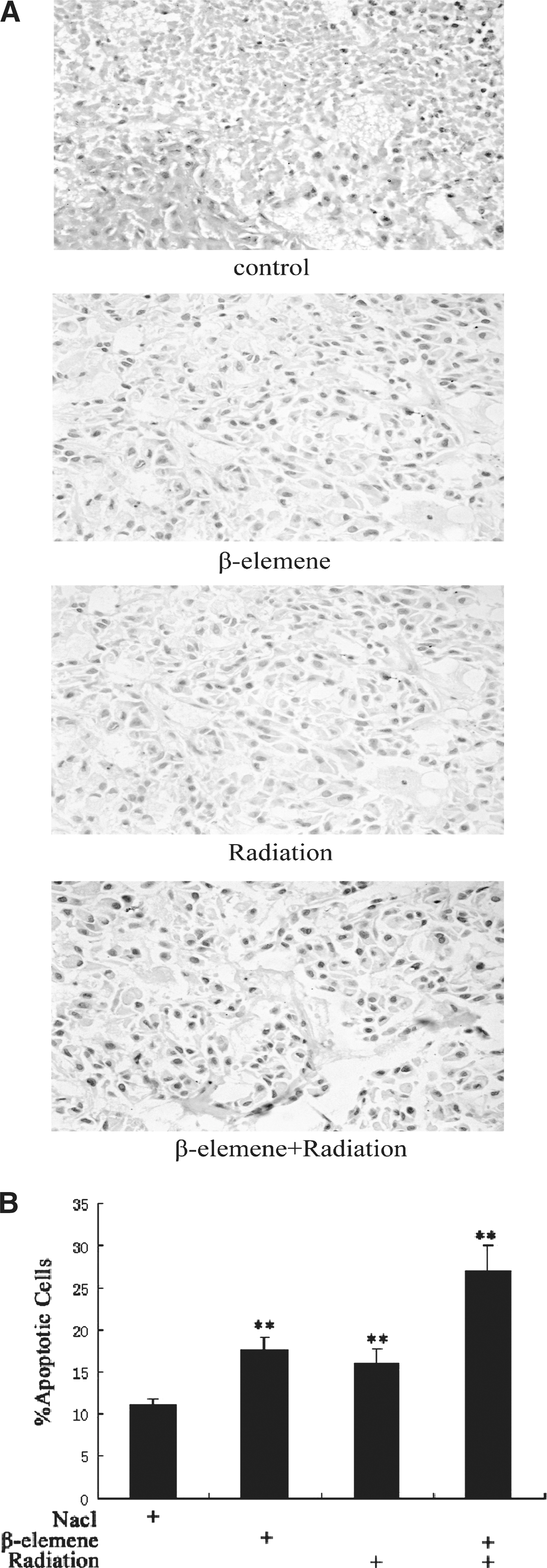
Apoptosis was determined with a TUNEL assay kit. Apoptotic cells were counted under a light microscope and reported as a percentage of the total cells presented for evaluation.
Discussion
It has been reported that β-elemene has antitumor activities against many types of cancer cells in vitro and in vivo. 30 –34 Our laboratory has shown that β-elemene enhanced the sensitivity of the A549 lung adenocarcinoma cell line to radiation in vitro. 35,36 In this study, we explored the radiosensitizing effect of β-elemene on A549 xenograft tumors in vivo for the first time. We showed that β-elemene had antitumor activity in a dose-dependent manner, and it augmented tumor response to radiation when dose levels were higher than 45 mg/kg. β-elemene at the 100 mg/kg dose level had significant antitumor activity when used alone and was synergistic when administered before radiation. Because the main purpose of this study was to investigate the radiosensitizing mechanisms of β-elemene, however, this dose was ultimately not selected in order to reduce the influence of direct antitumor activity.
It is interesting to find that the EF of β-elemene at 25 mg/kg was less than 1, which may suggest an antagonism effect with radiation. Theoretically, at the 25 mg/kg level, at which dose an antitumor action has been demonstrated for β-elemene alone, at least an additive effect with radiation might be expected. β-elemene is a novel cytotoxic agent, and its interaction with radiation is far from clear. While it is possible that a low dose of β-elemene may antagonize the effect of radiation, it is also conceivable that the EF has limitations as a measure under certain conditions. This warrants further investigations in the near future. β-elemene is currently being tested in clinical trials in China, and the best dosage for patients has not been determined. Although the doses used in this study were derived from animal tumor model systems, the results may still provide a valuable reference to the clinic.
It has been demonstrated that β-elemene enhances the radiosensitivity of cancer cells to radiation partly by inducing apoptosis. Previous data have shown that β-elemene could influence the expression of apoptosis-related molecules such as caspase-3, Bcl-2, and Bax. 10,11 β-elemene has also been shown to enhance tumor radioresponse through induction of cell cycle arrest at the G2/M phase, but the exact mechanism is still unclear. Therefore, having demonstrated that β-elemene enhances tumor sensitivity to radiation in vivo and having determined the optimal experimental dose, we sought further to identify potential novel targets of β-elemene.
Survivin has been reported to be overexpressed in non–small-cell lung cancer, 12 and patients with higher survivin levels have been shown to have significantly shorter survival in comparison with patients with low survivin levels. 37 Cancer patients with high levels of survivin exhibit increased resistance to radiotherapy. 22 –24 Survivin has been regarded as a radioresistance factor in colorectal and pancreatic cancers. 38 –40 Thus, survivin is a suitable molecular target for radiosensitization. The possible underlying mechanisms by which survivin contributes to tumor radioresistance remain to be understood and may involve suppression of apoptosis as well as cell cycle arrest and impaired DNA double-strand break repair. 38
Previous studies have shown that radiation significantly increased survivin mRNA/protein expression, 41,42 and the underlying mechanisms have not been clarified. Radiation can also increase HIF-1α activity, which has been suggested to be a main contributing factor for tumor radioresistance through multiple mechanisms. 43,44 The main mechanism is related to reactive oxygen generated by radiotherapy that leads to nuclear accumulation of HIF-1α and enhanced translation of HIF-1α-regulated proteins. The resulting increase in HIF-1-regulated cytokines has been shown to enhance endothelial cell radioresistance. HIF-1α is the oxygen-sensitive HIF-1 subunit, so some related studies were focused mainly on the association between HIF-1α and tumor vasculature. 45 –47 Because HIF-1α could directly bind to the survivin promoter and positively regulate the transcription of survivin, 19 it was considered possible that HIF-1α may exert a radioprotective effect by activating survivin.
In this study, our results demonstrated that radiation indeed increased survivin mRNA/protein expression in transplanted tumors, and for the first time, we showed that β-elemene decreased radiation-induced expression of survivin mRNA/protein in vivo. Because of the moderate antitumor effect of β-elemene at a dose of 45 mg/kg, it had a slight effect on the basal expression of survivin protein. β-elemene alone also induced apoptosis as indicated by the presence of more apoptotic cells in β-elemene treated tumors compared with the control group. Moreover, combination treatment of β-elemene plus radiation resulted in enhanced apoptosis compared with either of the monotherapies.
In addition, we found that although radiation enhanced HIF-1α mRNA/protein expression in the xenograft tumors, β-elemene plus radiation treatment could antagonize this induction and further decrease HIF-1α mRNA/protein expression. One model that could explain this result is that HIF-1α may be a target of β-elemene, which radiosensitizes tumor cells by inhibiting HIF-1α and survivin. Therefore, the HIF-1α/survivin pathway and its relationship to cell division and apoptosis merit further investigation.
Conclusions
Our results show that β-elemene significantly enhanced radiosensitivity of lung adenocarcinoma xenograft in vivo. β-elemene decreased radiation-induced expression of survivin and HIF-1α mRNA/protein, which may contribute to its ability to enhance radiation response. β-elemene may also promote apoptosis, which contributes to its radiosensitizing effect. Our study confirms that β-elemene is a promising drug to enhance tumor radioresponse, and survivin and HIF-1α are novel targets of β-elemene.
Footnotes
Acknowledgments
This research was supported by grants from the National Natural Science Foundation of China (NO.30770646) and the Innovative Team Project of the Liaoning Provincial Education Department (NO.2008T031).
Disclosure Statement
No competing financial interests exist.