
Abstract
The current study investigated the radioprotective effect of Ocimum sanctum on the salivary gland of rats administered radioiodine (131I) and compared its efficacy with a known radioprotectant, amifostine. The experimental rats were divided in four groups and sacrificed in three different batches at 1, 3, and 6 months of time interval after 18.5 MBq/100g (i.p.) 131I exposure. Six months duration batch received 131I exposure twice with the gap of 3 months. Two groups of experimental rats were presupplemented with O. sanctum (40 mg/kg for 5 days, orally) and amifostine (200 mg/kg, s.c) before 131I exposure separately. Increased Technetium-99m-pertechnetate (99mTcO4 −) uptake at 30 minutes post injection in salivary glands of only 131I exposed rats may imply delay in clearance at 6 months of exposure in comparison to their counterparts sacrificed at 1 month. Parotid gland histology showed atrophy with lipomatosis in only 131I exposed rats at 3 and 6 months of duration. O. sanctum and amifostine presupplemented and subsequently exposed to 131I rats at 3 and 6 months duration exhibited comparable histopathology with controls. Our study indicates possible radioprotective effect of O. sanctum and amifostine against high-dose 131I exposure.
Introduction
High dose radioiodine (131I) therapy is a standard procedure in the treatment of differentiated thyroid cancer to ablate remnant thyroid tissue post thyroidectomy or for metastasis. Due to the presence of sodium iodide symporter, iodide gets actively transported into the thyroid gland and a number of nonthyroidal tissues such as salivary gland, stomach, lacrimal gland, and lactating mammary gland. 1,2 Among these, salivary glands are known to exhibit irreversible radiation damage such as sialadenitis, xerostomia, and neoplasia in patients with differentiated thyroid cancer after high-dose 131I therapy. 3,4 Therefore, high-dose 131I therapy is generally recommended under salivary gland stimulation in order to minimize the salivary gland damage, however, with limited success. 5,6 Ionizing radiation damages DNA either directly or indirectly via the production of free radicals. Radioprotection can be achieved by use of free radical scavengers during 131I therapy. So far, amifostine is the only chemo and radioprotective drug available to reduce the side effects of radiation exposure in patients with cancer. 7,8 Amifostine is preferentially accumulated in certain tissues including the salivary gland, thus making these tissues less sensitive to radiation damage without protecting tumor cells. 8 –10 However, a major drawback of the use of amifostine is its severe adverse effects that sometimes result in discontinuation of the use of amifostine in some patients. 11 However, the use of amifostine as a radioprotectant is mainly reported in patients with head and neck cancer receiving external radiation exposure. 8,11 Limited studies have reported the usage of amifostine against internal radioiodine exposure in differentiated thyroid carcinoma and with controversial results. 7,12,13
At present, many traditional ayurvedic herbal formulations, plant extracts alone or in combination, are being explored for their efficacy against radiation-induced cell damage. Among them, Ocimum sanctum, a medicinal plant in India, is known to have various beneficial effects including radioprotection against 60Co gamma radiation. 14 –16 Our earlier work has shown better radioprotection by oral supplementation of O. sanctum as compared with other natural antioxidants in salivary glands of mice exposed to an internal therapeutic dose of 131I at 24 hours. 17 –19 Since irreversible salivary gland damage is known to occur at later stages, we thought of exploring the possible beneficial effect of O. sanctum on the salivary glands of rats after a long duration and the repeated therapeutic dose of 131I exposure and compared its efficacy with a known radioprotectant, amifostine.
Materials and Methods
Animals
Female adult Wistar rats weighing 250–300 g were obtained from Animal House Facility, BARC, Trombay, Mumbai. They were maintained in an air-conditioned room with free access to water and food. Animal studies were performed in compliance with BARC Institutional Animal Ethics Committee's guidelines.
Experimental design
Totally, 72 rats used for the experiment were divided into the following four groups with six animals each. Group I: Control. Group II: 131I exposed (18.5 MBq/100 g 131I, i.p). 131I as Na131I was obtained from the Board of Radiation and Isotope Technology. Group III: oral O. sanctum (40 mg/kg) for 5 consecutive days before 18.5 MBq/100g 131I exposure. O. sanctum used in the study was a generous gift from Dr. Yogendra Nayak, Department of Pharmacology, Manipal University, Manipal, India. Group IV: administration of amifostine (200 mg/kg, s.c.) 15 minutes before 18.5 MBq/100g 131I exposure. Amifostine was obtained from NATCO Pharma Limited. Totally, three such batches of 1, 3, and 6 months of duration receiving 131I exposure were studied.
The animals were weighed and sacrificed after 1, 3, and 6 months of duration of 131I exposure. Six-month experimental animals were exposed twice with 18.5 MBq/100g 131I with 3 months gap between the two exposures.
The experimental rats were supplemented with 1.5 μg/100 g of thyroxine (T4) and 1% calcium lactate in drinking water to maintain the euthyroid state.
Parameters
Biodistribution studies using 99mTc-pertechnetate
The 99mTc-pertechnetate (99mTcO4 −) uptake was studied by i.p. administration of 1.85 MBq of 99mTcO4 − to all the animals from the 1 and 6 months duration group, 30 minutes before sacrifice. 99mTcO4 − was prepared in-house from a 99Molybdenum - 99mTc generator.
The rats were sacrificed by cardiac puncture under ether anesthesia. Tissues excised for counting radioactivity included liver, stomach, kidneys, bladder, and salivary glands (parotid and submandibular from both the sides).The wet weights of the parotid and submandibular glands were recorded before measuring their 99mTcO4 − activity. In addition, blood was also collected and counted for its 99mTcO4 − activity. The percent uptake in blood was calculated by correcting it for the volume. The carcass was separately counted for its radioactive content.
The 99mTcO4 − activity was counted by using a broad, well-type scintillation counter designed for animal tissues, and the percent 99mTcO4 − uptake was calculated by considering injected activity as 100%. In case of salivary glands, the % uptake was calculated as percent injected dose/g tissue weight/kg body weight.
Histopathology
The parotid glands of 3 rats from each group at 1, 3, and 6 months of duration were fixed in 10% neutral buffered formalin. Sections at 3–5 μm were prepared and stained with hematoxylin/eosin for histopathology in a conventional manner.
Statistics
Statistical analysis was performed using ANOVA with Bonferroni correction. Within-group comparison for 99mTcO4 − uptake was done by using a paired t-test with a significance level <0.05, whereas between-group comparison was done by using Student's t- test with a statistical significance level <0.01(with continuity correction).
Results
Total body weight as well as parotid and submandibular weights in all the experimental rats did not show any significant change in comparison to control animals at 1, 3, and 6 months of duration (Table 1). After administration of 18.5 MBq/100g 131I, the rats from all the three groups exhibited an ablation of the thyroid gland as early as 1 month. This was evident by the absence of thyroid tissue at the time of sacrifice as well as the absence of 99mTcO4
Total number of animals=72 (mean±SD).
Gr. I, control; Gr. II, 131I; Gr. III, O. sanctum+131I; Gr. IV, amifostine+131I; SD, standard deviation.
Biodistribution studies were performed using 99mTcO4
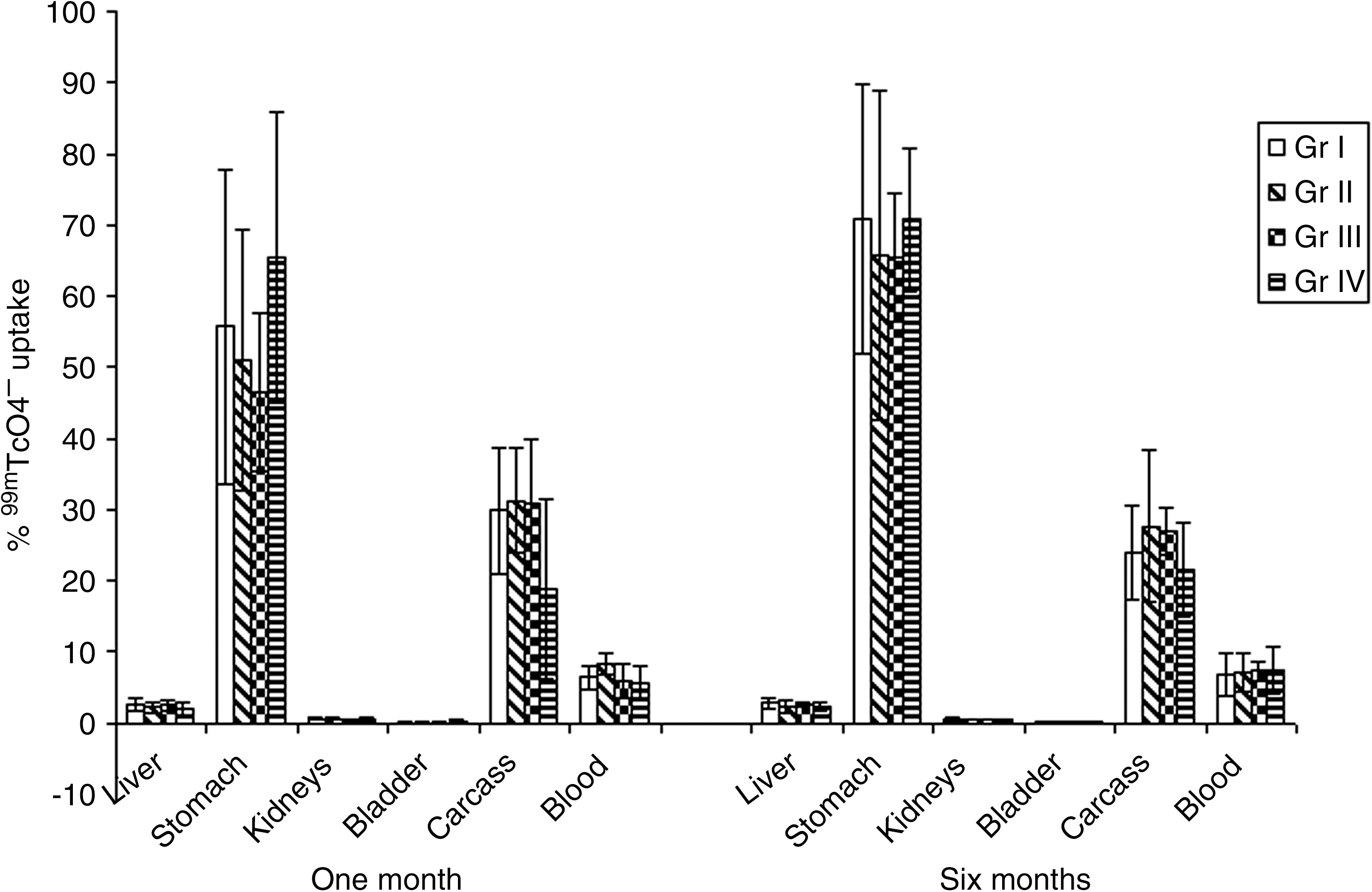
Biodistribution studies at 30 minutes using 99mTcO4 − in experimental rats (mean±standard deviation, n=6).
Table 2 shows percent 99mTcO4

Total number of animals=72 (mean±SD).
p=0.010 vs. Gr. II (1 month).
p=0.039 vs. Gr. III (1 month).
p=0.015 vs. Gr. III (1 month).
p=0.004 vs. Gr. I (6 months).
Discussion
Salivary gland dysfunction is a known complication of 131I therapy that can have a serious impact on the quality of life of a patient with thyroid cancer. 2,20 Due to the damaging effect of 131I on the salivary gland, high-dose radioiodine therapy is generally performed under salivary gland stimulation. Sialogogues are thought to have an important function in preventing the side effects of 131I therapy. 20 However, this approach has its limitations, as it requires continuous administration of sialogogues. 21 Lemon products are known to decrease the transit time of 131I through the salivary gland so as to reduce the amount of radiation exposure to the tissues, however, without much beneficial effect in the patient population. 5,6 In view of good prognosis of differentiated thyroid carcinoma after 131I therapy, it is important to avoid the salivary gland damage, ultimately improving the quality of life of these patients. Currently, amifostine is the only drug approved by the Food and Drug Administration for the purpose of radioprotection in patients with cancer. However, it is known to exhibit adverse effects that may lead to the discontinuation of the same in some patients. 11,22 In addition, its beneficial effects in patients with thyroid cancer undergoing 131I therapy are yet to be established. 12,13 Hence, the major focus of our experiment was to develop a better radioprotectant for the salivary gland against therapeutic 131I exposure that would be nontoxic and highly effective. Our earlier work in mice had given us encouraging preliminary results for salivary gland radioprotection against 3.7 MBq of 131I exposure for 24 hours by using O. sanctum presupplementation. 17,19
Our current experiment compares the effect of O. sanctum and amifostine presupplementation on the body weight and salivary gland weight of the rats. Biodistribution of 99mTcO4
The batch of experimental animals sacrificed at 6 months time interval receiving two 131I exposures showed 2.19 and 1.59 times increase in 99mTcO4 − uptake within groups in the parotid gland of Gr. I, Gr. III animals. However, in the submandibular gland, the increase was 1.92, 1.35 and 1.36 times in Gr. II, Gr. III, and Gr. IV, respectively, when compared with 1 month exposure. The observed increase was statistically significant in 131I exposed group in parotid as well as submandibular glands. O. sanctum pretreated and 131I exposed group also demonstrated a significant increase in 99mTcO4 − uptake in parotid glands. Trapping of 99mTcO4 − by the salivary gland generally begins at 1 minute and reaches a peak in about 5–10 minutes. At a later stage, it also provides the information regarding the excretion pattern. The observed increase in the uptake could be the indication of slow excretion due to the parenchymal damage, thus resulting in delayed 99mTcO4 − clearance ultimately increasing 99mTcO4 − uptake. In contrast, Bohuslavizki et al. 25 have observed progressive decline in 99mTcO4 − uptake from 4 to 24 weeks after 2 GBq of 131I exposure by acquiring sequential images of each up to 25 minutes in the same batch of animals. However, we have studied the percent uptake of 99mTcO4 − at 30 minutes of fixed time interval in parotid and submandibular glands of the experimental rats. We could not perform the sequential imaging in the same batch of animals, as they were sacrificed at fixed time intervals. Detailed sequential imaging studies in these groups particularly in O. sanctum pretreated (Gr. III) and amifostine (Gr. IV) pretreated groups are required to address the reason for the difference in clearance of the 99mTcO4 − in the salivary glands in these two groups.
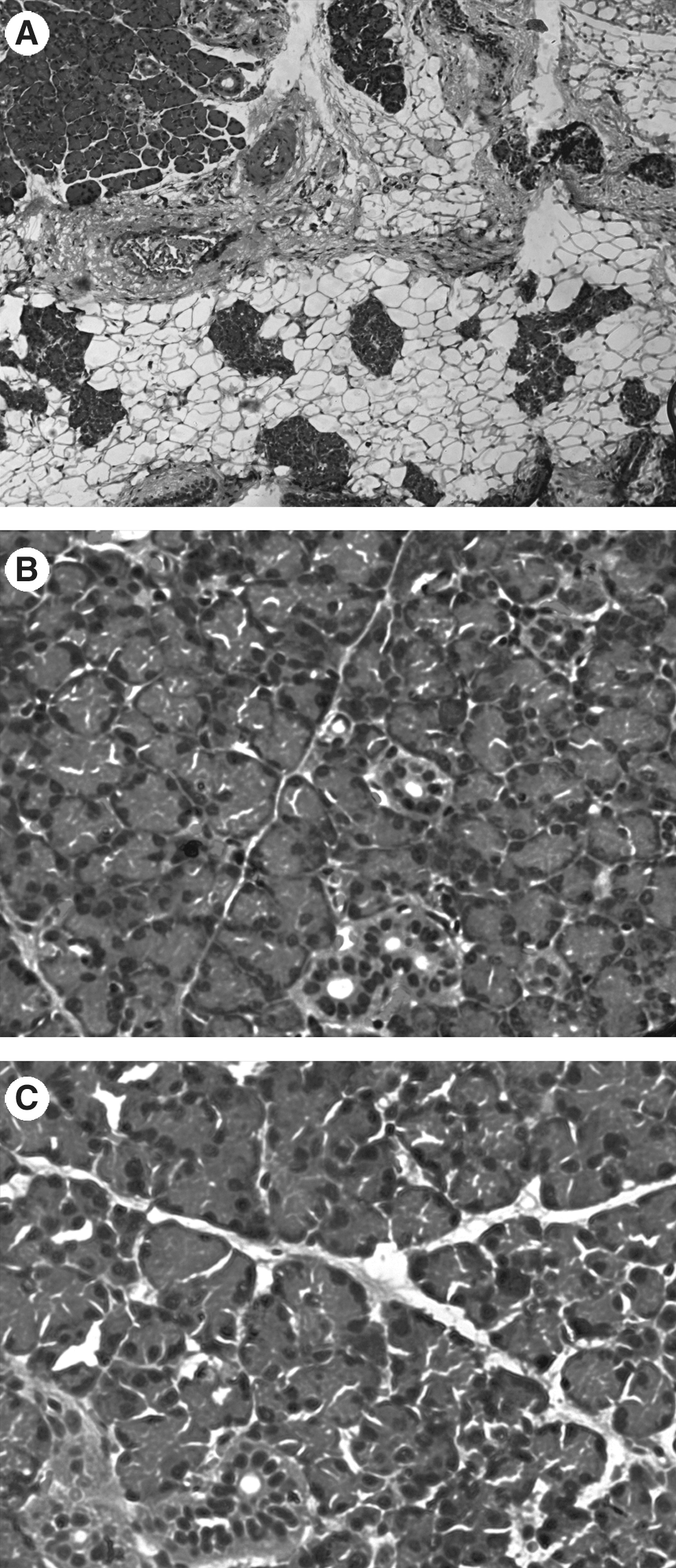
The histopathological studies have shown no significant changes in parotid gland architecture at 1 month. The lack of significant morphological alteration at 1 month is in agreement with many earlier reports. 26− 29 Our study differs from those with regard to the type of radiation used. They have externally irradiated the head and neck region of the experimental animals with 60Co, whereas in the present experiment, the rats were exposed to 131I in vivo. At later stages such as 3 and 6 months of duration, 131I exposed rats show cellular atrophy in the parotid gland accompanied by lipomatosis. On the other hand, rats pretreated with O. sanctum and amifostine showed comparable cellular histology to control animals. This is in agreement with the observation reported by Bohuslavizki et al., 25 wherein rabbits were exposed to a high dose of 131I in vivo to study the radioprotective effect of amifostine. Kutta et al. 30 studied the radioprotective effect of amifostine on the histology as well as electron microscopy of the salivary gland of rabbits after exposure to 131I. They have observed marked lipomatosis and apoptosis in the salivary gland of the rabbits exposed to 1 GBq radioiodine exposure at 3 and 6 months of duration. Similarly, Yan et al. have also observed severe salivary gland damage after external duel field irradiation at 16 weeks of time interval in the pig parotid gland in comparison to a single-field irradiation of the same dose. 31
The radioprotective effect of amifostine is attributed to its preferential localization in the salivary glands and ability to scavenge free radicals.
11
In the current study, the beneficial effect of amifostine was observed on the glandular histology of rats receiving 131I exposure as well as uptake studies done at 30 minutes after administration of 99mTcO4
In case of differentiated thyroid carcinoma, the usage of amifostine as a radioprotectant has not been unequivocally accepted. Controversial reports regarding its efficacy underline the need of exploring a better radioprotectant for the salivary gland protection after high-dose 131I therapy. In the light of this, our study reports the radioprotective ability of O. sanctum on the salivary gland in rats exposed to 131I. The use of O. sanctum should be explored in detail for its effect on the functional ability of the salivary gland as a safe radioprotectant that can be effectively used in patients with differentiated thyroid carcinoma receiving 131I therapy.
Footnotes
Disclosure Statement
The authors declare that there are no financial or other conflicting interests in the present work.