
Abstract
Aims:
The aim of this study was to investigate the safety and toxicity of biodegradable 32P–chromic phosphate–poly L lactic acid (32P-CP-PLLA) particles interstitially implanted into Beagle dog livers.
Methods:
Eighteen healthy Beagle dogs were randomly divided into 6 groups (n=3), and were treated with drugs of different formulations or doses, as well as controls. At different time points after surgery, the experimental dogs were weighed. Detection of indicators of blood chemistry and liver fibrosis, SPECT bremsstrahlung imaging, computed tomography, histological examination, continuous blood measurement, and counting of urine and fecal radioactivity were performed for these dogs.
Results:
SPECT imaging showed that after implantation of radioactive particles into livers, radioactivity continuously accumulated in the implanted sites, while no radioactivity imaging was found in the nonimplantation sites. The mean absorbance doses in the implantation sites were 89.8–178.7 Gy. Local spherical lesions were observed in tissues. The average effective half-life time of 32P-CP-PLLA was 11.8 days. Within 4 weeks after surgery, slight or moderate swelling and degradation of liver cells were detected, while in 8 weeks after surgery, they are normal. For the blood chemistry, liver fibrosis, and other indicators, no significant differences were found between the control groups and particle implantation groups (F=1.378, p=0.232).
Conclusions:
32P-CP-PLLA particles have advantages including good targeting, immobile, being degradable in vivo, easy to be protected, and so on. It is suitable for treating solid tumors with blood supply. 32P-CP-PLLA particles are a kind of safe, novel, radioactive implantation drug.
Introduction
Radioactive particle implantation is a kind of short distance–targeted radiotherapy, which implants radioactive microparticles into tumors using a minimally invasive method. These radioactive microparticles can kill tumor cells at a low-dose level by their long-term, continuous radiation. Early reports included radiotherapies using implantation of 226Ra and 192Ir into tissues, which emit α rays (4.777 MeV) and γ rays (400 keV), respectively. 1 However, radiation of these nuclides possesses high-level energies and strong penetration. Therefore, these nuclides are difficult to be prevented from affecting healthy tissues and often result in complications after implantation. Furthermore, implantation of these nuclides still has some technique limitations, which lead to slow development of the 226Ra and 192Ir methods.
Since 1980s, with the successful developments of three-dimensional computer treatment planning system, video surveillance system, and the low-energy nuclides (e.g., titanium metal particles 125I and 103Pd), implantation of radioactive particles was well-used clinically. The low-energy nuclides are highly compatible, with high cure rates of tumors in comparison to healthy tissues. Moreover, the low-energy nuclides can be easily operated and are less toxic. Currently, these low-energy nuclides are mainly used to treat prostate cancers, lung cancers, head and neck cancers, and liver cancers. 2 –6 Although 125I or 103Pd particles have good effects in treating malignancies such as prostate cancers, clinical applications showed that permanently implanted metal particles can migrate to other organs such as lung, resulting in radioactive pulmonary embolism. Merrick reported that early stage prostate cancer patients with 103Pd implantation have a pulmonary embolism rate of 22.2% (16/72), 7 causing many inconveniences and potential hazards to patients. 32P is an ideal, therapeutic radionuclide, which emits β-rays with an average energy of 0.695 MeV, a physical half-life of 14.28 days, and an average range of 3–4 mm (maximum, 8 mm) in tissues. The interstitially injected 32P–chromic phosphate (32P-CP) has obvious advantages in treatments of solid tumors. 8 However, interstitially delivered drugs may distribute to all over the body by the lymphatic and blood circulation systems, especially liver, causing increasing systemic side-effects with increased doses.
In this article, our research team develops 32P–chromic phosphate–poly L lactic acid (32P-CP-PLLA) particles, which can be degraded in vivo, using 32P-CP and biocompatible PLLA. 32P-CP-PLLA was implanted into organs with ample blood supply, such as livers of healthy Beagle dogs, with doses and methods that will be used clinically in the future. The radiation damage-time effects in implanted sites, systemic toxicity, and metabolic degradations of particles in vivo are observed, with the use of interstitially injected, colloidal 32P-CP as control. Our studies provide strong scientific data for clinical application safety of 32P-CP-PLLA.
Materials and Methods
Materials
32P-CP-PLLA particle preparation 9,10 is described as follows. 32P-CP-PLLA particles are biodegradable, radioactive particles that are made by mixing and pressing highly radioactive, colloidal 32P-CP (provided by the HTA CO., LTD) and PLLA together. After grounded by using planetary ball milling instrument to the nanometer or micrometer level, PLLA materials were mixed together with colloidal 32P-CP. Using ethanol as a dispersant and surfactants to regulate the affinity between PLLA and 32P-CP, the mixture was stirred for 1 hour and kept in 60°C oven for 9–12 hours. By using 1% magnesium stearate as the lubricant and interface blocker, the mixture was pressed into 32P-CP-PLLA particles using an advanced machine made by ourselves. The 32P -CP-PLLA particles are light green, physically stable cylinders with a diameter of 0.8 mm, a length of 2.0 mm, the quality of 1.3 mg, and the hardness of 5–7 N. The particles have evenly distributed radioactivity (37.0 MBq or 74.0 MBq each). These particles can be implanted in vivo via acupuncture and are well compatible to tissues. Major laboratory equipments used in this study include SPECT (Model, FORTE; Philips Company), γ Counter (USTC Chuangxin Co., Ltd.), BS-110S electronic balance (Sartorius Company), and CRC-15R enhanced β live meter (Capintec Company).
Experimental grouping and methods
All animal experiments were approved by the governing Animal Welfare Committee and conducted in accordance with the regulations of the institution. Eighteen healthy Beagle dogs (9 male dogs and 9 female dogs) recruited in this experiment were provided by the Sifang Experimental Animal Science and Technology Company, Yangzhou, China, with a certificate of No. SCXK (Su) 2008–0006. These dogs, with an average weight of 9 kg, were randomly divided into 6 groups as shown in Table 1 (n=3) using the random-number-table method. The PLLA particles implanted into dogs in group 6 and group 3 have the same chemical content.
P-CP-PLLA, 32P–chromic phosphate–poly L lactic acid.
Drug administration was described as follows. Prior to implantation, intravenous anesthesia was performed with 5% sodium pentobarbital, followed by conventional hair removal, disinfection, and towel usage. Upon laparotomy, needles (parallel to the lobe) were punctured into the left or right lobe of liver with a depth of 6 cm. When the needles were taken out, 32P-CP-PLLA particles, PLLA particles, or colloidal 32P-CP were interstitially implanted or injected into lobes. The particles (37 MBq or 74 MBq/each) were linearly implanted with a space of approximately 5 mm between two of them. After surgery, breathing and pulse rate of the dogs were carefully observed. The dogs were then kept at room temperature (25°C), with additional measures to assure their body temperature stable. Within the 3 days after surgery, the dogs were intramuscularly injected with penicillin (800,000 U/each dog/bid).
SPECT and computed tomography examination
Bremsstrahlung imaging was performed for Beagle dogs immediately after surgery, in day 2 (d2), d4, or d6. The imaging conditions were as follows: low-energy, general-purpose collimator; energy peak, 70 keV; window width, 20%; acquisition count, 300K. SPECT and computed tomography (CT) images were fused to observe the in vivo distribution of 32P-CP-PLLA and 32P-CP particles. The radioactivity count rates in the blood of the dogs were determined at time points including 0, 5, 10, 15, 30 minutes, 1, 6, 12 hours, 1, 2, 3 days, 1, 2, 4, 6, 8, 10, and 12 weeks after surgery. CT scans on the abdomen of the dogs were conducted before and after experiments or before sacrifice.
Measurements of plasma concentration and in vivo excretion
At 0, 5, 10, 15, 30 minutes, 1, 6, 12 hours, 1, 2, 3 days, 1, 2, 4, 6, 8, 10, and 12 weeks after surgery, 1 mL of venous blood was collected for counting radioactivity by liquid scintillation. Each dog was placed in a special metabolic cage after surgery. Urine and feces were collected every 24 hours. Feces were weighted and urine volume was measured. Radioactivity counts (cpm) in unit mass feces and unit volume urine were calculated. The total radioactivity excreted by the feces and urine within 24 hours was converted into activity value (MBq). Urine and feces were continuously collected using the same method within 30 days. The ratio (%) of the total excreted radioactivity to the total implanted radioactivity in 30 days was calculated.
Calculation of half-life time and estimation of absorbed doses by major organs
After euthanasia, 32P-CP-PLLA and 32P-CP particles in dog livers were taken out for radioactivity determination. The half-life time of 32P-CP-PLLA was calculated according to the following formula: It =I 0e−0.693t/Teff, 11 in which I 0 is the radioactivity at t=0, It is the radioactivity after time course t, and e is base of natural logarithms (2.718). The half-life time of 32P-CP was calculated according to the following formula: T eff=T 1/2 Tb /T 1/2+Tb , 12 in which T 1/2 is physical half-life time (14.28 days), and Tb is biological half-excretion time. The absorbed dose at d90 was calculated according to the following formula: D β=1.443 (ΣΔ i ) C 0 T eff[1−e−(0.693/Teff)t ] (cGy), 13 in which D β is the absorbed dose at hour t, cGy is unit, Δ i is the absorbed dose equilibrium constant of ray i emitted by radioactive nuclides (the Δ i of 32P is 3.99573×10⁻⁵ cGy/[Bq·h]), C 0 is β radionuclide concentrations (Bq/g) when t=0, T eff is the half-life time of 32P, t means time, and e=2.71828183.
Laboratory tests
In each week, blood routine tests and liver or renal function tests were performed for the Beagle dogs. Indicators of liver fibrosis were also examined, including procollagen III, hyaluronic acid, Cholyglycine, procollagen, and laminin.
Histological analyses
In week 1 (w1), w2, w3, w4, w6, w8, and w10, liver biopsy was performed for the experimental Beagle dogs, followed by conventional light microscopy. In d90, the live dogs were euthanized, followed by autopsies for histological examination of vital organs.
Statistical methods
Data were analyzed with statistical software (SPSS 13.0; SPSS an IBM's company). Measurement data were expressed as mean value plus standard deviation
Results
General data
After drug administration, all Beagle dogs in groups 1–6 were in good conditions, showing normal breathing rhythms, good movements, and normal body temperature (39°C±0.5°C), and no uncomfortable symptoms were detected. All dogs had good wound healing, without swelling and effusion observed. Two weeks later, the suturing lines were removed. Except dogs in group 5, which ate slightly less than before, all dogs ate and drank similarly to before surgery. Their urine and feces waste volumes, as well as sex behavior, were not changed either. At d23 after surgery, the mean weight increase for group 1 to group 6 was 1.6, 1.3, 1.2, 0.8, −1.7, and 1.8 kg, respectively. Dogs in group 5 died of liver failure at d23, d29, or d45 postsurgery. All other dogs were euthanized, followed by autopsy.
Anatomy
Slight adhesions were observed between liver and diaphragm of dogs in groups with particle implantation. With blunt dissection, yellow hard ellipsoidal fibrous structures were found at the implantation sites within the right lobe of livers. The outer ranges of these structures were obviously limited by the peripheral normal liver tissues. These structures were about 8 mm away from the liver surface, with sizes positively correlated with numbers of the implanted particles and range of the implanted targets. In the longitudinal sections, different numbers of dark green, incompletely degraded particles were found to be linearly localized along the long axis. These crunchy particles were wrapped by the peripheral tissues, not easy to be separated from them. Cross sections showed yellow, hard circular fibrous tissue rings with the implanted particles as their centers. These fibrous rings were composed of two concentric circles (inner and outer) with different radii. The inner structures consisted of particles and light red loose damaging foci. The outer layer comprised normal liver tissues. Between the two concentric circles, there were yellow fibrous, connective tissues (Fig. 1a–c). Radioactivity in these tissues was detectable by detector.
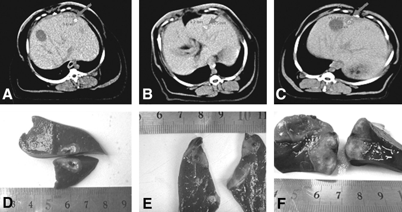
Upper row: 3 months after 32P-CP-PLLA particle implantation into Beagle dog liver, CT imaging showed limited damage foci in the implantation site. The foci images consisted of slightly low-dense center and circular high-dense area. Edemas were found in peripheral area. The neighboring liver tissues were normal. Damage foci sizes are related to doses and scopes of implantation.
For dogs in groups with colloid injection, ridge-like, streak-like uplifts were found in surfaces of the injected liver lobes. There was no detectable color difference between these uplifts and the peripheral healthy liver tissues. In the longitudinal sections along the ridge, a sinus with smooth wall, but without contents, was found in the inner side. A background-level radioactivity was detected in these areas. For dogs in group 5, extensive adhesions were found between liver and peritoneal. With blunt dissection, it was found that the livers were becoming larger and the boundaries between lobes turned to be unclear. Extensive hepatic fibrosis was observed. Altogether, these results suggested that no significant pathological alterations were found in the hearts, spleens, lungs, kidneys, and other organs of these experimental dogs.
SPECT imaging and CT scanning
One dog was randomly selected from every group for SPECT imaging and image fusion immediately after surgery, or at d2, d4, and d6 postsurgery (Fig. 2). The SPECT imaging indicated spherical or ellipsoidal radioactive regions in the implanted sites. The radioactivity in the implanted sites became less and less after surgery. No radioactive imaging was found in nonimplantation sites of livers, spleen, and other organs within 6 days after surgery. Imaging of the whole livers of dogs in the groups with colloid injection showed uneven radioactivity distribution. The average radioactivity count rate in the regions of interest of the injected lobes to the uninjected lobes was 5 to 3. Radioactive imaging faded within the time course postsurgery. No radioactive imaging was found in spleens.
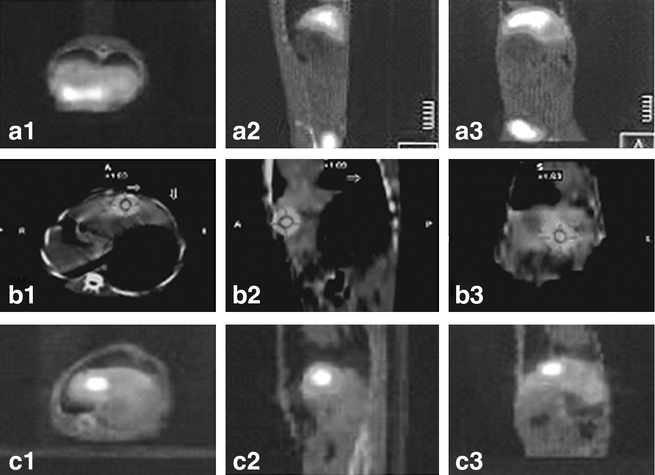
SPECT three-dimensional fault and ECT/CT image fusion of Beagle dogs postsurgery:
The CT scan results of all Beagle dogs before surgery were normal. Prior to euthanasia, CT scanning (Fig. 1d–f) of dogs in groups with particle implantation indicated that the liver surfaces are smooth. The overall size and shape of livers were not significantly changed in comparison to before surgery. At the implantation sites, small pieces of high-density imaging were found, with small cystic shadows observed inside and different degrees of edema surrounded. With increasing implantation doses and ranges, sizes of the damaged foci developed accordingly.
Drug concentration in blood and metabolism
The radioactivity count rates in blood were as follows over time. For dogs in groups with particle implantation, the peaks (698–1371 cpm/mL) appeared in d3 postsurgery, followed by declining multipeaks. No radioactivity was detected after 70 days. For dogs in groups with colloid injection, the peaks (4538–10066 cpm/mL) appeared in d1 postsurgery, followed by exponential decrease over time. No radioactivity in the blood was found after 50 days postsurgery. Differences in peak values among particle groups or colloid groups were both statistically significant (F=2.756, p<0.001).
For dogs in groups with particle implantation, radioactivity count rates in urine and feces decreased by ladder-like multipeak curves over time. In d30 postsurgery, the cumulative percentages of radioactive counts in urine and feces were 7.3% and 9.9%, respectively. For dogs in groups with colloid injection, radioactivity count rates in urine and feces appeared as single-peak, skewed curves. In d30 postsurgery, the cumulative percentages of radioactive counts in urine and feces were 23.7% and 8.9%, respectively.
Calculation of half-life time and estimation of absorbed doses
After euthanasia, 32P-CP-PLLA and 32P-CP particles in dog livers were taken out for radioactivity determination. The mean half-life time of 32P-CP-PLLA was calculated as 11.8 days, while the mean half-life time of 32P-CP was calculated as 6.8 days. In d90 postsurgery, the absorbed doses by major organs were estimated and given in Table 2.
Hematology and blood biochemistry
Within 3 months postsurgery, blood routine tests and blood biochemical indicator tests were performed weekly (Table 3). In the first week after surgery, when compared with the control group (group 6), dogs in groups 1–5 had transient increases in WBC levels and decreases in PLT levels. Except dogs in group 5, WBC and PLT of all dogs became to be normal within 2 weeks postsurgery. Upon comparisons of WBC, BUN, or Cre among the experimental groups (groups 1–5) or between the experimental groups and the control group (group 6), no statistical differences were found (F WBC=1.465, P WBC=0.207; F BUN=1.110, P BUN=0.374; F Cre=1.656, P Cre=0.128). The only significant differences were the significantly decreased Hb and PLT values and the abnormally increased ALT and AST values of group 5 during the survival periods of the dogs, when compared with the other groups (F Hb=12.465, P Hb<0.001; F PLT=5.897, P PLT b<0.001, F ALT=11.218, P ALT b<0.001; F AST=30.583, P AS b<0.001).
↓, decreased; ↑, increased.
Dynamic observation of five indicators of liver fibrosis
Five indicators of liver fibrosis were given in Table 4. Differences between groups 1–4 and the control group (group 6) were not statistically significant. The mean values of five indicators in group 5 were significantly higher than those in the other groups, among which the differences in PC III, HA, CG, and PC IV were statistically significant (F PCIII=3.727, P PCIII b<0.001; F HA=9.856, P H<0.001; F CG=18.988, P CG<0.001; F PCIV=4.598, P PCIV<0.001), but the differences in LN were not statistically significant (F LN=1.378, P LN=0.232).
↓, decreased; ↑, increased; PC III, procollagen III; HA, hyaluronic acid; CG, Cholyglycine; PC IV, procollagen IV; LN, laminin.
Histology
Microscopy observation showed that within 4 weeks postsurgery, some liver cells of dogs in groups 1, 2, and 4 became larger, with lightly stained cytoplasm, balloon-like degenerations, and visible lipid vacuoles. Tissue structures in hepatic lobules and portal areas were normal. No liver cell necrosis and fibrous tissue hyperplasia were found. After 8 weeks, in comparisons with the control group, dogs in experimental groups showed no abnormalities.
When compared with those in group 1 and group 2, dogs in group 3 had more serious liver cell swelling and degradation within 4 weeks postsurgery, accompanied by some fuzzy structures found in the hepatic lobule and portal areas. Mild fibrous tissue proliferation and slight liver cell necrosis were observed. Between week 4 and week 8, the liver cell swelling became more seriously, with sinus expansion, congestion, expansion of the portal area, fibrous tissue hyperplasia, and some mononuclear cell infiltration observed. However, no obvious formation of fibrous septa (grade 2) was found. After the 8 week postsurgery, in comparisons with the control group, dogs in experimental groups showed no abnormalities.
Within 4 weeks postsurgery, dogs in group 5 had more serious balloon-like liver cell degeneration when compared with those in group 3. Furthermore, some tissue structures in the hepatic lobule and portal areas became fuzzy, with a small amount of liver cells dying. Between week 4 and week 6 postsurgery, the scope of visible necrosis enlarged and dual-core liver cells proliferated in some areas. After the dogs in group 5 died, and the Beagle dogs in the other groups were euthanized, light microscopic examination of the hearts, lungs, spleen, kidneys, and other vital organs beyond the liver tissue was performed. No tissue damages were found.
Discussion
The biggest advantage of internal radiotherapy with radionuclide, in comparison with general radiotherapy, is its continuous exposure at low dose levels, which not only effectively kills tumor cells, but also maximizes the protection of normal tissues. 14 When doses are increased, interstitial colloid administration of 32P-CP induces increased systemic side-effects, which significantly limits its clinical application. Studies of a kind of strongly targeted, 32P-CP–carrying vector are the key to become conducive to standardization of clinical applications and reduce systemic side-effects. In 1990s, Order 15 has reported an improved technology, with which large human serum albumin particles (MAA, 10,000 units) are directly injected as vectors for transduction of radioactive cell toxins, resulting in thrombosis in tumors. And radioactive drugs such as 32P-CP are then injected. By this method, radioactive drugs will continue to be retained in solid tumors up to at least 24 hours. Following studies suggest that this method allows the use of increased doses in order to improve tumor therapy efficacy. Although this approach overcomes the overflowing of colloidal 32P-CP out of tissues, but its distribution in tumors is still uneven.
It has been recently reported that BioSilicon and 32P-CP can be mixed to make into 32P-BioSilicon particles. BioSilicon is a nanoporous cellular rubber, which can pack 32P-CP by the use of their different sizes of pores to extend the lifetime of 32P in the lesions. This technique has achieved significantly inhibitory tumor effects in animal experiments and clinical trials for liver cancer treatment. 16,17 BioSilicon is manufactured by the pSiMedica Company in United Kingdom. However, the in vivo slow releasing and tissue compatibility of drugs are still in the stage of researching. Poly L lactic acid is a kind of polymer material that is biocompatible and can be absorbed in vivo. It has been approved by the U.S. FDA for the use of surgical sutures and in vivo drug carrier material. In this article we use PLLA as a carrier of 32P-CP for the development of in vivo–degradable 32P-CP-PLLA particles. The safety and feasibility of 32P-CP-PLLA particles implanted into tissues were observed.
Livers are just less sensitive to radiation than bone marrows, lymphoid tissues, and kidneys. Studies have shown that radiation tolerance doses of the whole liver, 2/3–1/3 liver, and 1/3 liver were 30 Gy/3–4 weeks, 48–52.8 Gy/3–4 weeks, 66–72.6 Gy/4–5 weeks, respectively. About 75% of patients, who have whole liver irradiation (>40 Gy), develop liver dysfunction. Liver cancer is generally considered as a kind of tumor relatively resistant to radiation, with a lethal radiation dose of 60 Gy/6 weeks. As liver tumors have higher radiation tolerance doses than the normal liver tissues, conventional radiotherapy has poor efficacies of liver cancers. 12 In this study, effects of the cumulative liver absorbance doses on liver functions of the Beagle dogs in every experimental group are as follows. Upon liver injections of 185 MBq of 32P-CP, the absorbed dose of whole liver is 31.4 Gy, without significant liver damage observed. Therefore, this is a tolerable dose. When 370 MBq of 32P-CP is injected, the whole liver absorbed dose is 56.4 Gy, with obviously abnormal detections of the blood routine tests, blood biochemistry indicators, five indicators of liver fibrosis, and averages of weight increases, indicating a strong liver toxicity and systemic side-effects. Within 45 days postsurgery, all Beagle dogs in group 5 died of liver failure. Therefore, Beagle dogs with liver interstitial injection of 32P-CP have a lethal dose of 370 MBq. Our results show that Beagle dog's liver can tolerate radiation of 32P-CP-PLLA particles at levels that are 2-fold of the lethal dose of 32P-CP. In the implantation sites, the absorbed 32P-CP-PLLA radiation doses by liver tissues are up to 89.8–178.7 Gy, while in nonimplantation sites the absorbed 32P-CP-PLLA radiation doses by liver tissues are very low. For dogs in groups with 32P-CP-PLLA implantation, no abnormal detections of the blood routine tests, blood biochemistry indicators, five indicators of liver fibrosis, and averages of weight increases are found. This indicates that implantation of particles only damages local tissues, but not the targeted organs or other parts of the body. Studies have shown that implantation of radioactive particles is a key technique that increases drug dosage, but does not increases the systemic side-effects. In this study, changes in animal body's stress and possible infection caused by anesthesia and surgery may be the major reason why transient increase of WBC occurred after surgery. With anti-inflammatory treatments, WBC levels returned to normal soon. Inevitable bleeding and physical damages such as the pull and puncture of livers and particle implantation led to slight decreases of PLT, and slight transient increases of ALT and AST in every experimental group. However, all of these increases are not beyond the normal range.
With gross anatomy and morphological observation of experimental dogs, it is further confirmed in this study that the radioactive colloid injected into livers is easily diffused into the blood vessels, causing widespread damages of the liver microcirculation and functions, thereby resulting in hepatic lobule local fibrosis. Moreover, when doses are increased, these damaging effects are even more apparent. However, particles implanted into livers localize at the implanted sites, wrapped by the peripheral tissues and not displaced. The local damage foci caused by radiation of the implanted particles is clear. Light microscopy of dogs in the 740 MBq particle groups showed local cell necrosis and fibrosis in livers. Dogs in the remaining particle groups and low-dose colloid groups had transient liver cell congestions and edema degeneration. The liver lobe structures around the implanted particles are normal. The damage sizes in dogs of particle groups are consistent with the range of 32P β-ray in tissues and the scope of particle implantation, indicating that dose distribution of 32P-CP-PLLA particles in normal liver tissues steeply drops within a short distance. The consistency between damage sizes and the range of 32P β-ray in tissues leads to facts that tissues in a limited area around the implanted particles received most of the energy. However, nearby and distant tissues received very few of radioactivity doses. Therefore, no significant damage effects on adjacent normal tissues are detected. CT and SPECT imaging results further confirm that 32P-CP-PLLA particles have excellent target localization and tissue compatibility, which can effectively improve ratio of the targeted to nontargeted areas. This dose distribution has important practical significance in the targeted therapy of solid tumors with rich blood supply.
Radioactivity count rates in urine and feces indicate that the implanted 32P-CP-PLLA particles are degraded in the body, with excretion of noncolloidal forms of 32P-CP and tiny fragments through feces slightly more than from urine. The radioactivity count rates from urine and feces decreased by a multipeak descending curve in a time course, indicating that the particles were slowly degraded over time. Therefore, radionuclides in particles were successively released into the blood, not one-time released. Particles do not migrate with the blood, with the degraded debris absorbed by tissues or excreted. Therefore, no residues are finally left in body. Calculation indicates that more than 80% 32P radionuclide of the implanted particles are localized in the implantation zone, while about 20% of the 32P are slowly released into the blood and lymph circulation, which can kill metastatic tumor cells. This degradation characteristic can effectively improve the targeting therapeutic efficacies of radionuclide, with a potential of treating lymphatic metastasis. 18 For dogs in groups with interstitial injection of colloidal 32P-CP, more radioactive 32P was excreted in the urine than in feces. Through traditional interstitial injection, about 50% 32P-CP are localized in the implantation zone. A large number of drugs are retained in body, resulting in clear liver toxicity and systemic side-effects. Therefore, the authors suggest that 32P-CP interstitial therapy should not be used for treating solid tumors with rich blood supply, especially liver cancers.
32P-CP-PLLA particles have advantages including pure β-ray emissions, middle-level energy, short range, suitable half-life time, good targeting, being degradable in vivo, immobile, easy to be protected, good for treating all types of solid tumors with blood supply, good for treating lymphatic metastasis, and so on. Use of 32P-CP-PLLA particles overcomes problems of other drugs, such as permanent stay in the body after implantation, poor localization, and easy migration that may cause blood clots. In the experiment using the 32P-CP-PLLA particle implantation to cure human pancreatic cancers, Yang et al. found that tumor volume was reduced in a dose-dependent manner when compared with the control group. 19 Our study shows that 32P-CP-PLLA particles implanted into the liver greatly enhance the continuous 32P-CP gathering at the implanted site. Very little 32P-CP is absorbed by normal tissues. Therefore, it is a novel, safe radioactive implant. Other studies have also shown that 32P-CP-PLLA particles implanted into tumors have potentials to further increase the effects of curing malignant solid tumors.
Footnotes
Acknowledgments
This work was supported by Chinese National High-tech Research Development Plan (863 plan) (grant No. 2007AA02Z471).
Disclosure Statement
No competing financial interests exist.