
Abstract
Ulinastatin is a broad-spectrum enzyme inhibitor extracted from urine. Previous data from our group suggested that ulinastatin could significantly inhibit proliferation of human breast MDA-MB-231 cells, growth of tumor xenograft in nude mice, and expression of interleukin (IL)-6 and IL-8. In the present study, we investigated whether there is an additive effect of ulinastatin and docetaxel on growth of breast cancer xenografts in nude mice and its possible mechanisms. Nude mice and primary human breast cancer cells were treated with phosphate buffered saline (PBS), ulinastatin, docetaxel, or ulinastatin plus docetaxel, respectively. Their effects on xenograft growth; expressions of cyclooxygenase-2 (COX2), prostaglandin E2 receptor 2 (EP2), IL-10, and IL-2; and secretion of prostaglandin E2 (PGE2) were examined using variety of methods, including semi-quantitative reverse transcription–polymerase chain reaction (RT-PCR), Western blot, enzyme-linked immunosorbent (ELISA) assay, and immunohistochemistry SP method. The treatment with ulinastatin, docetaxel, or ulinastatin plus docetaxel could significantly (1) inhibit COX2 and IL-10 expression in primary tumor cells at both mRNA and protein levels, (2) reduce PGE2 secretion in culture supernatant (p<0.05), (3) inhibit COX2, EP2, and IL-10 protein levels in primary xenograft of nude mice, and (4) increase IL-2 expression (p<0.05) in primary xenografts of nude mice. In addition, ulinastatin and docetaxel had additive effects. We suggest that ulinastatin had similar effects of docetaxel and can enhance docetaxel's anticancer effects possibly by inhibiting COX2 expression, reducing PGE2 and EP2 expression and their binding, upregulating IL-2, and downregulating IL-10.
Introduction
Our previous study found that breast cancer tissues present Th2-type immunodominance, suggesting that shift of Th1/Th2 immunobalance to Th2 may be one of the reasons for tumor metastasis. 1 Ulinastatin, a broad-spectrum protease inhibitor, can significantly inhibit proliferation of human breast MDA-MB-231 cells, growth of tumor xenograft in nude mice, and expression of interleukin (IL)-6 and IL-8. In addition, supplementing ulinastatin can enhance antitumor effects of chemotherapeutic drug cyclophosphamide. 2,3 Another study previously found that cyclooxygenase-2 (COX2), an inducible enzyme, is responsible for the synthesis of prostaglandins (PG). One of the products of this enzyme is prostaglandin E2 (PGE2), which exerts suppressive actions through interaction with prostaglandin E2 receptor 2 (EP2) or prostaglandin E4 receptor 4 (EP4) receptors. The Th1/Th2 balance reflects the immune status. IL-2 is an immune-enhancing factor produced by Th1 cells, and IL-10 is an immunosuppressive factor produced by Th2 cells. IL-10 and IL-2 are also regulated by PGE2. Thus, in this study, we further explored whether ulinastatin have additive inhibitory effect to docetaxel, a widely used antitumor drug, on in vivo breast cancer xenograft in nude mice, and its possible mechanisms, including its expression of COX2, IL-10, IL-2, PGE2, and EP2 in cultured primary breast cancer cells and in tumor tissue.
Materials and Methods
Reagents
Ulinastatin was from Techpool Biochemical Pharmaceutical Co., Ltd. Docetaxel was from Hangzhou Sanofi-Aventis-Minsheng Pharmaceutical Company. They were prepared at 100-fold of peak plasma concentrations and used at 800 UI/mL for ulinastatin 3 and 3.7 μg/mL for docetaxel, 4 respectively. Rabbit antihuman COX2 and IL-10 monoclonal antibodies were from Bioworld Technology Company. Mouse antihuman breast cancer-associated antigen (CA153) monoclonal antibody, DAB staining kit, and SP immunohistochemistry kit were from Beijing Zhongshan Golden Bridge Biological Technology Co., Ltd. PGE2 ELISA kit was from Takara Bio-Engineering Co., Ltd. Fluorescein isothiocyanate (FITC)-conjugated goat antimouse IgG, HRP-conjugated goat antirabbit IgG, and rabbit antihuman COX2, IL-10, IL-2, and EP2 polyclonal antibodies were from Wuhan Boster Biological Engineering Ltd. Fetal bovine serum (FBS) was from Hangzhou Evergreen Biological Engineering Materials Co., Ltd.
Cell extraction and culture
A breast cancer sample was collected by surgery under sterile conditions from a 72-year-old female patient who admitted to the Second Clinical College, Chongqing Medical University, and pathologically found to have invasive breast ductal carcinoma before operation, and sent to our laboratory within 2 hours. The patient showed ER and PR positive, CerbB-2 negative, and did not take any chemotherapeutic drugs. The specimen was sliced to 1 mm3 and digested with 0.05% type II collagenase at 37°C for 20–30 minutes. After static stay at room temperature for 12–16 hours, supernatants were collected and centrifuged at 800 r/min for 5 minutes. The precipitated tumor cells were diluted to 105/mL and cultured in the RPMI 1640 medium supplemented with 10% FBS, 100 U/mL penicillin, and 100 mg/L streptomycin.
Experimental animals
Forty-five female BALB/c (nunu) mice (age, 4–6 weeks; weight, 18–21 g) from Animal Research Center of Chongqing Medical University (Production license number: SCXK [Beijing] 2005-0013; Use license number: SYX [Chongqing] 2007-0001) were kept in SPF-grade environment at 22°C–25°C and 50%–65% humidity. Drinking water, food, and other experimental materials were sterilized, and all experimental operations were complied with sterile principle.
Animal experiments
The above cultured primary breast cancer cells at logarithmic growth phase were collected by digestion with 0.25% trypsin. After checking the cell viability with trypan blue (≥85%), cells were adjusted to about 5×10⁷/mL. A 0.2-mL cell suspension was subcutaneously inoculated into the right breast of each nude mouse. Tumor development and size were observed and measured every 3 days. Twenty-one days after inoculation, 32 out of 45 (71%) mice had tumors. Among them, 28 had tumor volume ≥500 mm3 and were randomly assigned into four groups with seven mice in each group. Mice in the control group were intraperitoneally injected 0.2 mL saline/day for 20 consecutive days. Mice in the ulinastatin group were injected 0.2 mL of 8000 U/mL ulinastatin once a day for 20 consecutive days. 3 Mice in docetaxel group were injected 0.2 mL of docetaxel solution at 20 mg/kg at days 1, 7, and 14 of the experiment. 4 Mice in the ulinastatin plus docetaxel group were injected 0.2 mL of 8000 U/mL ulinastatin once a day for 20 consecutive days and 0.2 mL docetaxel at 20 mg/kg body weight at days 1, 7, and 14 days. At day 21, all mice were sacrificed by cervical dislocation and the xenografts were collected. After measuring the maximum diameter and minimum diameter, xenografts were fixed in 10% formaldehyde solution and prepared as paraffin samples.
Tumor volume (V) and tumor inhibition rate (E) were calculated based on the following equations: V=(L 2 ×D)/2 and E (%)=[1−(V 0−V 1)]/(V c0−V c1), where L and D are the maximum and minimum diameter of xenograft, respectively; V 0 and V 1 are the tumor volumes in drug treatment group before and after injection, respectively; and V c0 and V c1 are the tumor volumes in control group before and after injection, respectively.
To explore the additive effect of ulinastatin and docetaxel, q value of King's formula was calculated as: q=E docetaxel+ulinastatin/[(E docetaxel+E ulinastatin)−E docetaxel×E ulinastatin]. q>1.15 was considered as a having synergistic effect, 1.15>q>0.85 was considered as having an additive effect, and q<0.85 was considered as having an antagonistic effect.
Immunostaining of human breast cancer-associated antigen CA153 in cultured primary breast cancer cells
About 1×10⁵ cultured breast cancer cells were seeded on a coverslip and cultured to 80% confluency. Cells were washed with phosphate buffered saline (PBS) for three times, fixed with 4% cold paraformaldehyde for 10 minutes, washed again with PBS, and permeabilized with 1% Triton X-100 for 10 minutes. After incubating 5 minutes with PBS for three times, cells were incubated in turn with normal goat serum for 30 minutes, mouse antihuman CA153 monoclonal antibody or PBS as negative control for overnight at 4°C followed by 20 minutes at room temperature, PBS for 5 minutes for three times, FITC-conjugated goat antimouse lgG antibody at 37°C for 1 hour, and PBS for 5 minutes for three times. Cells were then mounted on a slide with 95% glycerol, and observed and imaged with an inverted fluorescence microscope (Olympus).
mRNA levels of COX2 and IL-10 measured by reverse transcription–polymerase chain reaction in cultured primary breast cancer cells
Primary breast cancer cells at 1×10⁵ cells/mL were inoculated into culture flasks, and treated with 800 UI/mL ulinastatin, 3 μg/mL of docetaxel, or ulinastatin plus docetaxel. Forty-eight hours after treatment, culture media were discarded and cells were used to isolate total RNA using the RNAiso Plus kit according to the manufacturer's protocol. After examining concentration and purity, RNAs were used to synthesize cDNAs using RT reagent kit based on the manufacturer's instruction. The amount of COX2 and IL-10 was examined by semi-quantitative reverse transcription–polymerase chain reaction (RT-PCR). The primers used in polymerase chain reaction (PCR) for COX2, IL-10, and β-actin were designed based to the sequences with GenBank No. NM-000963.2, NM-000572.2, and NM-001101, respectively, using Primer 5.0 software. They were COX2 forward primer 5′-TCCTCCTGTGCCTGATGATTG-3′ and reverse primer 5′-GGCGCAGTTTACGCTGTCTAG-3′; IL-10 forward primer 5′-CCGAGATGCCTTCAGCAGAGT-3′ and reverse primer 5′-GGAGTTCACATGCGCCTTGAT-3′; and β-actin forward primer 5′-CTGGGACGACATGGAGAAAA-3′ and reverse primer 5′-AAGGAAGGCTGGAAGAGTGC-3′. The corresponding predicted products were 281, 202, and 564 bp, respectively. The PCR conditions were 30 seconds at 94°C followed by 30 cycles of 30 seconds at 57°C, 58°C, and 59°C for COX2, IL-10, and β-actin, respectively, and 30 seconds at 72°C. PCR products (5 μL) were subjected to 1.8% agarose gel electrophoresis and analyzed with automatic gel image analysis system. Relative levels of COX2 and IL-10 were calculated as optical density of COX2 product/optical density of β-actin product and optical density of IL-10 product/optical density of β-actin product, respectively.
Cellular protein expression of COX2 and IL-10 determined by Western blot
Primary breast cancer cells at 1×10⁵ cells/mL were inoculated in culture flasks, and treated with 800 UI/mL ulinastatin and 3.7 μg/mL of docetaxel alone or together. Forty-eight hours after treatment, cells were washed with PBS twice and then lysed with 100 μL cell lysis buffer on ice for 30 minutes. Cell lysates were collected by centrifuge at 12,000 rpm for 15 minutes at 4°C, and protein concentration was measured using the Bradford method. Seventy micrograms of protein was subjected to reducing sodium dodecyl sulfate–polyacrylamide gel electrophoresis and transferred onto the polyvinylidene fluoride membrane. After blocked with a blocking solution at room temperature for 1 hour, the membrane was incubated overnight with 1:500 diluted primary antibodies against COX2 or IL-10, respectively, at 4°C and subsequently with 1:100 diluted HRP-conjugated goat antirabbit antibody for 2 hours at room temperature. After washing with PBS, signals were observed by incubation with ECL luminescent substrate and detected with Universal Hood2 Chem GelDocxR Gel Imaging System (Bio-Rad).
Secreted PGE2 measurement with ELISA
Primary breast cancer cells at 4×10⁴ cells/mL were inoculated into a six-well plate with 2 mL in each well. After refreshing the medium at 80% confluency, cells were treated with 800 UI/mL ulinastatin, 3.7 μg/mL of docetaxel, or 800 UI/mL ulinastatin plus 3.7 μg/mL of docetaxel. Twenty-four hours after treatment, culture supernatants were collected to measure secreted PGE2 levels with an ELISA kit based on the manufacturer's instruction.
Expression of COX2, IL-10, IL-2, and EP2 in mouse xenografts with immunohistochemistry SP method
Paraffin samples of tumor tissues were prepared as 5-μm-thick slices after dehydration with gradient ethanol and dewaxing with xylene. The slices were then mounted on slides and incubated with 50 μL primary antibodies against COX2, IL-10, IL-2, and EP2 at l:50 dilution, respectively, or with PBS as control. After washing with PBS, the slices were incubated with 50 μL of biotin-conjugated second antibody followed in turn by 50 μL of streptomycin against avidin/peroxidase solution and 100 μL DAB solution for 10 minutes. After counterstained with hematoxylin, the samples were dehydrated with ethanol step by step, treated with xylene, sealed with neutral balsam, and observed under a light microscope. Cells with brownish yellow granules in cytoplasm were considered as positive and analyzed using Image-Pro Plus 6.0 image analysis software to calculate integrated optical density.
Statistical analysis
All data were expressed as mean±standard deviation and analyzed with statistical analysis software PASW 18.0. Differences between groups were compared using analysis of variance. A p-value less than 0.05 was considered statistical significance.
Results
Breast tumor cell growth, morphology, and identification
Among the isolated cells, most were tumor cells showing elongated, spindle-shaped, adherent morphology; some were stromal cells showing polygonal-shaped, extended morphology; and a few were cell debris or impurities. Stromal cells were removed by repeated adhesion and medium refresh. About 2 weeks after inoculation, cells were in a stable growing and proliferating status with more 90% viable cells in trypan blue staining. Cytoplasm glycoprotein was positive for antihuman breast cancer-associated antigen (CA153) fluorescent staining, indicating that the cultured cells were indeed breast cancer cells (Fig. 1).
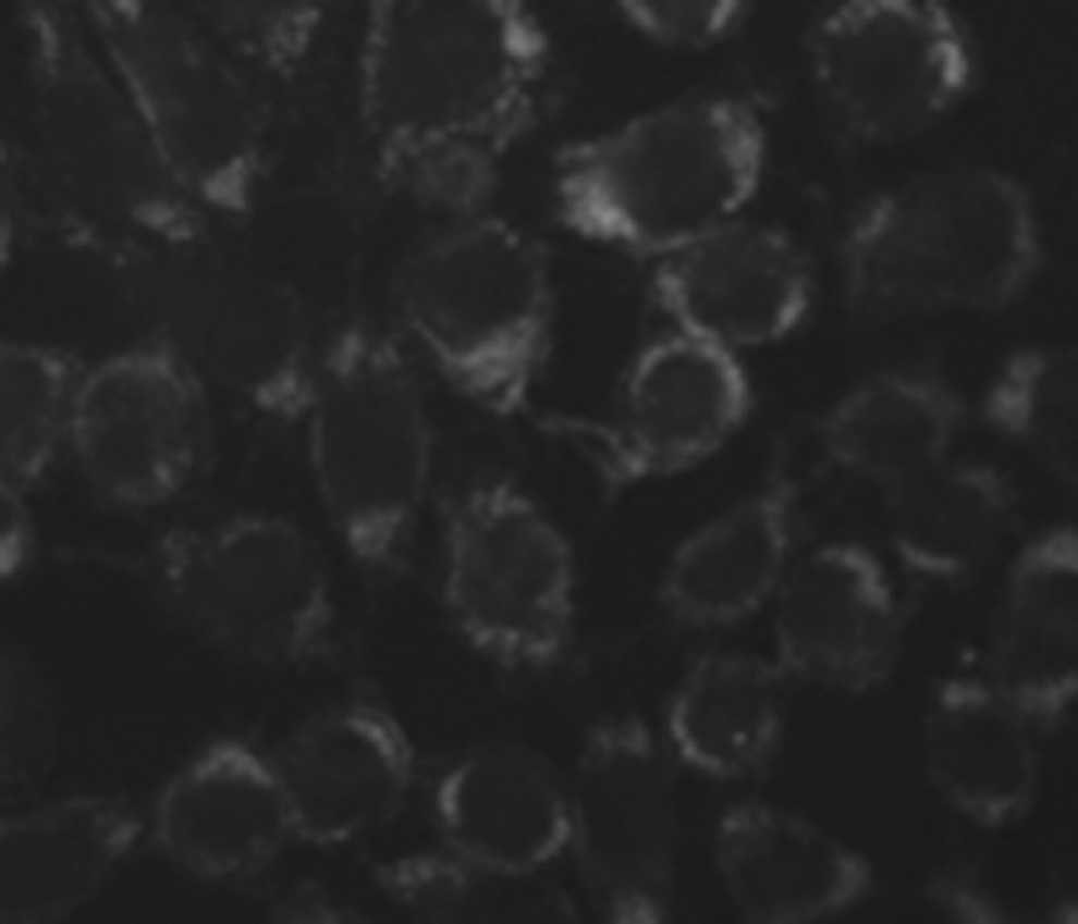
Positive expression for CA153 protein (400×).
Effects of ulinastatin and docetaxel on protein and mRNA levels of COX2 and IL-10 in primary breast cancer cells
As shown treatment of primary breast cancer cells with docetaxel and ulinastatin alone for 48 hours significantly decreased protein (Fig. 2) as well as mRNA (Fig. 3) abundance of COX2 and IL-10 compared with control. In addition, treatment with ulinastatin plus docetaxel further significantly reduced both protein (Fig. 2, F COX2=0.270, p<0.05; F IL-10=0.129, p<0.05) and mRNA (Fig. 2, F COX2=0.231, p<0.05; F IL-10=0.270, p<0.05) abundance of COX2 and IL-10 compared with treatment alone.
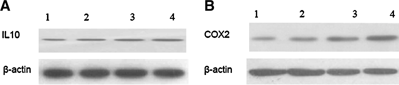
Western blot analysis of COX2 and IL-10 protein expression.
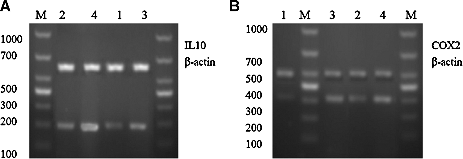
Effect of ulinastatin and docetaxel on mRNA levels of COX2 and IL-10 in the primary breast cancer cells.
Effects of ulinastatin and docetaxel treatment on secretion of PGE2 in primary breast cancer cells
As shown in Figure 4, secreted PGE2 was 48.62±4.46 ng/mL in control cells. After 48 hours of treatment with ulinastatin or docetaxel, its amount decreased significantly to 26.84±3.00 ng/mL and 15.21±1.86 ng/mL, respectively. In addition, treatment with ulinastatin plus docetaxel further decreased the PGE2 level to 9.91±1.06 ng/mL, which was significantly different from that in cells treated with ulinastatin or docetaxel alone (F=0.165, p<0.05).
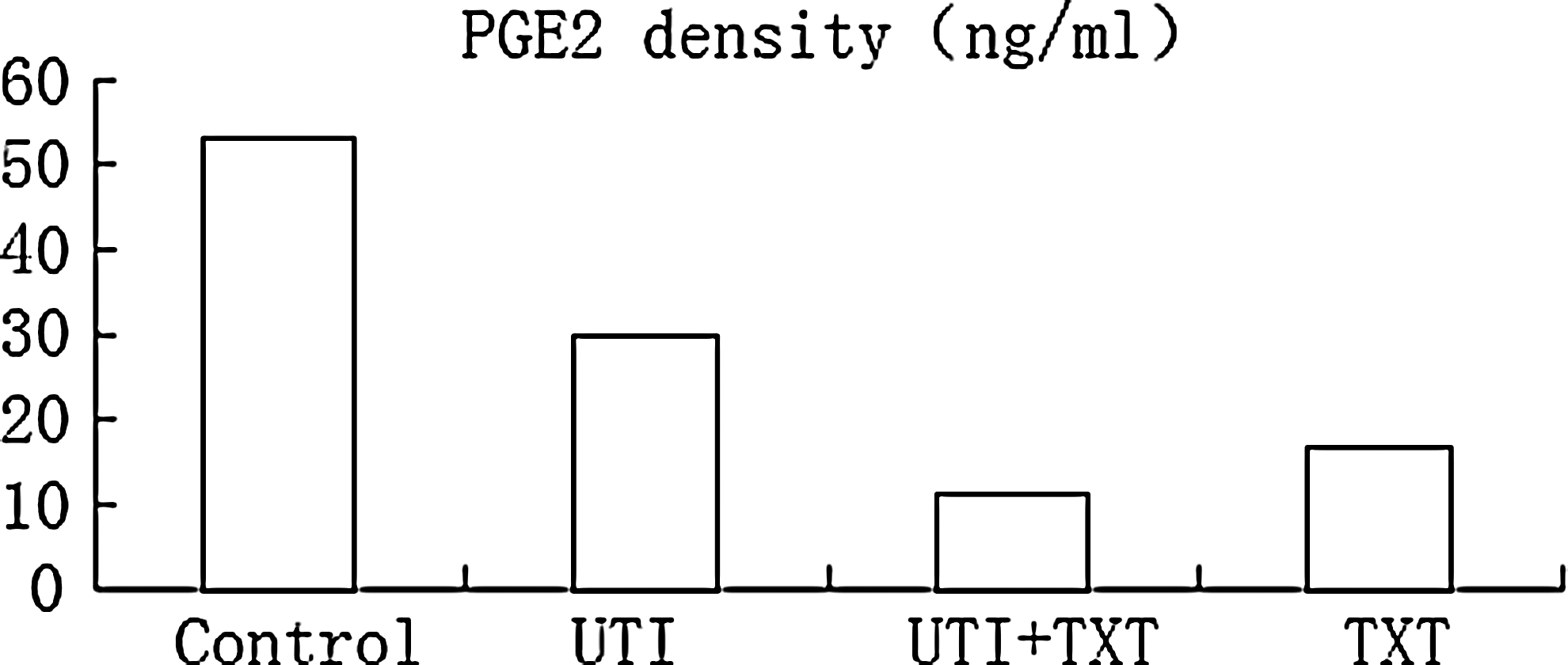
Enzyme-linked immunosorbent assay analysis of prostaglandin E2 secreted by the primary breast cancer cells.
Effects of ulinastatin and docetaxel on xenograft growth in nude mice
Two mice died during the experiment, one in the control group and the other in the ulinastatin group, possibly because of tumor consumption-induced cancerous cachexia. Before treatment, tumor volumes in all groups were similar (0.548±0.144 cm3). At day 21, tumor volume increased to 4.406±0.358 cm3 in the control group, but only to 3.547±0.029, 1.626±0.049 and 1.026±0.027 cm3 in mice treated with ulinastatin, docetaxel, and ulinastatin plus docetaxel, respectively. The tumor inhibition rates were 22.78%, 59.067%, and 88.783% for ulinastatin, docetaxel, and ulinastatin plus docetaxel, respectively. In addition, the inhibition rate of ulinastatin plus docetaxel was significantly higher than that of either ulinastatin or docetaxel treatment alone (p<0.05). Figure 5 shows the growth curves of xenografts in nude mice treated with control, ulinastatin, docetaxel, and ulinastatin plus docetaxel. In control and ulinastatin-treated mice, the average tumor volume increased dramatically and continuously overtime, whereas that in ulinastatin-treated mice increased gradually, but significantly slower than that in control mice (p<0.05). In docetaxel and ulinastatin plus docetaxel-treated mice, the tumor volume first slightly increased and then decreased overtime. Compared to all other groups, tumor growth in mice treated with ulinastatin plus docetaxel was significantly slower (p<0.05), indicating that treatment with docetaxel plus ulinastatin has stronger inhibitory effect on tumor than docetaxel alone. The calculated q was 1.088, meaning that ulinastatin and docetaxel had additive inhibitory effects in treatment of breast cancer xenografts in nude mice.
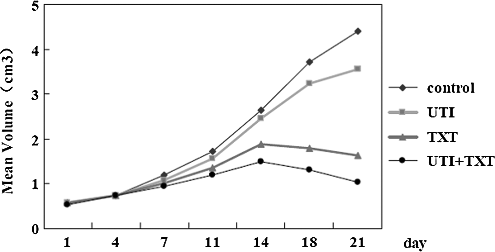
The growth curve of xenografts.
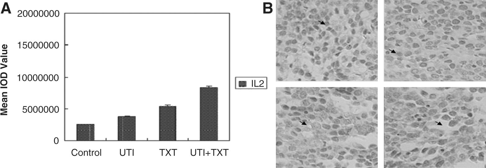
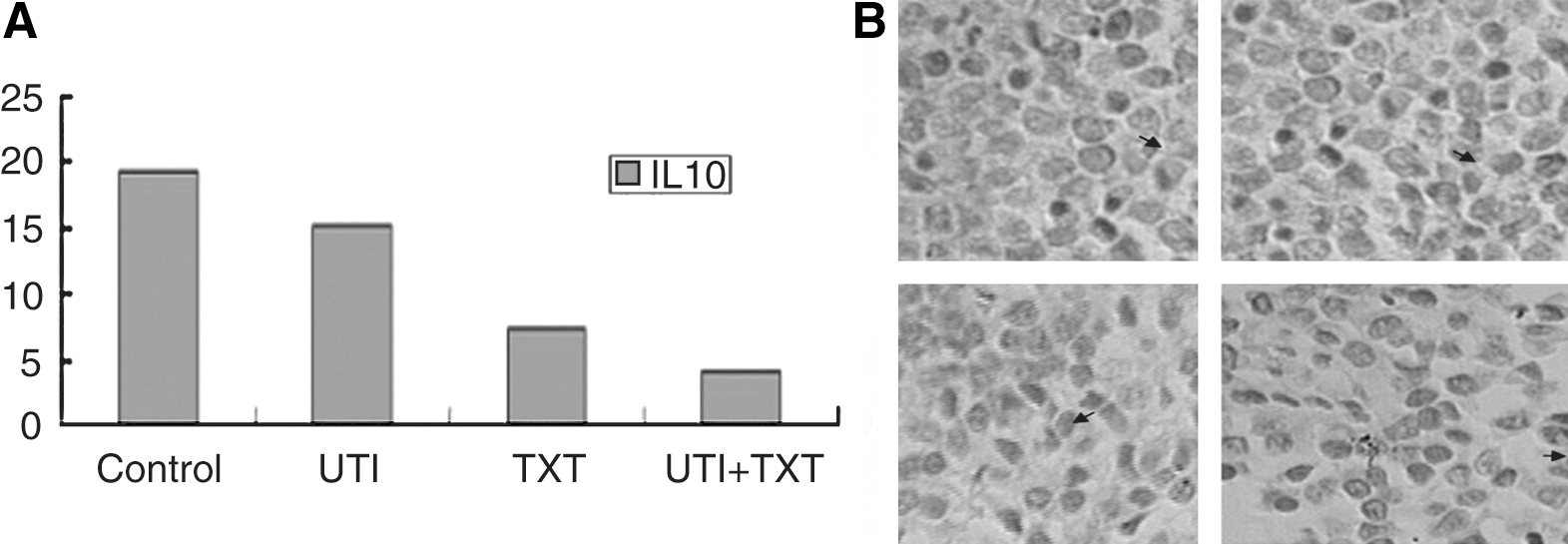
Effects of ulinastatin and docetaxel on expression of COX2, IL-10, IL-2, and EP2 in xenografts in nude mice
Pathological examination of xenografts in nude mice (hematoxylin and eosin staining) confirmed that the xenografts were of type human invasive ductal carcinoma. Figures 6 –8 show their relative expression levels. As shown, xenografts in mice treated with ulinastatin, docetaxel, and ulinastatin plus docetaxel had significantly lower expression of COX2, IL-10, and EP2 and higher expression of IL-2 compared with control mice. In addition, xenografts in mice treated with ulinastatin plus docetaxel had even lower COX2, IL-10, and EP2 and higher IL-2 levels than mice treated with ulinastatin and docetaxel alone (p<0.05).

Discussion
Breast cancer is one of the most common malignant tumors in women. Its incidence increases every year, and trends to increase in women at younger age. At present, it is mainly treated with surgery and chemotherapy. However, the treatment efficacy in some patients is poor due to the invasion and metastasis of tumor cells.
COX is a membrane-bound protein and the rate-limiting enzyme in PG synthesis from arachidonic acid. It has at least two COX isoenzymes, namely, COX1 and COX2, in mammals. COX1 is a constitutive enzyme and expressed in most normal tissues, whereas COX2 is expressed only in certain tissues such as brain, kidneys, and eyes under normal physiological conditions 5 and can be rapidly induced by growth factors, cytokines, endotoxin, hormones, and other cancer-promoting agents. Studies have found that COX2 is overexpressed in gastrointestinal cancer, lung cancer, and prostate cancer, 6 as well as in 40% patients with breast cancer 7 and involved in tumorgenesis, development, and metastasis including breast cancer invasion and proliferation through various mechanisms. Thus inhibition of COX2 has been an effective strategy in treatment of cancer.
Nonsteroidal anti-inflammatory drugs as nonselective COX inhibitors have been used to treat patients with breast cancers. Although these drugs exert anti-inflammatory and antitumor effects through COX2, they also inhibit COX1, causing mucosal and gastrointestinal bleeding and other adverse reactions. 8 Selective COX2 inhibitors can preserved their anti-inflammatory and antitumor effects while reducing gastrointestinal adverse reactions. 9 However, long-term use of high-dose selective COX2 inhibitors can increase thrombosis. 10,11 Thus, co-application of other proteases inhibitor to COX2 can be regarded as a good choice in cancer treatment.
Ulinastatin is a broad-spectrum enzyme inhibitor extracted from urine. It can inhibit varieties of enzymes, including proteases and hydrolases and mainly used for clinical treatment of pancreatitis. We have previously found that ulinastatin could reduce matrix metalloproteinase-9 expression, 3 inhibit growth of both human breast cancer MDA-MB-231 cells and tumor xenograft in nude mice, and reduce expression of IL-6 and IL-8. 2 In this study, we further found that treatment of primary human breast cancer cells as well as its xenografts in nude mice with ulinastatin could inhibit their growth and further enhance the efficacy of docetaxel on tumor growth in vitro and in vivo.
COX2 expression is regulated by binding of its cyclic adenosine monophosphate response element with transcription factors AP-1 and PEA3 activated by HER2/neu/PI3K/Akt signaling pathway. Inhibition of Akt phosphorylation by blocking EGFR-mediated signaling pathway could reduce COX2 expression and eventually inhibit breast cancer development. 12 Among downstream molecules of COX2, PGE2, PGI2, and TXA2 are closely related to tumor development. Inhibition of PGI2 can promote platelet aggregation and lead to thrombosis and stroke. 13 Thus, PGE2, which can promote tumor formation through its four ligands, EP1–4, becomes an important target downstream of COX2 signaling pathway in antitumor therapy. 14 In this study, we found that treatment of primary human breast cancer cells as well as their xenografts in nude mice with ulinastatin could inhibit and further enhance the inhibitory effects of docetaxel on levels of COX2 and EP2 as well as PGE2 secretion.
Th1/Th2 balance reflects the immune status. Our previous study has found that breast cancer tissues present Th2-type immunodominance, suggesting that shift of Th1/Th2 immunobalance to Th2 may be one of the reasons for tumor metastasis. 1 IL-2 is an immune enhancing factor produced by Th1 cells and can promote T cell proliferation and enhance cytotoxic activity of T cells and monocytes. 15 IL-10 is an immunosuppressive factor produced by Th2 cells and can inhibit activation and cytokine production of T cells and proinflammatory cytokine production of monocytes and granulocytes. IL-10 and IL-2 are also regulated by PGE2. 16 In this study, we explored the possible roles of ulinastatin and docetaxel treatment on expression of IL-10 and IL-2 and found that treatment of breast cancer cells and their xenografts in nude mice with ulinastatin can inhibit IL-10 expression at both protein and mRNA levels and enhance IL-2 expression, and more importantly enhance the effects of docetaxel on IL-10 and IL-2.
The study found that ulinastatin had similar effects of docetaxel and can enhance docetaxel's anticancer effects possibly by inhibiting COX2 expression, reducing PGE2 and EP2 expression and their binding, upregulating IL-2, and downregulating IL-10.
Footnotes
Acknowledgments
This project was supported by the Fund of Chongqing Science and Technology Commission (CSCT, 2008AC5082).
Disclosure Statement
The authors declare that none of the authors have any conflicts of interests.