
Abstract
The aim of this work was to determine the potential of 99mTc carbonyl-labeled dextran-cysteine-mannose (DCM20) as a mannose receptor targeting agent for sentinel lymph node (SLN) detection using biological in vitro and in vivo assays. 99mTc labeling of the previously reported DCM20 ligand was carried out via the [99mTc(H2O)3(CO)3]+ synthon. High-performance liquid chromatography (HPLC) showed >99% radiolabeling yield using 50 μg of the ligand. In vitro cell uptake studies performed in RAW 264.7 mouse macrophage precursor cells showed a specific uptake of the preparation. In vivo distribution and scintigraphic imaging were studied in the Wistar rat model. Appreciable uptake and retention of the radiolabeled conjugate was observed in the SLN (4.53%±0.29% at 15 minutes and 3.35%±0.72% at 180 minutes postinjection [p.i.] for 2.5 μg/animal) with a high percentage of popliteal extraction (≥98% at all time points studied), and negligible activity in other nodes as well as blood and nontarget organs. The radiolabeled conjugate also exhibited rapid clearance from the injection site (from ∼39.1% clearance at 15 minutes to ∼56.5% clearance at 180 minutes p.i.), comparable to current clinically employed agents for SLN detection. These results suggest that [99mTc(CO)3]DCM20 could be a potentially useful receptor-based SLN detection agent.
Introduction
Sentinel lymph nodes (SLNs), the nodes receiving lymphatic drainage directly from a region of primary tumors, 1 are widely regarded as providing useful prognostic information for several cancers, including melanomas and cancers of the breast, lung, colon, head and neck, and gynecological origin. 2 –7
There may be more than one SLN for a primary tumor, and they may not necessarily be the geographically most proximal nodes. Hence, they should be detected by agents that target their location by tracking lymphatic drainage from the tumor region. In the clinical scenario, dyes, 99mTc-labeled colloidal agents, or a combination thereof are increasingly relied on for the selective identification of SLN's for subsequent biopsy examination in order to detect the presence of cancer cells. 8 The desired in vivo properties for an ideal SLN detection tracer include rapid clearance from the site of injection, selective uptake in the SLN, and minimal spread to other nodes/nontarget regions. Currently, 99mTc-labeled sulfur colloid and human serum albumin (HSA) nanocolloid are used in clinical practice for the detection of SLN. 9,10 These agents have particle size as the single most important differential factor dictating the kinetics and selectivity of their uptake in SLN. Since particle size is inversely proportional to the rate of transport of the tracer from the site of injection to the lymphatic system and its further spread beyond the sentinel node, the currently employed agents of this class have a significant degree of retention at the site of injection and slower kinetics of transport thereon to the lymphatic network.
To alleviate the reliance on particle size and the compromise involved therein, mannose receptors expressed by macrophages located in the lymph node have been identified as a target for developing mannose-bearing receptor-specific ligands for SLN detection. 11 –13 It is envisaged that specific affinity to the mannose receptor will lead to the high uptake of a 99mTc-labeled mannose-bearing tracer by the SLN macrophages, allowing for the design of ligands with a significantly smaller particle size. Such ligands may be expected to exhibit a high rate of clearance from the injection site, thereby resulting in a lower radiation background, without an adverse impact on the selectivity of uptake in the SLN. The first ligand of this type is Lymphoseek, a [99mTc]DTPA mannosyl dextran complex that has been patented and is in clinical trials for SLN mapping in different cancer types. 11 There are also reports on the use of 99mTc-labeled mannosyl HSA 14 for the same purpose. The International Atomic Energy Agency (IAEA), Vienna, initiated a multi-nation coordinated research project toward the development of novel mannose receptor targeting 99mTc-labeled tracers that could be used for SLN detection. A preliminary biological evaluation study of dextran-pyrazolyl-mannose derivatives 15 performed as a part of this project showed encouraging results toward the application in SLN detection. Based on the principle just mentioned, newer derivatives were reported by Tsotakos and coworkers, 16 constructed using dextran as a backbone, S-derivatized cysteine as the chelator for labeling with the [99mTc(H2O)3(CO)3]+ precursor, and mannose residues for binding to mannose receptors on the lymph node. Of these, dextran-cysteine-mannose (DCM20) was synthesized by the reaction of dextran (MW 11800) with allyl bromide to give allyl dextran, followed by the formation of dextran-S-cysteine, which is subsequently mannosylated to give the final product. DCM20 (MW 22 kDa) could be quantitatively labeled with the [99mTc(H2O)3(CO)3]+ core. Here, we report the results of the biological evaluation of [99mTc(CO)3]DCM20 with a view toward its possible use as an SLN detection agent in the clinic.
Materials and Methods
DCM20 ligand (molecular weight ∼22 kDa) used in these studies was synthesized at the Institute of Radioisotopes and Radiodiagnostic Products, NCSR “Demokritos,” Athens, Greece. IsoLink kit® vials for the preparation of [99mTc(H2O)3(CO)3]+ core were a gift from Mallinckrodt-Covidien, Petten, Holland. 99mTcO4 − was eluted using isotonic saline from an in-house 99Mo/99mTc generator at the Radiopharmaceuticals Division, Bhabha Atomic Research Center. HPLC-grade solvents were degassed before use for all HPLC analyses. Dulbecco's modified Eagle's medium (DMEM), Eagle's modified minimal essential medium (MEM), Hank's balanced salts solution (HBSS) used for cell culture, and in vitro experiments were procured from Sigma. Fetal bovine serum (FBS) for use as a growth supplement in cell culture was procured from Hi-Media, India. RAW 264.7 mouse macrophage precursor cell line and BRL3A rat liver cell line used for in vitro cell uptake studies were procured from the National Center for Cell Science, Pune, India.
Radioactivity measurements during labeling studies were carried out using a well-type NaI (Tl) detector (ECIL). HPLC analyses were performed on a JASCO HPLC system (JASCO) coupled to a PU 1575 UV/visible detector (JASCO) and an NaI (Tl) radioactivity detector (Raytest). The system was equipped with a C-18 reversed phase HiQ Sil (5 μm, 4×250 mm) column. Radioactivity measurements for in vivo distribution studies were performed on an integral line flat-bed NaI (Tl) Scintillation Detector (Harshaw). Scintigraphic imaging was performed on the Millennium MPS Scintigraphic Imaging System (Wipro-GE Healthcare Systems).
Radiolabeling of DCM20 via [99mTc (H2O)3(CO)3] + synthon
DCM20 was labeled with 99mTc using the procedure of Tsotakos et al. 16 Briefly, [99mTc (H2O)3(CO)3]+ synthon was prepared by adding 0.5 mL of 99mTcO4 − (∼370 MBq) to the contents of the IsoLink kit vial and allowing for a reaction in a water bath at 100°C for 20 minutes. The pH of the prepared synthon was adjusted to ∼7.5 using 0.1 N HCl, and 500 μL of it was added to 50 μg DCM20 and reacted at 70°C in a water bath for 30 minutes. The diagrammatic representation of [99mTc(CO)3]DCM20 is as given in Figure 1. The formation and yield of the [99mTc (H2O)3(CO)3]+ synthon and the labeled product [99mTc(CO)3]DCM20 were monitored by HPLC using H2O (solvent A) and CH3OH (solvent B) with 0.1% trifluroacetic acid in a gradient mode (at 0–1 minutes 0% B, at 1–9 minutes 0%–70% B, at 9–19 minutes 70% B, at 19–25 minutes 70%–95% B, at 25–35 minutes 95%–0% B) at a flow rate of 1 mL/min. For assessment of the labelling yield, measured activity of the reaction mixture was injected into the HPLC. The eluate of the complete HPLC run was collected in fractions, measured, and the total was compared with the injected activity to assess the quantitative recovery of 99mTc from the HPLC column as well as to rule out any reduced 99mTc colloid.
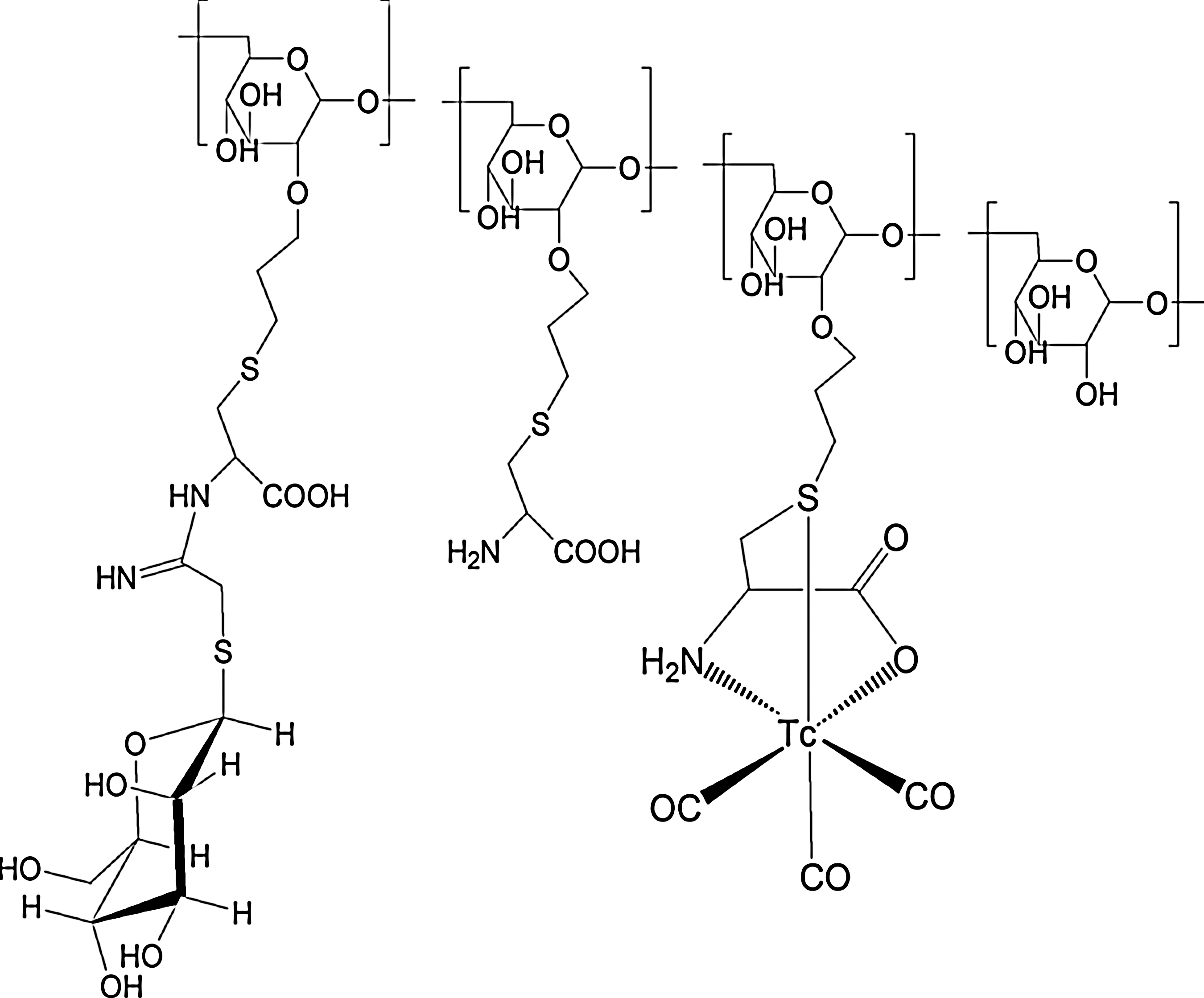
Diagrammatic representation of [99mTc(CO)3]DCM20. DCM20, dextran-cysteine-mannose.
In vitro biological evaluation studies
The aim of the in vitro studies was to ascertain the specific uptake of [99mTc(CO)3]DCM20 by mannose receptor-bearing cells. The RAW 264.7 mouse macrophage precursor cell line has been observed in the literature as bearing mannose receptors 17 and was selected as a suitable type among the cell lines available to us to assess the in vitro uptake of the mannosylated ligand. Previously reported in vitro binding studies of mannosylated ligands have used homogenized liver tissue 18 where the ligand was expected to bind with the mannose receptors located on the membrane of the macrophages associated with the liver (Kupffer cells). Hence, to determine nonspecific uptake, a rat liver cell line BRL3A was chosen as the control. RAW cells were cultured in DMEM, while BRL3A cells were cultured in MEM. The cells were incubated at 37°C with 5% CO2 and, in both cases, 10% FBS was added as a growth supplement. For harvesting, the cells were removed without any trypsinization, by the flushing of media and the scraping with a cell scraper.
For the experiment, RAW and BRL3A cells were plated in 24-well tissue culture plates at 106 cells/well in the respective culture media containing 1% FBS. They were incubated overnight at 37°C with 5% CO2 to allow for adherence and monolayer formation on the well surface. Before the addition of [99mTc(CO)3]DCM20, the media in each well were replaced with 450 μL of fresh culture media containing 1% FBS. Different concentrations of [99mTc(CO)3]DCM20 in a 50 μL volume were added to the wells. Each reaction was carried out in triplicate. The cells were incubated for 2 hours at 37°C in an orbital shaker incubator (30 rpm). At the end of the incubation period, the media were aspirated, and the wells were washed twice with ice-cold HBSS to remove unbound activity. Finally, 1 mL of warm 8 M NaOH was added to each well to dissolve the cell layer, and the contents were transferred to radioactivity counting tubes. Activity measurements were performed in a well-type NaI (Tl) counter with an energy window for 99mTc. Specific uptake of the [99mTc(CO)3]DCM20 conjugate in the mannose receptor-bearing RAW cells at each concentration was determined by subtracting the corresponding uptake value in the nontarget BRL3A rat liver cells.
In vivo biological evaluation studies
To assess the impact of changing ligand concentration on the in vivo localization of the preparation, animal studies were performed with two concentrations (200 μg/mL and 50 μg/mL) of [99mTc(CO)3]DCM20. All the animal experiments performed herein followed the guidelines of the institutional animal ethics committee. Studies were carried out in Wistar rats (female, 200–250 g) as described in Morais et al.
15
Briefly, the animals were anesthetized with ketamine:xylazine (10:1) before the administration of activity. Each animal was injected with ∼1.8 MBq of [99mTc(CO)3]DCM20 in ∼50 μL volume subcutaneously in the footpad region, followed by a gentle massage for ∼1 minutes with a gauze pad. The rejection criteria followed was the observation of any bleeding at the site of injection or measurements of more than 0.5% of administered dose on the gauze pad. Postinjection (p.i.), the animals were kept in separate sets (n=4 per set) for incubation periods of 15, 60 and 180 minutes under normal conditions, and water was provided ad libitum. Five minutes before the end of each incubation period, the animals under anesthetized condition were given a subcutaneous administration of ∼50 μL Patent Blue Dye (1%w/v in saline) in the same region as the labeled preparation. At the end of incubation, the animals were sacrificed, and the relevant organs and tissues, including the popliteal node (which serves as the SLN in this protocol) and secondary nodes, were excised for the determination of in vivo distribution of 99mTc activity. Radioactivity measurement was performed on a flat bed-type NaI (Tl) detector. The activity retained in each organ/tissue was expressed as a percentage of the total injected dose (%ID). Popliteal extraction (PE) was calculated using the formula given next (Vera et al. 2001):
For scintigraphic imaging, using the same techniques as just referred to for anesthesia and the administration of [99mTc(CO)3]DCM20, ∼37 MBq of the radiolabeled preparation (in 50 μL volume) was administered to each animal (n=3). Blue dye tracking is not required in this study. For image acquisition, the animals were placed with their dorsal side facing the collimator. Planar static images were acquired at 15, 60, and 180 minutes p.i. using the Genie Acq Image Acquisition software (Release 3.0). Acquisition parameters were as follows: Matrix 256×256, Zoom 1.33, 5 minutes acquisition time. The site of injection was masked with lead shielding during acquisition. Subsequent image processing was achieved with the Xeleris image processing software (Version 1.0272).
Results and Discussion
99mTc-carbonyl labeling of DCM20 conjugate
Greater than 99% yield on labeling with 99mTc could be obtained using 50 μg of the DCM20 ligand as shown by HPLC. In the given HPLC system, while 99mTcO4 − and the [99mTc(H2O)3(CO)3]+ synthon were eluted at 3.3 and 6.3 minutes, respectively, [99mTc(CO)3]DCM20 had a retention time of 13.6 minutes (Fig. 2). It was observed from the radioactivity measurement of the collected fractions of HPLC eluates that injected activity was quantitatively recovered (>99% from the column). No activity was retained on the column, indicating the absence of any reduced 99mTc colloid. The presence of a single peak corresponding to the labeled product indicates the absence of any unbound forms of 99mTc, obviating the need for any further purification.
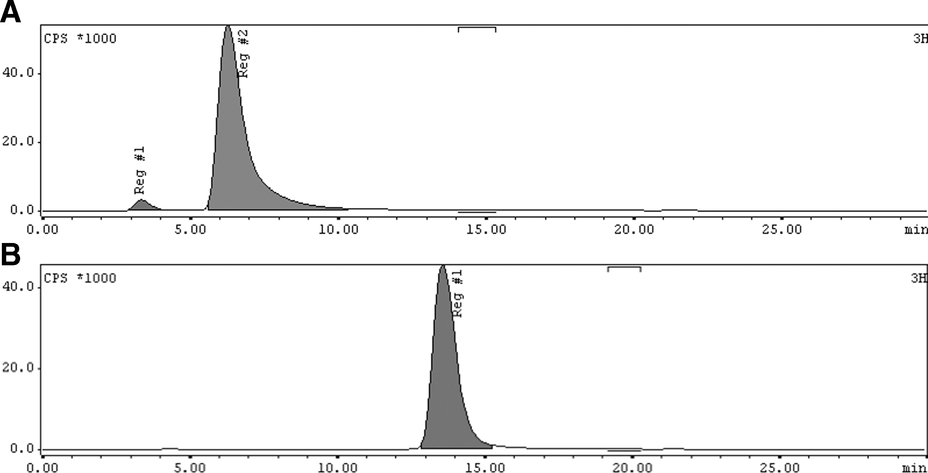
HPLC pattern of
In vitro studies
The result of the in vitro cell uptake studies is provided in Figure 3. Specific uptake of [99mTc(CO)3]DCM20 in RAW cells was calculated after subtracting the uptake obtained at each concentration to the nonspecific cell line BRL3A, and expressed as a percentage of the total activity added. Under the conditions of the reaction, [99mTc(CO)3]DCM20 showed specific concentration-dependent uptake in the mannose receptor-bearing cell line.
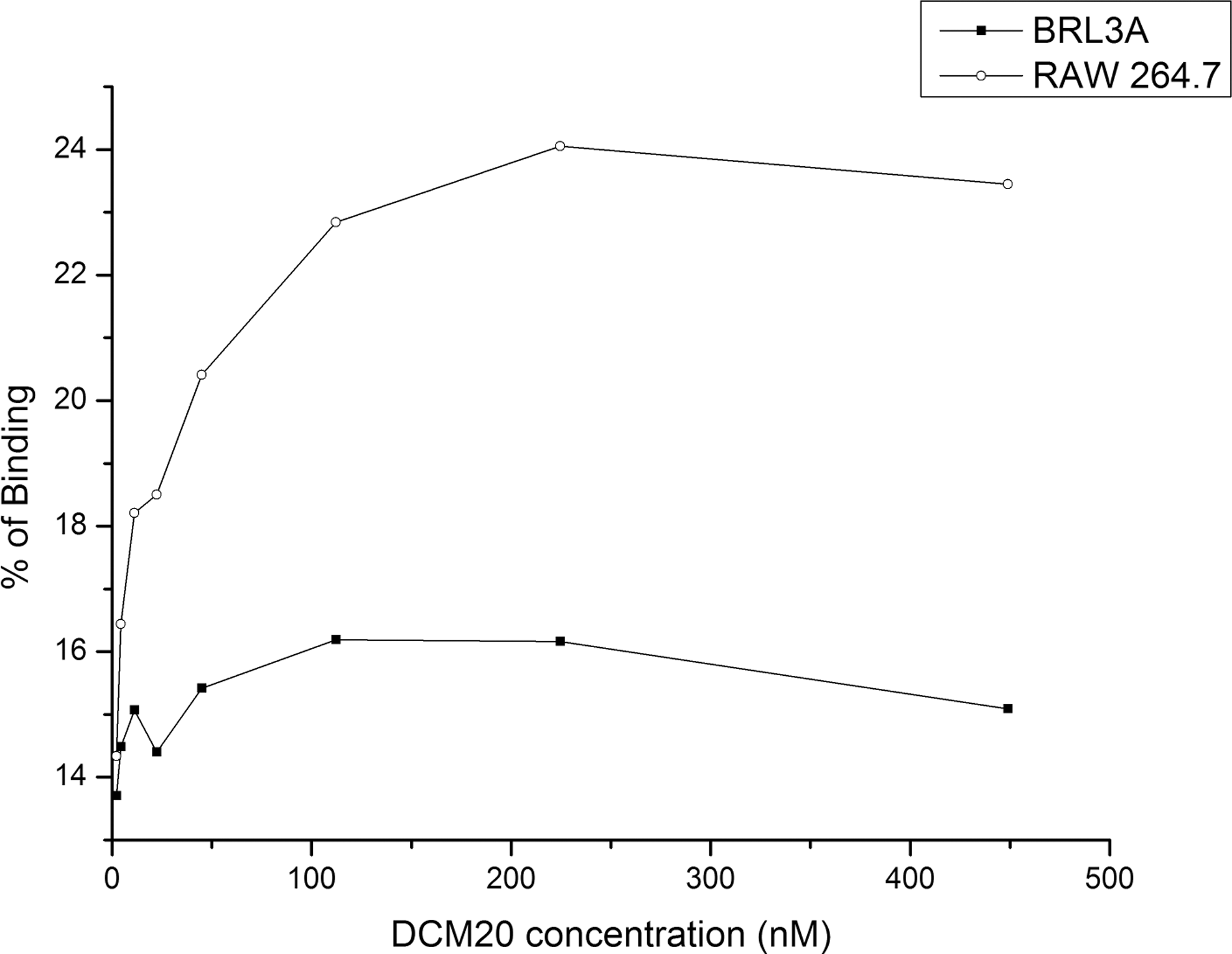
In vitro uptake curve showing specific uptake of [99mTc(CO)3]DCM20 in mannose receptor-bearing RAW 264.7 mouse macrophage cells.
In vivo studies
For the purpose of studying the effect of the variations of ligand concentration on the pattern of in vivo uptake, the animal studies were performed using two different concentrations of [99mTc(CO)3]DCM20. Initially, [99mTc(CO)3]DCM20 at a concentration of 200 μg/mL was used. At 50 μL per animal, this would translate to 10 μg ligand per animal at the site of injection. The results of in vivo biodistribution studies for this ligand concentration are given in Table 1. At 60 minutes, ∼7.5% of injected activity was localized in the sentinel node. However, there was also a significant accumulation of activity in the secondary node, which resulted in a low PE value of ∼53%. At 180 minutes, the PE increased to ∼74%, mainly due to the washout of [99mTc(CO)3]DCM20 from the secondary node. An appreciable amount of activity was also observed in the liver. It may be hypothesized that the mannose receptors of the SLN macrophages are saturated by the concentration of the mannose-bearing ligand used, thereby causing the spread of unbound [99mTc(CO)3]DCM20 to further nodes in the lymphatic system and, subsequently, to the blood circulatory system, leading to the accumulation of activity in the liver. These instances of nontarget accumulation indicated that [99mTc(CO)3]DCM20 administered at this concentration is not entirely suitable toward use as a tracer for SLND, at least in the tested animal model (From the measurements made in our study, typical rat lymph node weight was 5–7 mg). Larger animals with a greater mass of the lymph node may show a better distribution profile.
DCM20, dextran-cysteine-mannose; PE, popliteal extraction.
In the second set of experiments, biodistribution studies were carried out using 50 μg/mL of DCM20 conjugate, delivering 2.5 μg of tracer per animal in 50 μL at the site of injection. This made a significant difference to the in vivo distribution pattern as shown in Table 2. As early as 15 minutes p.i., ∼4.5% of the injected activity accumulated in the sentinel node. This is lesser than %ID value for sentinel node accumulation at the higher concentration, but appears to remain largely constant, with a gradual fall at 180 minutes. The importance of using a lesser amount of tracer was seen from the negligible activity accumulation in the secondary node at all the tested time points up to 180 minutes. As a result, the PE value remained very high throughout the period of the experiment, showing a high selectivity of uptake in the sentinel node. In addition, there was rapid clearance of activity from the site of injection (footpad). The activity at the site of injection reduced from ∼60% at 15 minutes to ∼43.5% at 180 minutes. This would suggest a reduced radiation background and improved sensitivity of detection in actual clinical practice. There was also no appreciable accumulation of activity in the blood, liver, or any nontarget regions. These are highly desirable properties for a proposed tracer in SLN detection in the clinic.
The scintigraphic images reflect the results of the biodistribution studies. A comparison of the images (Figure 4) obtained for different concentrations (10 μg ligand/animal and 2.5 μg ligand/animal) of [99mTc(CO)3]DCM20 administered for the various time points shows a clear improvement with the use of a lower concentration (2.5 μg ligand/animal), as the sentinel node is clearly demarcated throughout the period of study without appreciable counts in any other node or nontarget region.
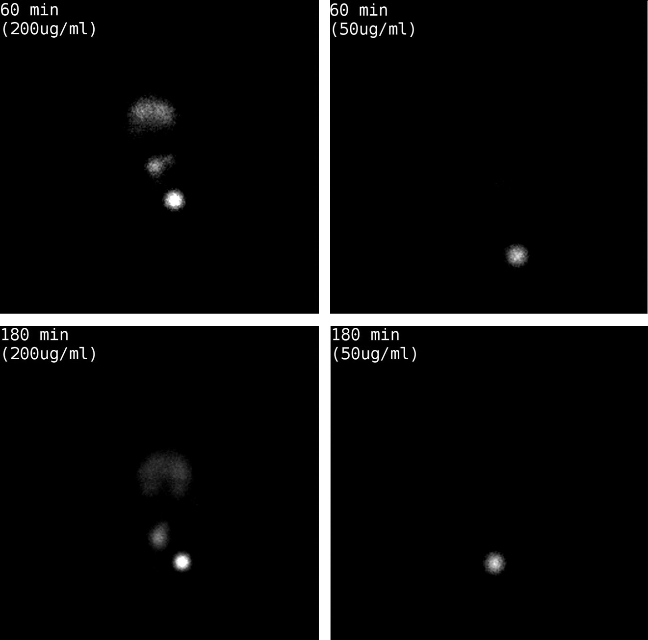
Scintigraphic images showing comparison of in vivo distribution of different concentrations of [99mTc(CO)3]DCM20 in Wistar rat model at corresponding time-points.
Our data cannot be directly compared with the reported literature for other reported radiotracers for SLN detection such as filtered 99mTc-labeled sulfur colloid or [99mTc]DTPA-mannosyl-dextran, as these compounds were evaluated using different animal models. However, in terms of the PE parameter (60 minutes p.i.), [99mTc(CO)3]DCM20 displays a higher or comparable PE value to filtered 99mTc-labeled sulfur colloid (78.8%±6.5%) and [99mTc]DTPA-mannosyl-dextran (90.1%±10.7%) 8 . In addition, the clearance pattern of [99mTc(CO)3]DCM20 from the injection site (both 50 and 200 μg/mL concentrations at 60 minutes p.i.) was superior to that reported for 99mTc-sulfur colloid (70.4%±11.0%), 99mTc-HYNIC-NMA-tricine2 (67.57%±8.29%), and 99mTc-DTPA-mannosyl-dextran (52.6%±10.5%). 11,13 The in vivo studies conducted on the Wistar rat model earlier reported with the 99mTc-labeled dextran-pyrazolyl-mannose complex 14 (as referred to in Morais et al. 15 ) were performed with 20 μg ligand administered per 50 μL per animal, but showed better specific uptake properties than those obtained with [99mTc(CO)3]DCM20 at 10 μg ligand per animal. It is now aimed at performing additional biological studies with different concentrations of the 99mTc-complex 1415 just mentioned in order to study the impact on its in vivo distribution profile in the tested animal model and make relevant comparisons to the results obtained herein for DCM20.
Conclusions
The in vitro biological studies performed with [99mTc(CO)3]DCM20 in this work indicate its ability to be specifically taken up in the macrophages of the lymph node. The in vivo studies demonstrate a differential distribution pattern that is dependent on the concentration of the labeled complex, which may be indicative of the ability of the complex to bind with and saturate the mannose receptors of lymph node macrophages. At the lower concentration used, excellent retention in the sentinel node and appreciable clearance from the site of injection were obtained. All these results suggest that the [99mTc(CO)3]DCM20 conjugate could serve as a useful tool for SLN imaging in the clinical scenario.
Footnotes
Acknowledgments
This work was carried out as a part of the IAEA Coordinated Research Project (CRP) on “Development of 99mTc radiopharmaceuticals for sentinel node detection and cancer diagnosis.” The authors are grateful to Dr. A. Duatti, Scientific Secretary, IAEA, and Dr. R. Pasqualini, CISBIO (France), for their crucial scientific input and direction given to this work. The authors express their sincere thanks to Dr. M.R.A. Pillai, Head, Radiopharmaceuticals Division, Bhabha Atomic Research Center, India, and Dr John Zaknun of IAEA for initiating the coordinated research project.
Disclosure Statement
There are no existing financial conflicts.