
Abstract
Hepatocellular carcinoma (HCC) is one of the most common malignancies over the world. Alpha-fetoprotein (AFP) is an oncofetal protein during HCC development that could generate weaker and less reproducible antitumor protection, and it may serve as a target for immunotherapy. Therefore, it is imperative to enhance its immunogenicity and develop therapeutic vaccines to eliminate AFP-expressing tumors. In this study, by way of glutaraldehyde cross-linking, we constructed a potential therapeutic protein complex vaccine, heat shock protein 72 (HSP72)/AFP. Our results demonstrated that AFP and HSP72 synergistically exhibited significant increases in AFP-specific CD8+ T cell responses and impressive antitumor effects against AFP-expressing tumors. Priming mice with the reconstructed vaccine, we elicited robust strong protective immunity. Our study suggests that a tumor vaccine by cross-linking tumor antigen and HSP72 is a promising approach for cancer therapy.
Introduction
Hepatocellular carcinoma (HCC) is one of the most common malignancies over the world and accounts for as many as 1.2 million deaths annually. 1 The highest incidence is in Southeastern and Eastern Asia, with a rate of 18.3–35.5 per 100,000 populations. 2 It is also rising rapidly in China because of hepatitis B and C infections. 1,2 Established treatments of HCC, such as chemo- and radio-therapy, usually fail to prevent the disease progress due to the proliferation of residual malignant cells that have escaped control by these methods. Although surgery and liver transplantation are the effective therapy, most patients still lose a chance due to diagnosis at a late stage or underlying liver insufficiency in the setting of cirrhosis. 3 The most common causes of mortality in patients with HCC include recurrence, metastasis, and the development of new primary tumors. 3 Novel therapies for HCC should be developed. A combined therapy is likely to prolong patients' life and living quality. In the recent years, immunotherapy with therapeutic vaccination, which could manipulate and enhance the host's immune system to elicit antigen-specific responses to the tumors, has attracted attention widely.
Much attention is paid to tumor active immunity whose purpose is to induce the hosts' immune attack on tumor cells. About 80% of HCC have a high expressing rate of alpha-fetoprotein (AFP), which may serve as a target for immunotherapy. 3,4 AFP is an oncofetal protein during HCC development that could generate weaker and less reproducible antitumor protection. Therefore, a vaccine specifically targeting AFP is particularly desirable. AFP oncogenic proteins are critical to the induction and maintenance of cellular transformation, which represent ideal target antigens for therapeutic AFP vaccine development. 5 Many therapeutic strategies that enhance the pre-existing antitumor immunity have been examined, including recombinant proteins, 6 peptides, 7 plasmid DNA, 8 chimeric virus-like particles, 9,10 viral or bacterial vectors expressing AFP proteins, 11 and adoptive transfer of tumor-specific T cells. 4,5 However, therapeutic vaccination has been restricted by inadequate antigen-specific immune responses. Therefore, ways to develop more potent immunotherapy aimed at initiating very robust antigen specific immune responses need to be thoroughly explored.
Heat shock proteins (HSPs) have been verified to act as potent immunoadjuvants to enhance antigen-specific tumor immunity. 12 HSP play an important role, not only in the process of protein folding, transport, and degradation, but also in participating in directing more efficient antigen presentation to CD8+ T cells through the MHC-I pathway. 12 This central role for HSP in therapeutic vaccine is facilitated by their dual functions in both chaperoning antigens and serving as an adjuvant. HSP are classified into families according to their size, structure, and function, including small HSP, HSP60, HSP70, HSP90, gp96, and calreticulin (CRT). They have different structures that are adapted to different immunoadjuvant functions. 12,13 Specifically, HSP90 and HSP70, the most abundant cytosolic chaperones, have been proposed to transport peptides to the heterodimeric transporters associated with antigen processing (TAP1 and TAP2); whereas gp96 and CRT, the major chaperone of the lumen of the endoplasmic reticulum (ER), have been presumed to facilitate assembly of the MHC I—β-2 microglobulin—peptide complexes in the ER. 13,14 Furthermore, HSP-based tumor vaccine strategies have been highly successful in animal models and are undergoing testing in clinical trials. 10,15,16 Therefore, an innovative approach that combines with families of HSP will likely generate more potent antitumor effects.
A number of groups have shown that superior levels of T-cell immunity could be generated using a heterogeneous prime-boost strategy, in which animals are primed and boosted with a palsmid vector encoding the stimulating molecule and targeted peptides.
8
–10
In many of these vaccine models,
17,18
HSP combined with certain antigen prime enhanced immunogenicity, presumably through processing and presenting the antigen to host antigen presenting cells (APCs
In the present study, we investigated whether the immunogenicity of AFP could be improved by HSP72 molecules and whether HSP72 could synergistically enhance the potency of the AFP therapeutic effect and further evaluated the immune responses induced by HSP72/AFP. We constructed a protein vaccine containing the molecule chaperon-HSP72 and AFP protein by way of glutaraldehyde cross-linking. Our results demonstrated that AFP and HSP72 synergistically exhibited significant increases in AFP-specific CD8+ T cell responses and impressive antitumor effects against AFP-expressing tumors. Primimg mice with the reconstructed vaccine, we elicited robust strong protective immunity. Therefore, cross-linking the HSP72/AFP protein holds promise for treating HCC through a combination of antigen-specific immunotherapy.
Materials and Methods
Mice and tumor cell lines
Six- to eight-week-old female BALB/C mice were purchased from the Experimental Animal Center at Fourth Military Medical University. All animals were maintained under specific-pathogen-free conditions, and all procedures were performed according to approved protocols and in accordance with recommendations for the proper care of laboratory animals. The investigation was approved by the Ethics Committee on animal Study at the Shaanxi University of Chinese Medicine (2004-4B). mAFP-producing H22 mice HCC cells were kindly provided by College of Biomedicine, Xi'an Jiaotong University, and maintained in RPMI 1640 (Gibco-BRL) with 10% fetal bovine serum, 100 U/mL penicillin, and 100 μg/mL streptomycin (Invitrogen Corp.) at 37°C under a humidified atmosphere of 95% air and 5% CO2. Lewis lung carcinoma (LLC) cells were purchased from the Institute of Biochemistry and Cell Biology of Chinese Academy of Sciences and cultured in DMEM (Gibco-BRL) with 10% fetal bovine serum at 37°C in 5% CO2.
AFP, HSP72, and conjugation
Mouse AFP was purchased from the GenWay Biotech, Inc. Lyophilized material was resuspended in sterile distilled water at 10 mg/mL, aliquoted, and stored at −70°C until use.
Mouse HSP72 expressing Escherichia coli colony was provided by Dr. X. Sun in North Dakota State University. The HSP72 expression vector contained an Isopropyl β-D-1-Thiogalactopyranoside (IPTG) inducible T7 promoter and was in frame with the C-terminal 6-His-tag (Invitrogen Corp.). E. coli cells transformed with the plasmid were grown until the optical density (OD) reached 0.5, and then, IPTG (1 mM final concentration) was added. After induction with IPTG at 37°C for 3 hours, cells were disrupted by sonication in a lysis buffer containing a nonionic detergent and lysozyme. Recombinant HSP72 protein was purified using nickel nitriloacetic acid (Ni-NTA)-agarose (Qiagen) column. Protein purity was assessed using sodium dodecyl sulfate-polyacrylamide gel electrophoresis (SDS-PAGE) stained with Coomassie blue. The identity of the purified protein was verified by Western blotting with a polyclonal antibody (Santa Cruz Biotechnology, Inc.). Protein concentration was determined using a BCATM protein assay kit (Pierce, Inc.).
One milligram AFP was coupled to 1 mg HSP72 in the presence of 0.2% glutaraldehyde in phosphate buffer saline (PBS) for 2 hours, and then dialyzed against PBS overnight. Aliquots of each conjugate were stored at −70°C until use.
Purification and Western blot analysis
The HSP72/AFP complex was purified by HSP72 polyclonal antibody-affinity chromatography and AFP polyclonal antibody-affinity chromatography sequentially. Protein purity was assessed using SDS-PAGE stained with Coomassie blue. The identity of the purified protein was verified by Western blotting with polyclonal antibodies (Santa Cruz Biotechnology, Inc.). Protein concentration was determined using a BCATM protein assay kit (Pierce, Inc.).
After centrifugation, the purified protein complex was denatured with SDS sample buffer (62.5 mM Tris-HCL, pH 6.8, 25 g/L SDS, 50 mL/L β-mercaptoethanol, 100 mL/L glycerol) at 100°C for 5 minutes. The purified protein complexes were then analyzed by 90 g/L SDS-PAGE. Proteins were transferred to a nitrocellulose membrane (Bio-Rad) and detected by immunoblotting with HSP72 rabbit antimouse polyclonal antibody (1:100) or AFP rabbit antimouse polyclonal antibody (1:100) at 4°C overnight, respectively. After a complete wash in PBS, the membranes were treated with horseradish peroxidase-labeled goat antirabbit antibody (1:100) for 45 minutes at 37°C. After a complete wash in PBS, the membranes were developed in 0.5 g/L freshly prepared 3, 3′-diaminobenzedine solution (DAB; Sigma Co.) for 8 minutes. Purified HSP72 and AFP were used as positive controls to determine the specificity of polyclonal antibody. β-actin was used as a negative control and internal reference.
Mice immunized with HSP72/AFP complex
Female BALB/C mice were randomly divided into the HSP72/AFP group, AFP group, HSP72 group, PBS control, and empty group. Every group had 10 mice. Before the injection, each group was diluted in saline to 100 μg/100 μL. Various groups of mice were injected into the left flank subcutaneously (s.c.). Priming and boosting was performed with 10 μg HSP72/AFP, AFP, or HSP72, whereas PBS was used as controls. A 0.3 insulin syringe with a 25-gauge 0.5-inch-long needle was used for the s.c. injections. Mice were boosted subcutaneously with the proteins just mentioned twice at 2 week intervals after the first priming. Two weeks after the last immunization, splenocytes were harvested and diluted to different concentrations.
ELISPOT assay
The ELISPOT was used to measure the frequency of cells producing the cytokine IFN-γ in splenocytes harvested from the immunized mice just mentioned. BD ELISPOT Plates (BD PharMingen) were coated with 5 μg/mL rat antimouse IFN-γ antibody in 100 μL of PBS. After overnight incubation at 4°C, the wells were washed and blocked with RPMI-1640 culture medium containing 10% fetal bovine serum. 1×10⁶ splenocytes were added to the ELISPOT plate wells along with 5 μg/mL of AFP containing 10% fetal bovine serum, 10 U/mL of human interleukin (IL)-2 (PEPRO Tech ET Ltd.). After being cultured at 37°C for 24 hours, the plate was washed and then followed by incubation with 2.5 μg/mL biotinylated IFN-γ antibody in 100 μL PBS containing 10% fetal calf serum (FCS) at 4°C overnight. After washing, avidin-HRP in 100 μL PBS was added and incubated for 1 hour at room temperature. After washing five times, spots were developed by adding 100 μL 5-bromo-4-chloro-3-indolyl phosphatase/Nitro Blue Tetrazolium (Boehringer Mannheim). The color spots, representing cytokine producing cells, were counted using an ELISPOT Reader System.
ELlSA assay
To determine the level of anti-AFP antibody in mice, we examined the serum of mice tail vein after the last immunization by ELISA. Ninety-six wells of a microplate were coated with 100 μL of 5 μg/mL AFP and incubated at 4°C overnight. The wells were then blocked with PBS containing 5% bovine serum albumin (BSA). Sera were prepared from the mice on day 14 postimmunization, serially diluted in PBS, added to the ELISA wells, and incubated at room temperature for 2 hours. After washing with PBS-T containing 0.05% Tween-20, the plate was incubated with 1:3000 dilution of a HRP-conjugated goat antimouse IgG antibody (Sigma-Aldrich Corp.) at room temperature for 1 hour. The plate was washed five times, developed with O-phenylenediamine away from light at 37°C for 15 minutes, and stopped with 50 μL of 2 M H2SO4. The ELISA plate was read with a standard ELISA reader at 490 nm. The quantity of antibody was measured in comparison with standard sample diluents.
Cytotoxic T-lymphocyte assays
BALB/C mice were immunized s.c. as just described. Two weeks after the last boost, 2.5×10⁷ splenocytes were cocultured with 5 μg/mL of AFP containing 10% fetal bovine serum, 10 U/mL of human IL-2 in RPMI 1640 supplemented with 10% FCS at 37°C in 5% CO2. After 5 days of stimulation, the viable splenocytes were recovered and used as effector cells, and the H22 cells or LLC cells were used as target cells. The Nonradioactive Cytotoxicity Lactate Dehydrogenase (LDH) release assay Kit (Promega, 249 USA) was performed to measure the effector cells against H22 or LLC tumor cells in the ratios of 10:1, 20:1, and 40:1, according to the manufacturer's protocol. Specific lysis was calculated according to the formula: percent specific lysis=([experimental release value − effector spontaneous release value − target spontaneous release value]/[target maximum release value −target spontaneous release value])×100. Results shown are representative of experiments repeated thrice.
In vivo tumor therapeutic experiments
To test the ability of HSP72/AFP vaccination to inhibit the growth of established tumors, BALB/C mice in every group were injected s.c. in the left flank with 5×10⁵ H22 cells per mouse on day 0, and then injected s.c. in the right flank with different vaccination treatments on day 3, 10, and 17 as just described. H22 tumor cells were washed after enzymatic digestion and resuspended in 0.2 mL of PBS per animal, then injected s.c. into the left flank, while PBS was used as control. The tumor growth was monitored every day. Tumor size was measured in two dimensions with calipers every 3 days 1 week after tumor inoculation. At each time point, tumor size was determined by measuring the smallest diameter (a) and the biggest diameter (b). Tumor volume was calculated using the formula: V=(a 2 b)/2. Tumor size measurements were performed by the mean value of each group and executed in duplicate to confirm the results. Percentage of tumor-free mice was recorded, and the survival of mice was monitored for 8 weeks from the day of tumor challenge.
Statistical analysis
All data were expressed as means±S.D. Comparisons between individual data points were made using a Student's t-test. The frequencies of IFN-γ-producing splenic cells were valued using χ 2-test. The Student's t-test was performed to analyze the significance of differences between final tumor volumes of different groups of animals. p<0.05 was considered statistically significant.
Results
Identification of purified HSP72/AFP complex
To obtain purified HSP72/AFP complex, a sequential polyclonal antibody-affinity chromatography was used. By HSP72 polyclonal antibody-affinity chromatography and AFP polyclonal antibody-affinity chromatography, respectively, the nonconjugated AFP and HSP72 were eluted. The purification of HSP72/AFP complex was confirmed by SDS-PAGE and Western blot (Fig. 1). The purified HSP72/AFP complex was identified by HSP72 antibody and AFP antibody via Western blot. The results confirmed that HSP72/AFP complex was constructed and produced successfully.
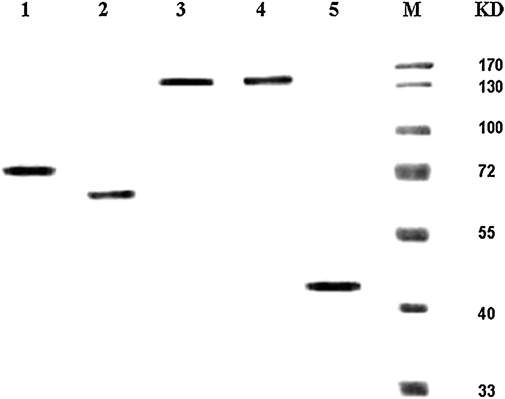
Analysis of HSP72/AFP complex by Western blot. The identity of the purified protein was verified by Western blotting with polyclonal antibodies. The relative molecular weights of HSP72, AFP, HSP72/AFP, and β-actin were ∼72, 69, 140, and 42 kDa, respectively. Lane 1, purified HSP72; Lane 2, AFP; Lane 3, purified HSP72/AFP complex with HSP72 antibody; Lane 4, purified HSP72/AFP complex with AFP antibody; Lane 5, β-actin; M: protein molecular weight markers. HSP72, heat shock protein 72; AFP, alpha-fetoprotein.
Prime-boost vaccines generate AFP-specific CD8+ T cells in vivo
Since cytotoxic T-lymphocytes (CTLs) have been known to play a critical role in tumor immunity, we examined AFP-specific CD8+ T cell frequency after HSP72/AFP vaccination by ELISPOT assays. Two weeks after the last immunization, splenocytes were harvested and stimulated with AFP. As shown in Figure 2, immunization of BALB/C mice with HSP72/AFP vaccine elicited much stronger T-cells responses than those with AFP immunization, whereas a vaccination with HSP72 or AFP alone produced a small one. Six hundred fifty IFN-γ spot-forming CD8+ T cells specific for AFP protein were detected per 1×10⁶ splenocytes derived from the HSP72/AFP vaccinated mice, compared with only 28 of those derived from AFP vaccinated mice. Therefore, the numbers of IFN-γ-producing CD8+ T cells in the splenocytes from mice immunized with HSP72/AFP were 20 times more than those from mice immunized with the HSP72 or those from mice vaccinated with AFP (p<0.01). Our data suggested that HSP72/AFP vaccine generated significantly higher AFP-specific CD8+ T cells response compared with the other vaccinated groups.
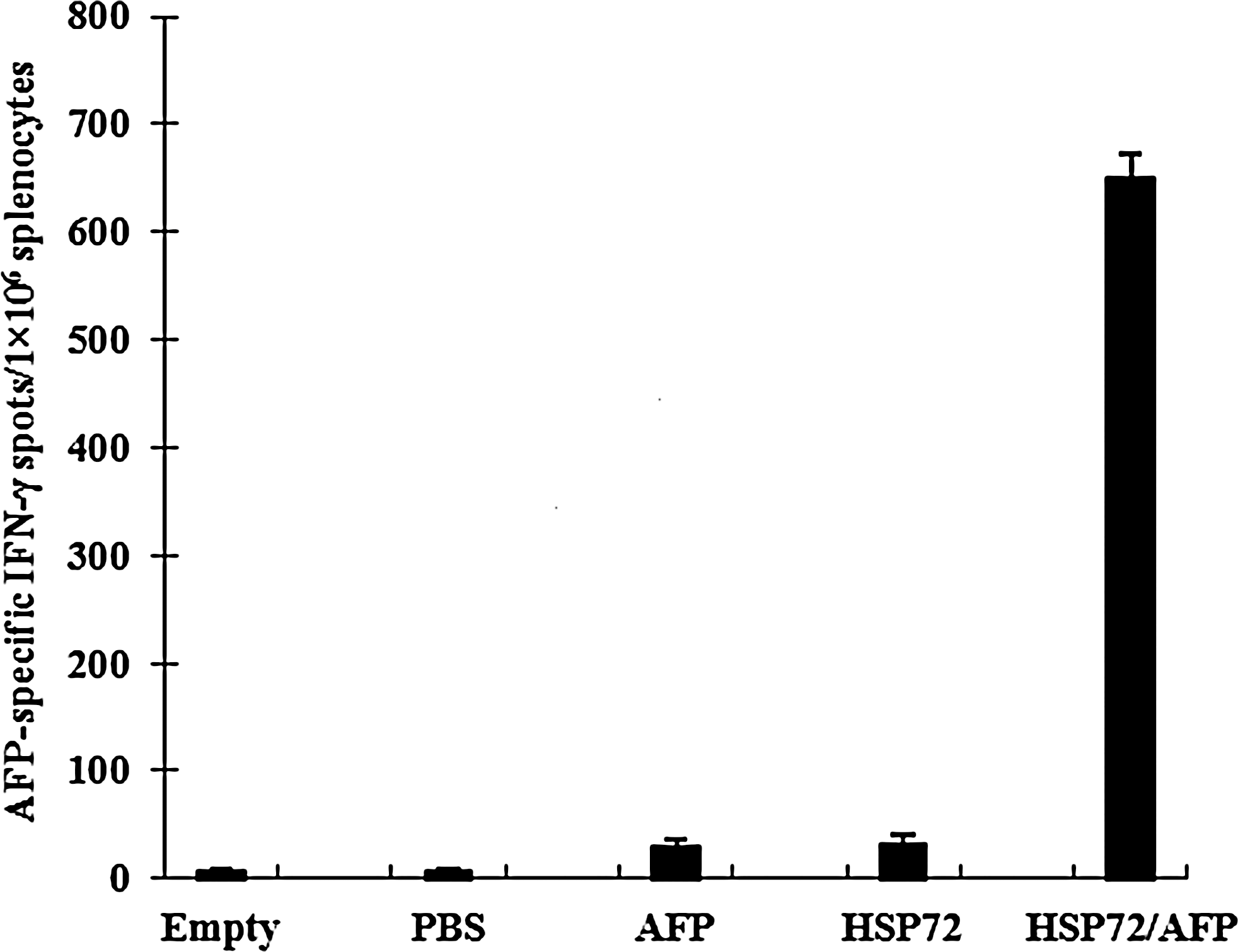
The quantity of AFP-specific CD8+ T cell secreting IFN-γ in the immunized mice spleen cells was tested by ELISPOT. AFP-specific IFN-γ spots counted. The spot numbers were the mean of triplicates±SE in each vaccinated group at 1×10⁶ cell number. Results shown here are AFP-specific spot-forming cells from five groups. Each group was tested in triplicate. PBS, phosphate buffer saline.
HSP72/AFP induced AFP-specific antibodies in vivo
The quantity of anti-AFP antibodies in the sera of the vaccinated mice was determined by ELISA. Fourteen days after the last vaccination, HSP72/AFP immunized mice produced a higher level of AFP-specific antibody than those immunized with AFP, while HSP72 and PBS groups produced a lower one. The concentration of anti-AFP antibody from mice immunized with HSP72/AFP was 30 times more than those from mice immunized with AFP (p<0.01) (Fig. 3). This result suggested that the modification of AFP vaccines definitely induced specific antibody responses.

AFP-specific antibody responses in immunized BALB/C mice. BALB/C mice were immunized with HSP72, AFP, and HSP72/AFP. PBS was used as a negative control. Serum samples were obtained from immunized mice 14 days after last vaccination. The presence of the AFP-specific antibody was detected by ELISA using serial dilution of sera. The results from the 1:20 dilution are presented showing the mean absorbance (A490 nm)±SE.
HSP72/AFP significantly primes potent AFP-specific CD8+ T cell responses
It has been previously demonstrated that the AFP/HSP70 DNA vaccine is able to induce the regression of AFP-expressing tumors in mice through either prophylactic or therapeutic treatment. 10,26 To examine whether HSP72/AFP vaccination is able to prime stronger AFP-specific CD8+ T cells than HSP72 or AFP immunization, we performed in vitro cytotoxicity assays of lymphocytes isolated from mice immunized with HSP72/AFP, HSP72, AFP, and PBS, respectively. Splenocytes were harvested 2 weeks after immunization and restimulated in vitro with AFP. Viable effector cells were assessed for cytotoxic activity against H22 cells or LLC cells (AFP-negative control). As shown in Figure 4, effector cells from mice immunized with HSP72/AFP showed much more stronger cytolytic effects on H22 target cells than those from mice vaccinated with HSP72, AFP, or PBS (p<0.01) (Fig. 4A). In addition, this cytolytic activity was specific for H22 cells, not for LLC cells (p<0.01) (Fig. 4B). Our results indicated that the cross linkage of AFP to HSP72 was required for enhancement of CD8+ T cell activity, as the administration of AFP or HSP72 only exhibited lower cytolytic activity. In contrast, splenocytes from the PBS control and empty group showed much lower levels of lysis of target cells.
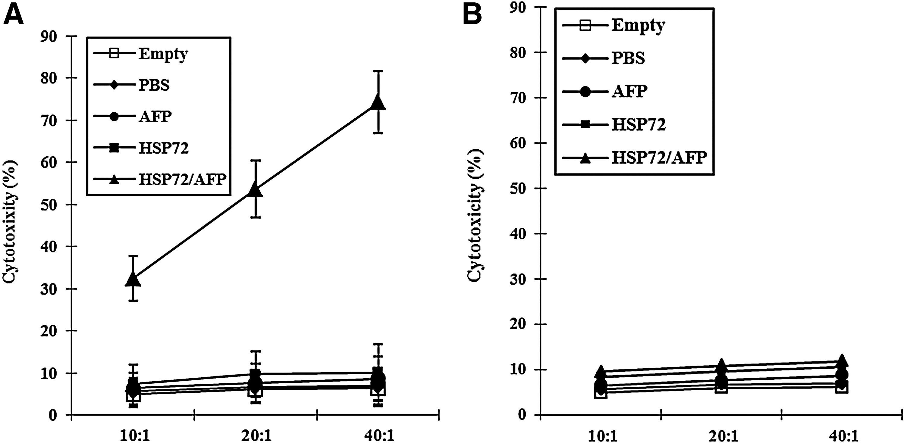
HSP72/AFP complex primed the strongest AFP-specific CD8+ T cell responses. Mice were immunized with HSP72/AFP, HSP72, AFP, and PBS, respectively. Mice were boosted subcutaneously with the proteins just mentioned twice at 2 week intervals after the first priming. Two weeks after the last boost, pooled splenocyte cultures were prepared and restimulated in vitro with AFP to generate effector cells separately. AFP-specific cytolytic activity was assayed against target cells at 10:1, 20:1, and 40:1 effector/target cell (E/T) ratios for H22 cells
HSP72/AFP vaccination induces therapeutic immunity against H22 tumors
In view of the AFP-specific CTL response elicited by the HSP72/AFP complex, we examined the therapeutic effect of different protein vaccines to induce regression of pre-existing AFP-expressing H22 tumors in vivo. In these studies, female BALB/C mice were injected with 5×10⁵ H22 cells s.c. in the left flank, and then vaccinated s.c. in the right flank with HSP72/AFP, AFP, and HSP72 on day 3, day 10, and day 17. PBS (100 μL/mouse) treatment was used as negative control. The growth of the tumor was monitored for about 4 weeks since day 7 post-tumor inoculation. As shown in Figure 5A, 100% of the mice immunized with HSP72/AFP had tumor-free survival for more than 60 days. In contrast, some of mice receiving HSP72 or AFP developed tumor growth within 10 days after the tumor challenge. All of the mice receiving PBS developed a tumor growth on day 10 after the tumor challenge. Only 20% of the mice immunized with AFP or HSP72 remained tumor free 60 days after the H22 challenge. In addition, the tumor-free percentage of AFP immunized mice (20%) was significantly lower than that of HSP72/AFP immunized mice, indicating that cross linkage between AFP and HSP72 is essential for effective therapy. For comparison, the mean tumor volumes from various groups of mice were shown in Figure 5B; mice immunized with HSP72 or AFP alone demonstrated higher average tumor volumes than mice immunized with HSP72/AFP (p<0.05). There was a statistically significant difference in the therapeutic effect of the H22 tumor between the HSP72/AFP and AFP vaccinated group (p<0.01). However, there was no statistically significant difference in the tumor mass among the groups immunized with AFP, HSP72, and the group administrated with PBS. Different vaccinations greatly influenced the survival of mice. About 80% of the mice immunized with AFP or HSP72 all died before day 49, and all the mice immunized with HSP72/AFP still survived at day 60, whereas all the mice treated with PBS demised before day 35. These data suggested that HSP72/AFP immunization could significantly reduce tumor size and prolong the survival time of the tumor-bearing mice than AFP immunization alone, indicating that the vaccination of HSP72/AFP induced a desirable therapeutic effect against H22 tumor cells loaded in vivo. In summary, these results showed that vaccination with HSP72/AFP could eradicate previously inoculated AFP-expressing tumors in mice and inhibit the growth of tumors, inducing a stronger antitumor activity in vivo.
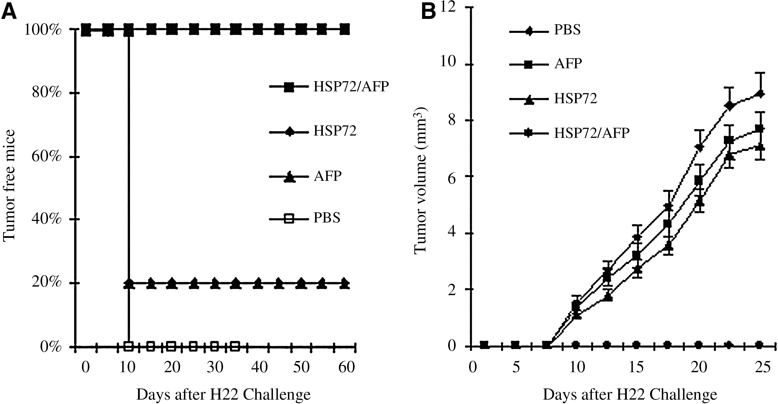
Therapeutic immunization of mice with HSP72/AFP vaccination.
Discussion
AFP always accompanies the growth of liver cells, and it is confirmed that AFP may be related to the proliferation of tumor or fetal cells. 27,28 The mechanism for growth-promoting activity of AFP is still unclear. Escaping from the surveillance of immune system is the primary cause for malignant growth of HCC cells. 29,30 Several investigations have showed that AFP could be individually synergistic with other growth factors to promote the growth of many tumor cells. 31,32 AFP receptors have been found anchoring on the membrane of various tumor cells. 33 –35 The receptor may mediate intercellular signal transduction, which influences the expression of genes related to proliferation. 36,37 AFP can stimulate the expression of some oncogenes that control cell cycle, and then enhance the proliferation of human HCC. 38,39 When the BEL-7402 cell line was treated with AFP, oncogene protein, such as c-fos, c-jun, c-ras, and mutant p53 and p21ras increased rapidly, which exerted an important function in modulating the growth and differentiation of the tumor cells. 38
Recent studies on the immunodominant epitopes of AFP have provided a solution to the obstacle of HCC immunotherapy. AFP is produced at low serum levels after birth throughout life. The majority of human HCC overexpress the oncofetal antigen AFP, Mr 64,000∼72,000 glycoprotein. Despite being exposed to high plasma levels of this oncofetal protein during embryonic development, the body still displays a low immunity to it. 39,40 Butterfield has found that four peptides of human AFP, processed and presented in the context of HLA-A0201, could be recognized by the human T cell repertoire, and could be used to generate AFP-specific CTL in human T cell cultures. 6 It was also found that the murine immune system could generate T-cell responses to this oncofetal antigen. Therefore, it may be an ideal target for immunotherapy. However, AFP immunization alone still results in lower levels of specific response and poorly reproducible protective immunity. 4,5
How to enhance a host's active immunity to AFP may be an interesting strategy for HCC therapy. Previous studies on AFP specific immunothereapy for HCC included AFP plasmid immunization, AFP-transduced dendritic cells (DCs) immunization, and AFP plasmid prime-AFP adenovirus boost immunization. 10 –14 AFP plasmid immunization produced detectable but low levels of AFP-specific T-cell responses and poorly reproducible protective immunity. 5,8 –11 Additional enhancement of the T-cell stimulatory effect is DCs engineered to express murine AFP, which demonstrated a powerful ability to generate tumor-specific immune responses. 41,42 However, the need for costly cell culture procedures limited their wide availability for clinical use, and the unstable culture technique might yield tolerating vaccine. 41 AFP plasmid prime-AFP adenovirus boost immunization could engender significant AFP specific T-cell responses and protective immunity in mice. 5,11 However, the miscellaneous procedures precluded their use. In the present study, we tested a novel strategy to induce antitumor immunity by a reconstructed vaccine conjugation AFP to HSP72 in mice via glutaraldehyde cross-linking. We found that the vaccine could elicit strong AFP-specific T-cell responses and produce a distinct protective effect on AFP-producing tumor, compared with other immunized groups. It is of interest to note that the cross-linking protein vaccine provoked not only the considerable stability of immunoprotective, but also a detectable level of anti-AFP antibody.
This study showed that HSP72/AFP could produce relatively strong CD8+ T-cell responses and antitumor effects, indicating that the antitumor effects of HSP72/AFP vaccines mainly depended on AFP-specific CD8+ T-cell responses. A high level of the anti-AFP antibody was also detected from HSP72/AFP immunized mice. Currently, the mechanism of antibody-dependent immune cell function is not well defined. We do not know whether humoral immunity mediated by B cells plays a primary role in the antitumor effect, but we could reckon that the anti-AFP antibody was likely to neutralize the AFP antigen or mediate the antibody-dependent immune cell cytotoxicity.
Immunogenicity and safety are two key factors that should be considered and balanced when developing an effective vaccine. Many researchers anticipate that HSPs can be used as an adjuvant to enhance immunogenicity of tumor therapeutic DNA vaccines. However, the safety of these types of vaccines should be considered not only in terms related to routine DNA vaccines, but also in terms of the potential risk of the adjuvant itself in humans. A concern of DNA vaccines is that they can potentially integrate into the host genome, leading to inactivation of antioncogenes or activation of proto-oncogenes and accordingly resulting in malignant transformation of host cells. Although this kind of integration frequency is low among host genes, 43 we cannot discount this risk entirely.
Protein-based vaccines have become an attractive approach to generate antigen-specific immunotherapy because of their simplicity, safety, efficacy, and capacity for repeated administration. Protein vaccines generate effective CTL and antibody responses by involvement of APCs that stimulate CD4+ and CD8+ T cells. Protein vaccines can be easily prepared on a large scale with high purity and stability and can also be repeatedly given to the same patient safely and effectively. 44,45 The features make protein vaccines a potentially attractive approach for cancer therapeutic vaccine development.
However, there is still a need to increase the potency of protein vaccines. Since tumor antigens are weakly immunogenic, researchers are currently emphasizing the use of versatile immunostimulatory molecules in the newer therapeutic cancer vaccines. Various HSP-based vaccine strategies have been described as enhancing fusion vaccine potency. In early studies from our group, we constructed an eukaryotic expression vector containing the molecular chaperon-HSP70 and AFP fragments, and then primed mice with the genetic vaccine, eliciting a robust strong protective immunity. 10 Another research group also confirmed that the linkage of HSP70 to the target antigen dramatically enhanced the potency of AFP fusion protein vaccines. 26 Therefore, an innovative approach that combines HSP with the tumor antigen will probably generate a more potent antitumor effect. HSP72 is one member of the heat shock protein family that has been shown to act as a potent adjuvant to enhance antigen-specific tumor immunity and innate immunity. 46,47 The use of HSP72-conjugated antigen vaccines represents a promising approach for enhancing antigen-specific T-cell-mediated immune responses as well as humoral responses for cancer vaccine development. 46,48
A few researches reported that HSPs could enhance the risk of autoimmunity due to cross-reactive T-lymphocytes primed by the HSP vaccines. 49 –51 However, the experiments that we have conducted showed that vaccination with the HSP72/AFP protein vaccine did not result in pathologic changes in the major organs of mice (data not shown). In this study, we described a novel AFP protein-based immunization strategy based on the vaccine by cross-linking HSP72 and AFP protein. Our data demonstrated that HSP72/AFP vaccination could elicit strong AFP-specific CD8+ T-cell immune responses, and induce established H22 tumor regression. Consistent with the in vivo results, the HSP72/AFP protein immunization induced specific CTLs that recognized H22 tumor cells in vitro, but not LLC cells. Our data also demonstrated that the HSP72/AFP protein vaccine generated the strongest therapeutic effects compared with other protein vaccines tested. Therefore, a novel strategy that combined linkage of HSP72 with antigen in the form of cross-linking protein cooperatively enhances the potency of antigen-expressing protein vaccines.
One of the potential explanations is that the linkage of HSP72 to AFP may lead to enhanced stimulation of AFP-specific CD8+ T cells in vivo via the so-called “cross-priming” mechanism. 52,53 Cross-priming is a complex process that requires enzymatic processing of the exogenous antigen and its trafficking through different intracellular compartments. 52 Dependent on the characteristics of the exogenous antigen and the functional capacity of the involved APCs, different processes may be adopted. 53 It is well established that complexes of HSP chaperoned synthetic peptides can direct peptides into the MHC class-I antigen-presentation pathways of APCs. By this process, chaperone-peptide complexes are thought to elicit tumor-directed CD8+ CTL. 54,55 CD91 serves as a receptor for HSP, including CRT, gp96, HSP70, and HSP90, and facilitates the cross-priming effects. 56 In addition, the HSP can also use different receptors to mediate their effects. 56 –58 It is conceivable that HSP72/AFP can interact with various receptors on APCs, leading to antigen cross-priming. These different receptor mechanisms may provide some explanations for the observed enhancement of AFP-specific CD8+ T cell response in mice vaccinated with HSP72/AFP, as compared with mice vaccinated with HSP72 or AFP alone.
In this study, we developed a strategy to construct an innovative protein vaccine. Cross-linking of HSP72 and AFP with lower concentration of glutaraldehyde not only established the HSP72/AFP complex but also preserved the activity of HSP72 immunoadjuvant and immunogenity of AFP antigen, which was confirmed by the strong ex vivo and in vivo antitumor effect. We attributed the successful AFP specific T-cell responses in mice to HSP72 through mediating APCs to efficiently take up and process AFP. Researches have shown that HSP72 itself has no antigenicity, and its immunogenicity has been attributed to the peptides chaperoned by HSP72. 46,47 It has been verified that HSP72 was a better molecule chaperon and adjuvant which could process and present a weak tumor antigen to MHC-I of host APCs, eliciting a specific T-cell response and CTL reaction. 46,48 Several studies have shown that HSP72-associated peptides could anchor an antigen on the cell membrane and directly present it to nature killer cells or γδ T cells as superantigen without being dependent on the stimulation of MHC-I molecules. 46,59 –61 In the present study, tumor rejection assay demonstrated that cross-linking protein vaccine HSP72/AFP elicited strong specific antitumor immunity against AFP-producing H22 cells than AFP vaccination. Results indicated that AFP immunogenicity could be improved greatly by HSP72, and vaccination with HSP72 could increase both humoral and T-cell proliferation responses to AFP.
Conclusions
Our results indicate that the HSP72/AFP protein vaccine can generate an impressive antitumor effect against AFP-expressing murine tumors through enhancement of AFP-specific CD8+ T-cell-mediated immune responses. Therefore, the tumor vaccine by cross-linking tumor antigen and HSP72 is a promising approach for cancer therapy that may be potentially applied to other cancer systems with known tumor-specific antigens.
Footnotes
Acknowledgments
This work is supported by Scientific Research Program Funded by Shaanxi Provincial Education Department (No. 2007JK233, 2010JK484), the Key Project of Ministry of Education of China (No. 205002), and the National Natural Science Foundation of China (No. 81172135).
Disclosure Statement
No competing financial interests exist.