
Abstract
Non-invasive methods for the assessment of distribution, homing, and retention of stem cells are desired for the successful demonstration of stem cell therapy. Cells labeled with 99mTc, 18F, and 111In have been reported for tracking the cells in vivo. However, they can be tracked only for a limited time due to the short half lives of these isotopes. In this context, stem cells labeled with 51Cr would be appropriate for tracking cells for a longer period of time owing to their half life of 27.7 days. Here, we have isolated mesenchymal stem cells (MSCs) from umbilical cord tissue, characterized them, and attempted to radiolabel them with 51Cr for mapping the fate of transplanted MSC cells after an intravenous injection via the tail vein in small animals.
Introduction
Stem cell therapy is a promising modality for the treatment of several clinical conditions in various areas such as cardiology, neurology, bone, and tissue regeneration as well as in oncology to replenish cells after radiotherapy and chemotherapy. 1,2
However, there are still many challenges for the translation of stem cell therapy to a viable clinical therapy. 3,4 The various factors that influence a successful demonstration of stem cell therapy are the optimal number of cells required for therapy, route of administration, cell migration, viability, differentiation, and engraftment of cells of interest after transplantation. Hence, a suitable method that could be used for monitoring homing as well as the fate of the delivered stem cells is essential. Several imaging techniques such as X-ray based imaging, optical imaging, ultrasound, magnetic resonance imaging (MRI), single photon emission computed tomography (SPECT), and positron emission tomography (PET) are reported for the tracking of survival and migration of cells. However, each method has its advantages and limitations. Among these, MRI and radionuclide imaging are more feasible and can be translated for clinical applications. 4 –6 Cells labeled with 99mTc, 18F, and 111In are helpful in tracking stem cells, but for a limited time owing to their short half lives of 6 hours, 110 minutes, and 12.3 hours respectively. 7 –9 However, it is important to follow the stem cells for a much longer duration, to know their fate post transplantation in vivo. 51Cr (Eγ=320 KeV, t ½=27.7 days) would be considered appropriate for the tracking of cells for a longer period of time due to its longer half life of 27.7 days.
Mesenchymal stem cells (MSCs) are multipotent, spindle-shaped plastic adherent cells that have the ability to differentiate to various lineages. Considering their ability to expand rapidly and their immunomodulatory properties, MSCs are an excellent candidate for allogenic cell therapy. 5,10 Human umbilical cord tissue is a comparatively newer source of adult MSCs (UCMSCs). Herein, we report the isolation and characterization of UCMSCs, the radiolabeling of cells with 51Cr, viability studies with time, and the fate of 51Cr-labeled stem cells after an intravenous injection in small animals.
Materials and Methods
Cell culture medium, biochemicals, and reagents were from Sigma-Aldrich (St. Louis, MO). EL4 cells were procured from the National Center for Cell Sciences, Pune, India. 51Cr was obtained from the Radiochemicals Section of our Division, BARC, India. The radioactivity was measured in a NaI (Tl) Scintillation counter from Pla Electro Appliances, Mumbai, India. Scintigraphic imaging was performed on the Millennium MPS Scintigraphic Imaging System (Wipro-GE Healthcare Systems).
Isolation and characterization of UCMSC
The UCMSCs were isolated as per our in-house published protocol. 11 The expanded UCMSCs were characterized at every passage by an immunophenotypic analysis. The cells (∼50,000) were stained with one or two of the monoclonal antibodies directly conjugated with FITC or PE directed against the antigens CD73, CD105, CD45, SSEA4, HLA-ABC, HLA-DR, CD31, CD29, CD44, CD14, and vWF. The stained cells were acquired on FACS caliber and analyzed using Cell Quest software (BD).
Radiolabeling of cells
For establishing a baseline and for the optimization of reaction conditions, EL4 (Human lymphoma) cells were initially radiolabeled with 51Cr. EL4 cells were cultured in DMEM with 10% fetal bovine serum at 37°C and 5% CO2. 0.92 MBq 51Cr was added to three different concentrations of EL4 cells, namely, 0.5×10⁶, 106 and 107 cells, maintaining the total volume at 1 mL and incubated at 37°C in CO2 incubator under sterile conditions for 1 hour. The cells were then centrifuged at 1500 rpm for 5 minutes, washed once with the DMEM medium, and the radioactivity associated with cells was measured. In a similar manner, the radioactivity retained with the cells at the end of 18 hours was also estimated. UCMSC cells were radiolabeled with 51Cr under similar conditions. 51Cr at various concentrations (0.92, 3.7, and 18.5 MBq) was added to 107 UCMSCs and incubated at 37°C for 1 hour. After centrifugation and washing of the cells, the radioactivity associated with cells was measured at 1, 3, and 18 hours to estimate the uptake as well as the % retention of activity in cells with time. The viability of cells was also studied using trypan blue at 3 hours post labeling.
Biodistribution studies
To map the fate of 51Cr-labeled UCMSC cells, bio-distribution studies were carried out in normal Swiss mice. Cells were labeled with 9.25 MBq of 51Cr activity, and an uptake of 27% was observed. ∼150 μL of radiolabeled cells (370 KBq/1.5×106) were injected in Swiss mice via the tail vein. Animals were sacrificed at 1, 3, 24, and 48 hours post injection, and % ID/organ was estimated after counting the various organs in the NaI (Tl) flat geometry counter. All animal experiments were carried out in compliance with the National Laws related to the conducting of animal experiments.
Imaging studies
An inflammation model was developed by injecting 150 μL of turpentine in the right thigh of Wistar rats. After 24 hours, inflammation was observed in the animals. Redness, swelling, and difficultly in mobility were observed in the right limb of all the rats. Rats were injected intravenously with 370 KBq/106 of 51Cr-labeled stem cells. For a comparison, 51Cr was also injected intravenously into another rat, and imaging studies were carried out after anesthetizing the animals. Images were captured at 2, 24, 48, and 96 hours using an MEGP collimator, Wipro GE SPECT Camera. Image acquisition was carried out for 150 kilo counts.
Results
Isolation and characterization of UCMSC
Stem cells were successfully isolated from the umbilical cord (UCMSC). The morphology of UCMSCs was spindle shaped (Fig. 1). The umbilical cord tissue-derived MSCs were characterized at every passage. The cells were checked for the expression of MSC markers, hematopoietic markers, cell adhesion molecules, and viability. Flow cytometry analysis of the UCMSCs showed typical MSC phenotype and expressed CD73 and CD105 but did not express CD45, CD31, CD14, and vWF antigens, as is expected of stem cells (Fig. 2). UCMSCs expressed HLA-ABC, but were negative for HLADR. These cells, however, expressed SSEA4, CD29, and CD44. The cells maintained >95% viability at all passages.
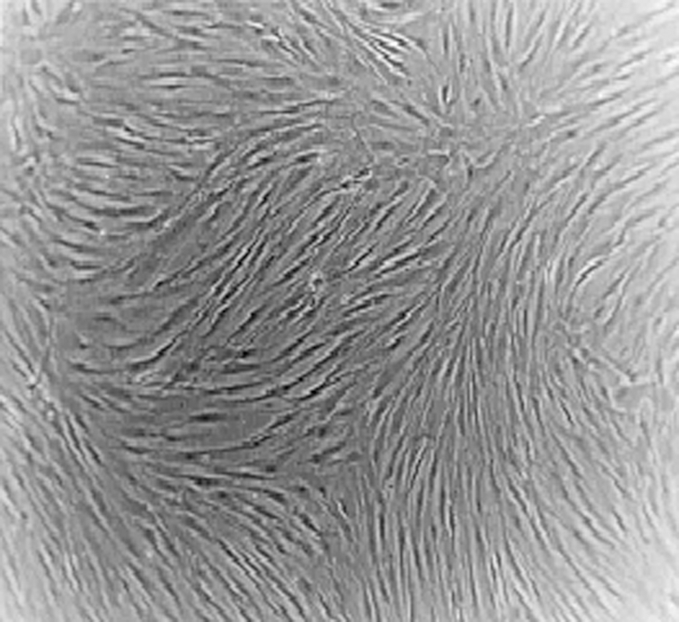
Morphology of UCMSCs. UCMSCs, umbilical cord mesenchymal stem cells.
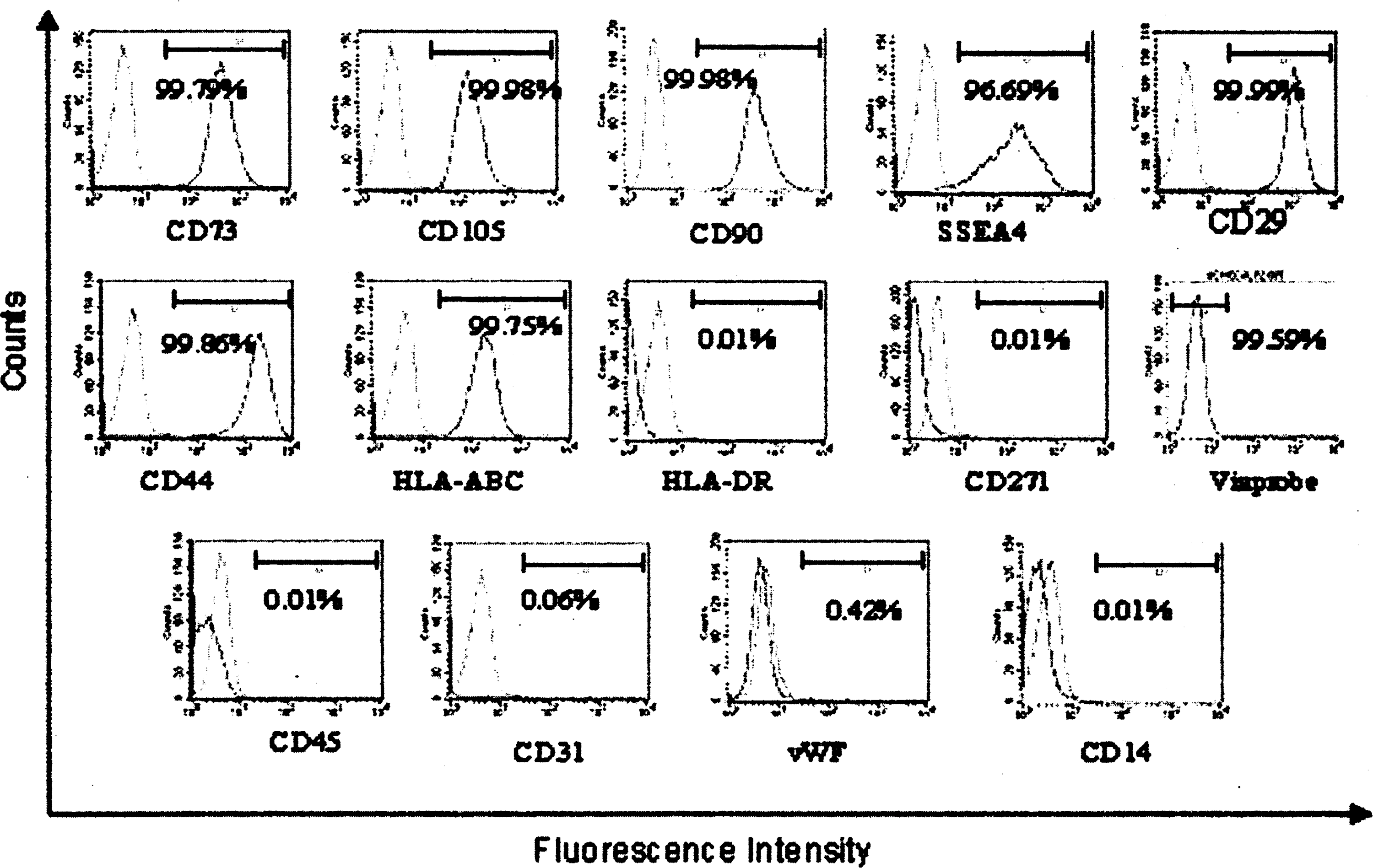
Immunophenotype analysis by flow cytometry. UCMSCs showed a typical MSC phenotype–CD73+/CD105+/CD45−/SSEA-4+/HLA-DR-/HLA-ABC+/CD31−/CD29+/CD44+/CD14−/vWF−/CD90+/CD271−.
Radiolabeling of stem cells
EL4 cells were initially used to optimize the labeling procedure. This was necessary, owing to the tedious procedure to grow the UCMSC in very large numbers, which are necessary to perform a series of experiments with adequate replication for statistical validity. In addition, this also provided an indication about the differences in labeling two types of proliferating cells, despite using similar reaction conditions. Figure 3 depicts the results of labeling EL4 cells with 51Cr. It was observed that the labeling of the cells was low when 1 million or lesser cells were used. At 10 million cells, an uptake of 30.83%±0.71% (n=4) was attained, which decreased to ∼17% after 18 hours. Based on these studies, UCMSC cells were labeled using 10 million (107) cells per experimental point. However, radiolabeling was attempted at radioactivity levels higher than those used in preliminary studies using EL4 cells, in order to achieve the highest activity possible, yet retaining the viability to a reasonable extent. Figure 4 depicts the results of the radiolabeling of stem cells. It is seen that at 0.92 MBq of 51Cr activity, the uptake was 37%. The activity at 18 hours was retained at 27%, which was higher than the corresponding figures for the EL4 tumor cells. It is also seen that when incubated with higher amounts of 51Cr, the uptake of activity was appreciable. At 3.7 MBq, the uptake as well as the retention of activity in cells after 3 and 18 hours was very similar to those observed with 0.92 MBq of 51Cr. When 51Cr was used at 18.5 MBq, the% uptake reduced to half the original value, but the decrease in retention followed a similar pattern with time. However, considering that the activity used is five times higher than 3.7 MBq, even a reduced uptake of 18% resulted in higher specific activity of labeled cells. Apart from the retention of activity in the cells, we considered the viability of the cells also as an important factor for studying the in-vivo behavior of the radiolabeled cells. Figure 5 shows the retention of viability in the radiolabeled cells, and it can be seen from the figure that the viability was retained to an extent of >90%, after a time span of 3 hours, which was almost as good as control cells when 51Cr activity used was 0.92 and 3.7 MBq and slightly lower at 18.5 MBq. Thus, it was ascertained that despite the amount of 51Cr activity used, the uptake as well as viability were good for approximately 3 hours. A few experiments that we could carry out to study the viability with time indicated that the cell viability decreased with time as expected, with nearly 50% of the viability being retained at 24 hours, for the 0.92 and 3.7 MBq levels; while it was far lesser when 18.5 MBq was used. However, radiolabeled cells when injected in vivo will get diluted and may not be affected much as they get exposed to activity in the in-vitro experiment.
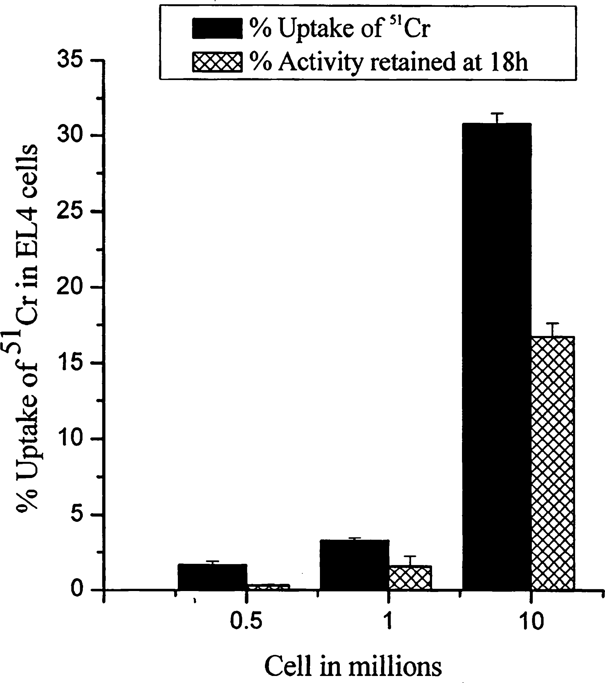
% uptake 51Cr in EL4 cells (n=4).
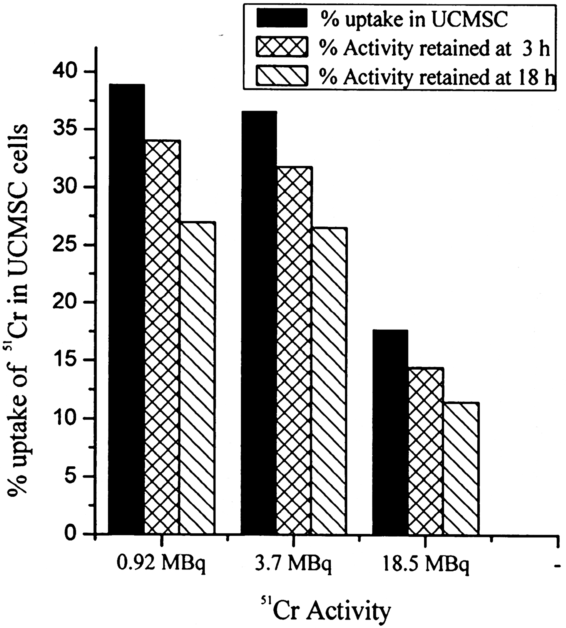
% uptake of 51Cr activity in 107 UCMSC cells (n=4).
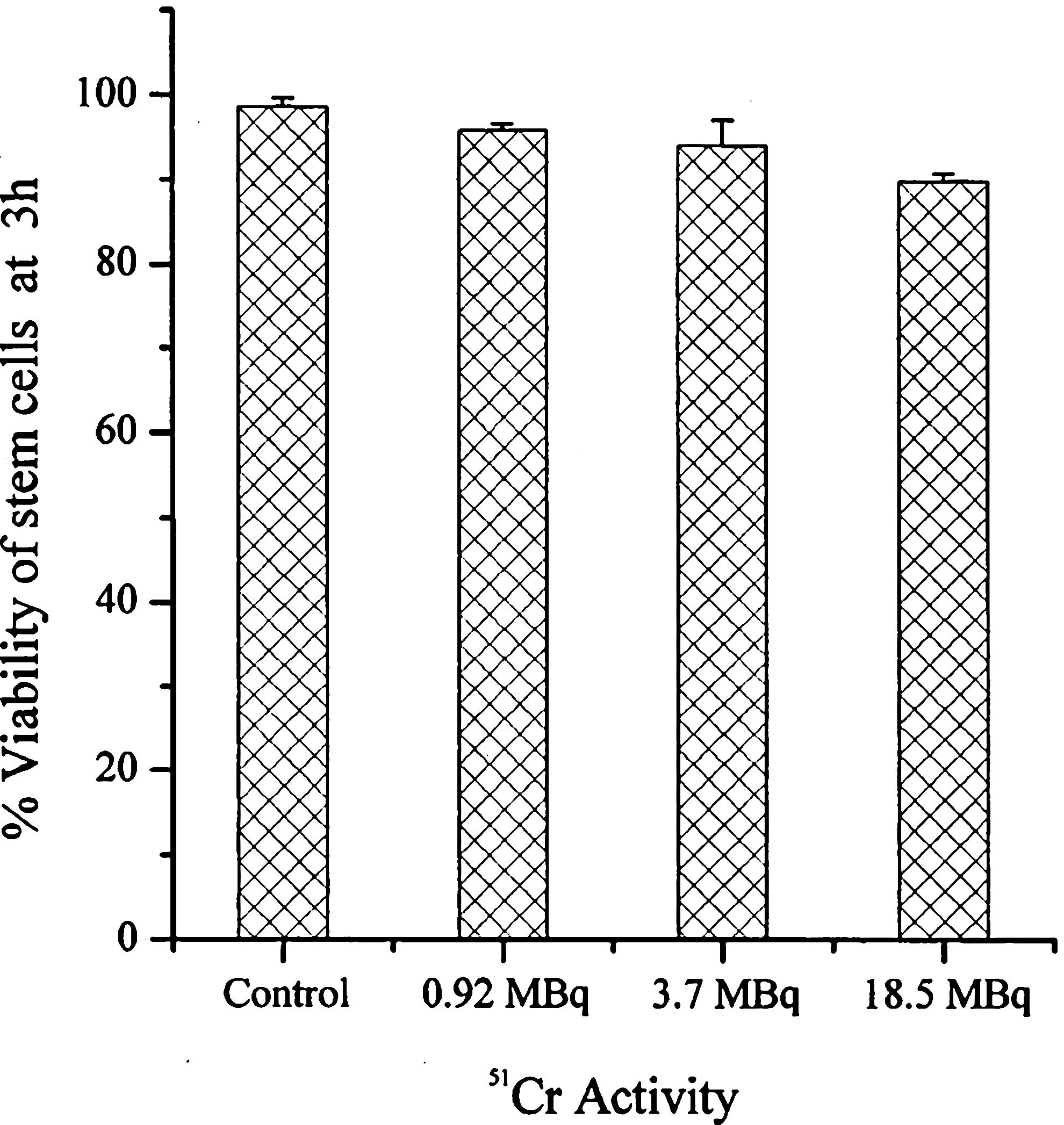
Viability of UCMSC at 3 hours post labeling with 51Cr (n=4).
Biodistribution studies
The injection of 51Cr-labeled UCMSC cells injected intravenously was well tolerated by the animals. Biodistribution studies revealed initial high uptake of activity in the lungs followed by accumulation in the liver (Fig. 6). The studies also indicated the clearance via the hepatobiliary and renal route (Fig. 7).
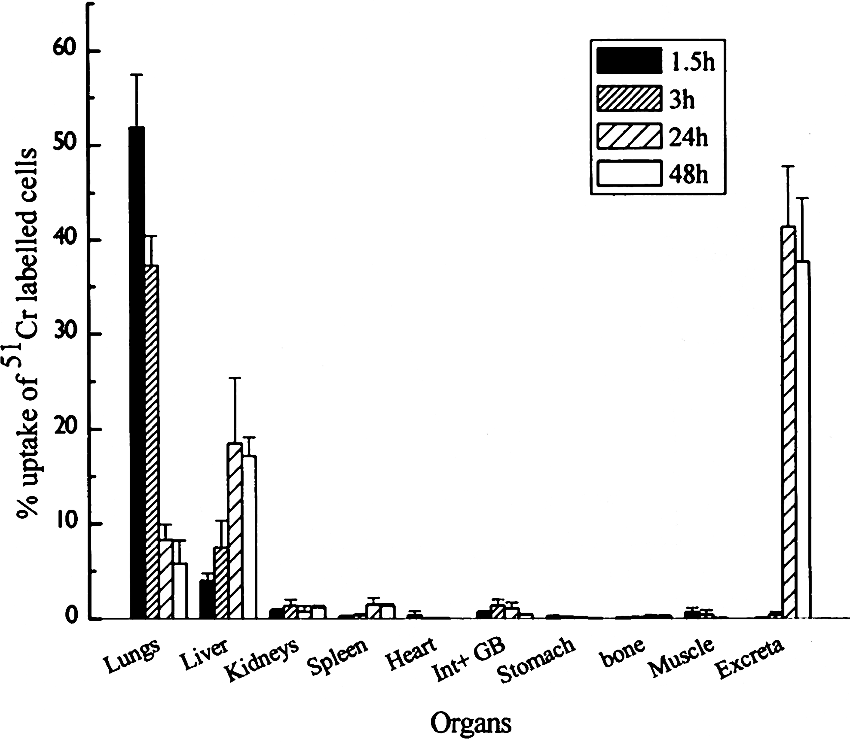
Uptake (%ID/organ) of 51Cr-labeled UCMSC in normal Swiss mice (n=4).
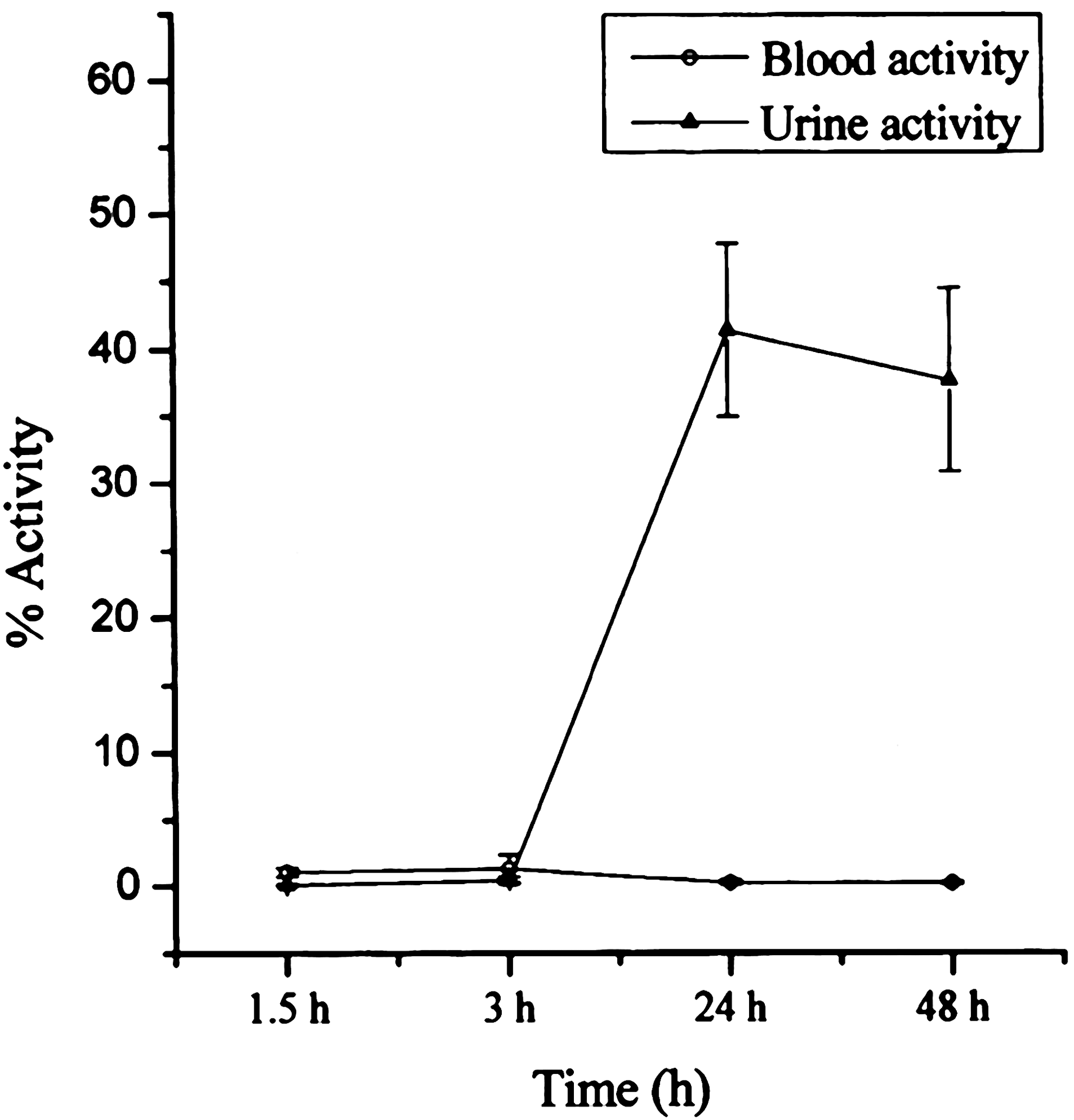
Clearance pattern of 51Cr-labeled UCMSC in normal Swiss mice.
Imaging studies
High uptake of 51Cr-labeled stem cells was observed in lungs with no activity in other vital organs of rats injected with labeled stem cells at 2 hours p.i. The initial uptake of activity in lungs on iv injection correlated with the biodistribution studies conducted on normal mice. No significant activity in the blood was observed. Activity was observed in inflamed muscle of the animal with T/NT (contra lateral muscle) ratio of 1.5 at 2 h p.i. At 24, 48, and 96 hours, slow clearance of activity from the lungs and uptake in the liver and GI tract was observed (Fig. 8A, B). In case of animals injected with 51Cr, at 2 hours p.i, considerable activity was observed in the whole body due to high blood activity. A significant uptake in the liver and kidneys was also observed. The apparent activity observed in inflamed muscle of the animal was mainly due to the high blood activity. At 24, 48, and 96 hours, retention of activity in the blood and high uptake of 51Cr in the kidneys was observed (Fig. 9A, B).
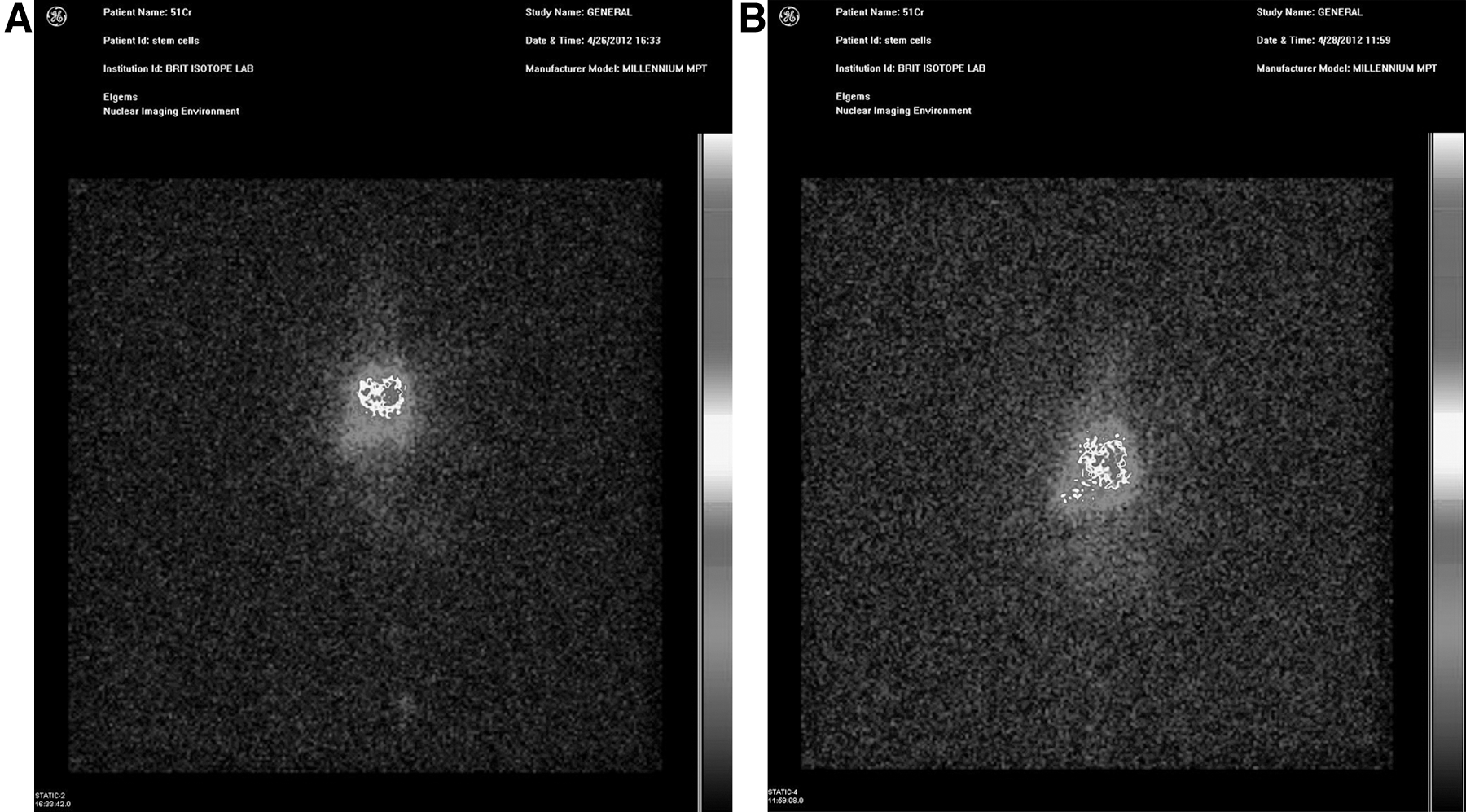
Imaging studies of 51Cr-labeled stem cells in Wistar rat at

Imaging studies of 51Cr in Wistar rat at
Discussion
Umbilical cord-derived MSCs are proposed to be used as an allogeneic product for treating various disease conditions in humans. In clinical situations, it is important to know the distribution of the cells in vivo. This study using 51Cr stem cells was carried out to understand the distribution of the UCMSCs post injection. 51Cr was considered suitable for labeling stem cells for various reasons. Long-term tracking of cells is possible with 51Cr due to its favorable emission of 320 KeV γ rays and a suitable t1/2=27.7 days. Moreover, due to 51Cr being a reactor produced isotope with amenable logistics, it is easily available at a reasonably low cost. 51Cr-labeled RBCs and lymphocytes have been reported since long for various applications. 12 –14 Further, both 99mTc and 111In are generally labeled in the form of metal complexes using chelating moieties such as DTPA, which could potentially change the behavior of the cells, particularly when the aspect studied is not just circulation. Hence, 51Cr has been labeled nonspecifically by passive uptake in cells and was found useful for the tracking of stem cells in-vivo.
For therapy, stem cells are injected loco regionally to the diseased site, however it is reported that when injected via i.v route stem cells will preferentially localize to the site of injury or diseased tissue. To provide proof of principle, biodistribution and imaging studies after i.v injection are desired in animal models of injury. Studies with 51Cr-labeled UCMSCs when injected via i.v. route revealed initial homing of cells in the lungs, which cleared with time. Initial high uptake in the lungs and clearance are in accordance with the previously reported results with other cell-labeling agents. 12 –14 A significant uptake in the inflamed tissue was not observed, probably due to reasons such as the stem cells may not be actively involved in repair during inflammation compared with other disease conditions such as infarction or injury caused by chemicals. However, these studies clearly indicate that 51Cr-labeled stem cells could be used for in vivo tracking. For preclinical studies in animal models of disease, better images are possible with high-resolution micro SPECT/nano SPECT cameras.
Conclusion
UCMSCs could be labeled with 51Cr in reasonably high yields without loss of viability. Preliminary biodistribution studies and imaging studies in small animals with 51Cr UCMSC gave an indication that the cells after an i.v injection predominantly localize in the lungs and liver and, subsequently, clear via both hepatobiliary and renal routes. In actual clinical situations, in which the delivery of stem cells is loco-regional, 51Cr-labeled cells may be useful for tracking the fate of stem cells with time.
Footnotes
Acknowledgments
The authors are thankful to Dr M.R.A. Pillai, Head, Radiopharmaceuticals Division, and Dr. V. Venugopal, Ex-Director, Radiochemistry & Isotope Group, BARC, for their constant encouragement and support toward this work. The authors are also thankful to Shri P V Joshi, Head, Radiochemicals Section, Radiopharmaceuticals Division, Bhabha Atomic Research Center, India, and his staff for the supply of 51Cr for this work. The authors are also thankful to the management team of Reliance Life Sciences Pvt Ltd, Navi Mumbai, for supporting the study.
Disclosure Statement
The authors have not received funding or grants from external agencies for this research and do not have any conflict of interest.