




Abstract
Bleomycin-glucuronide (BLMG) is the glucuronide conjugate of BLM. In the present study, BLMG was primarily enzymatically synthesized by using a microsome preparate separated from rat liver, labeled with 131I by iodogen method with the aim of generating a radionuclide-labeled prodrug, and investigated its bioaffinities with tumor-bearing Balb/C mice. Quality control procedures were carried out using thin-layer radiochromatography and high-performance liquid chromatography. Tumor growing was carried out by following Caco-2 cell inoculation into mice. Radiolabeling yield was found to be about 65%. Results indicated that 131I-labeled BLMG (131I-BLMG) was highly stable for 24 hours in human serum. Biodistribution studies were carried out with male Albino Wistar rats and colorectal adenocarcinoma tumor-bearing female Balb/C mice. The biodistribution results in rats showed high uptake in the prostate, the large intestine, and the spinal cord. In addition to this, scintigraphic results agreed with those of biodistributional studies. Xenography studies with tumor-bearing mice demonstrated that tumor uptakes of 131I-BLM and 131I-BLMG were high in the first 30 minutes postinjection. Tumor-bearing animal studies demonstrated that 131I-BLMG was specially retained in colorectal adenocarcinoma with high tumor uptake. Therefore, 131I-BLMG can be proven to be a promising imaging and therapeutic agent, especially for colon cancer in nuclear medical applications.
Introduction
Bleomycin (BLM) is a group of water- and methanol-soluble basic glycopeptide antibiotics isolated from fermentation products of the Streptomyces verticillus in 1966 by Umezawa in Japan. 1 The cytotoxic effect of BLM depends on the blocking of DNA synthesis, and in the presence of iron, oxygen, and a reducing agent, BLM breaks the DNA chains. 2 It is conventionally used for the treatment of squamous cell carcinomas, combined either with other chemotherapy or with radiation therapy 3 or as palliative treatment 4 , and also in the treatment of testicular cancers 5 and lymphomas. 6,7 The clinically used BLM is a mixture of three distinct isomers: A2 (the most abundant, 65%), B2 (30%), and DM, which is a demethylated form of A2 (5%) (Fig. 1).
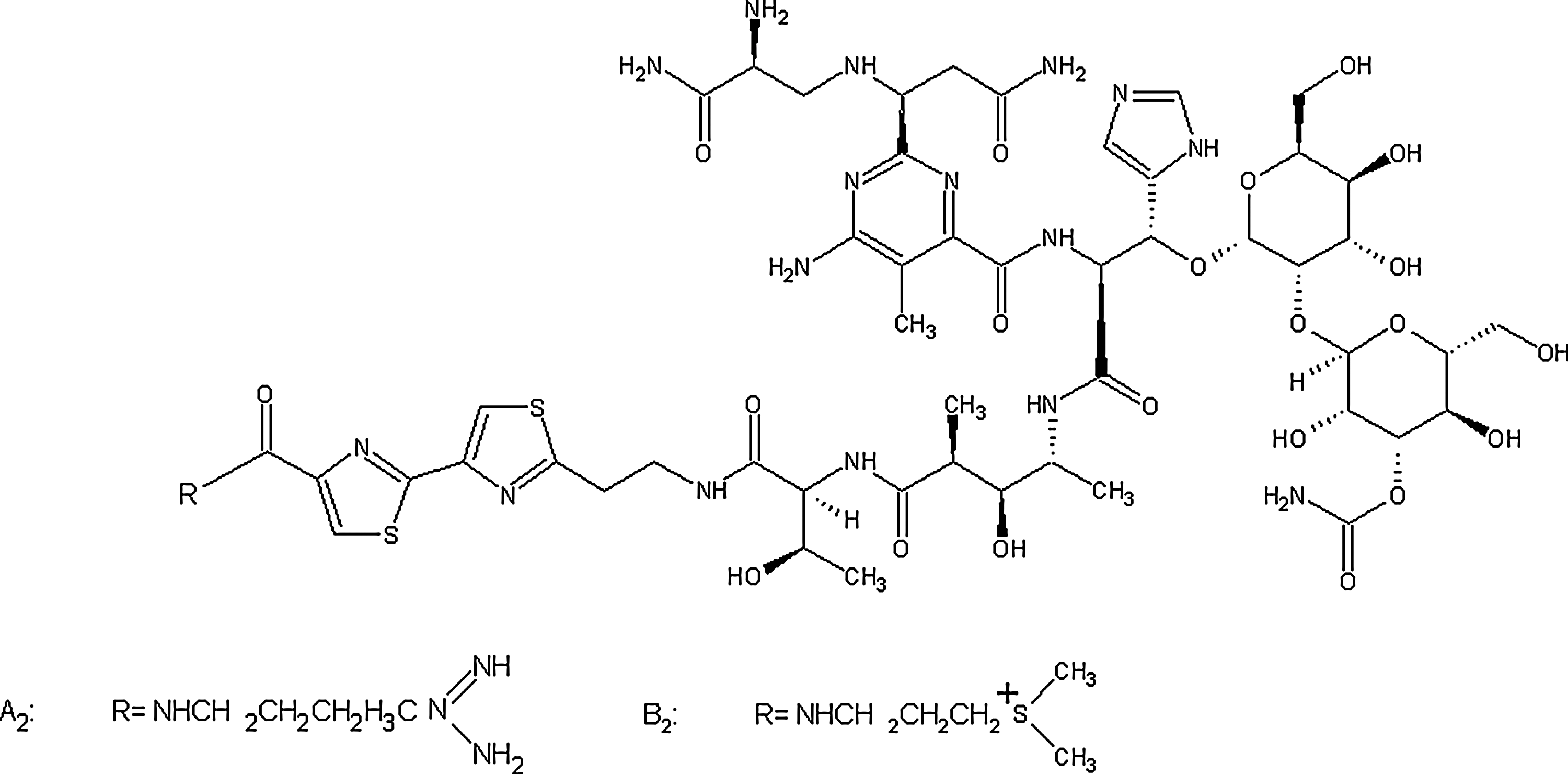
Chemical structures of BLM A2 and BLM B2 isomers.
Certain enzymes in tissues and body fluids may, through reversal of the detoxification process, influence the composition and availability of steroid hormones, toxins, and carcinogens. The enzyme β-glucuronidase, which hydrolyzes glucuronide conjugates, thereby reversing one of the main detoxification and excretion pathways, was found to vary in concentration in different cysts and tumor tissues over a 300-fold range. 8 It is known that breast cancer cells have high amounts of β-glucuronidase. Glucuronides of drugs often accumulate during a long-term therapy. The hydrolysis of glucuronides can be catalyzed by the enzyme β-glucuronidase, which has already been proven to be useful in tumor-specific bioactivation of glucuronide prodrugs of anticancer agents. 9 –11 Therefore, antibody-directed enzyme prodrug therapy and gene directed-enzyme prodrug therapy using glucuronide prodrugs, activated by the human enzyme β-glucuronidase, have been developed as an experimental approach to enhance tumor selectivity and to reduce systemic toxicity of anticancer agents.
BLM has been widely studied as a G-quadruplex interactive compound and telomerase inhibitor. 12 –20 G-quadruplexes are unusual DNA secondary structures based on planes of four guanines (G-tetrads) stabilized by Hoogsteen G–G pairings and monovalent cations. The central aromatic core of the perylene diimides is suitable for π–π stacking interactions with the terminal G-tetrad of DNA G-quadruplex, whereas the hydrophilic side chains interact with the DNA grooves. By means of these two kinds of interactions, these molecules are able to induce and stabilize G-quadruplex structures in G-rich single-stranded oligonucleotides. This is of the great pharmaceutical interest, since the terminal ends of eukaryotic chromosomes (telomeres) are characterized by the presence of a single-stranded G-rich overhang that represents the substrate of a reverse transcriptase enzyme, ribonucleoprotein telomerase, which is involved in the maintenance of telomere length. 21,22 This enzyme is not active in most somatic cells, but is active in most human tumors, and is therefore considered as being of high potential as a selective target for different antitumor strategies. 21
The aim of the current study was to synthesize a novel glucuronide derivative of BLM (BLMG) (Fig. 2) as a radiopharmaceutical that is able to be labeled with 131I, and to investigate its radiopharmaceutical potential using male Albino Wistar rats and adult male Albino rabbit in biodistribution and scintigraphic studies, respectively, and then to evaluate its accumulation in the human colorectal adenocarcinoma tumor- bearing female Balb/C mice.
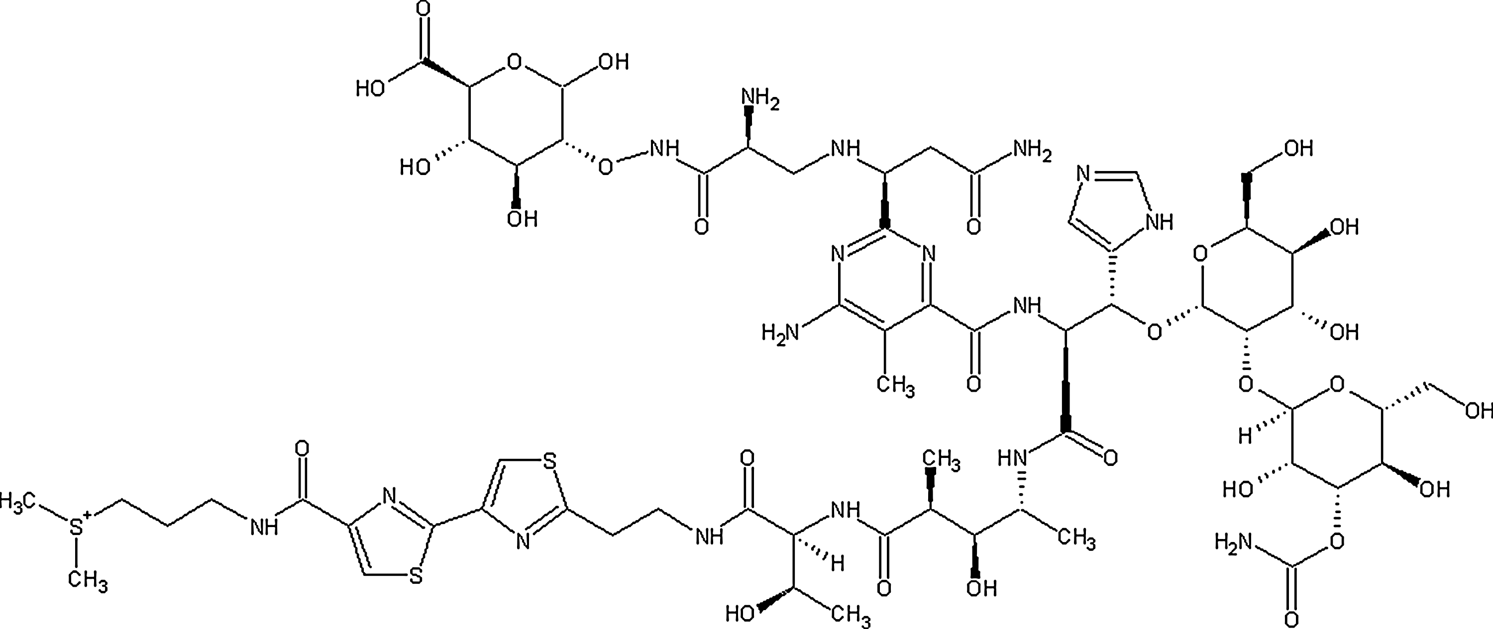
Chemical structure of bleomycin-glucuronide (BLMG).
Materials and Methods
BLM was purchased from Sigma Chemical Co. Radioiodination and its preliminary biological activities in the rabbit metabolism were examined using the gamma camera-imaging technique and biodistribution studies. Na131I (74 MBq) was obtained from the Department of Nuclear Medicine of Celal Bayar University. Iodogen (1,3,4,6-tetrachloro-3α,6α-diphenylglycouril), 4-(2-hydroxyethyl)-1-piperazineethane-sulfonic acid, sodium salt (HEPES), Tris buffer, UDP-glucuronic acid (UDPGA), and Triton X-100 were purchased from Sigma, and all other chemicals were purchased from Merck Co. Caco-2 (human colorectal adenocarcinoma) was obtained from American Type Culture Collection. Primary human intestinal epithelial cells (ACBRI 519) were purchased from Applied Cell Biology Research Institute. Minimum essential medium (MEM EAGLE) fetal bovine serum (FBS), Tripan blue (BIO. IND), phosphate-buffered saline (PBS), and Tripsin–EDTA were supplied from BIO. IND. Thin-layer radiochromatography (TLRC) and high-performance liquid chromatography (HPLC) chromatograms were obtained using a Cd(Te) detector equipped with a RAD 501 single-channel analyzer and HPLC (an LC-10ATvp quaternary pump and an SPD-10A/V UV detector and a syringe injector equipped with a 20-μL loop and 5-μm reversed-phase (RP)-C-18 column 250×4.6 mm I.D.; Macherey-Nagel), respectively. Liquid chromatography mass spectrometry (LC/MS/MS) chromatograms were taken using an LC/MS/MS instrument (Agilent HPLC 1100 binary pump, degasser, autosampler, and column oven) in Ege University ARGEFAR (Research and Application Center of Drug Development and Pharmocokinetics), and scintigrams were obtained using a double-headed gamma camera (Infinia, GE) in the Department of Nuclear Medicine of Celal Bayar University.
Synthesis of BLMG
Preparation of microsomal fraction from rat livers
Two male Albino Wistar rats were sacrificed by cervical dislocation. Microsomal fractions from the rat livers were prepared according to the procedure previously described by Zihnioglu. 23 Briefly, the liver was excised and placed in cold (0°C–4°C) 250 mM sucrose/5 mM HEPES 99%. It was then chopped using scissors and was blended and homogenized after adding 35 mL of 250 mM sucrose/5 mM HEPES, pH 7.4, in a teflon/glass homogenizer at 1700 g (1500 rpm) for 30 minutes.
Purification of UDP-glucuronyl transferase from rats
Homogenates were then centrifuged at 12,000 g (10,500 rpm) for 10 minutes, and the resulting supernatant was decanted through glass wool to trap fat particles and centrifuged at 105,000 g (31,500 rpm) for 1 hour. All further operations were performed at +4°C. Microsomal pellets were solved by resuspension in a volume (in 2 mL) of 0.2 M potassium phosphate, 2 mM mercaptaethanol, and 0.4% Triton X-100 (pH 7) buffer, equal to twice the wet weight (in grams) of the tissue. The suspension was stirred on ice for 30 minutes and centrifuged for 1 hour at 105,000 g to remove the insoluble material. The resulting supernatant was stored at −80°C until use.
Estimation of protein in microsomal samples
The protein content in microsomal samples was estimated using the Bradford method. This method is based on the observation of maximum absorption for a solution of Coomasie Blue G-250 at acid pH shifts from 465 to 595 nm when binding to protein occurs. Using this method, standard curves covering a range of protein concentration were constructed as follows: 0.02, 0.05, 0.01, 0.12, 0.15, 0.20, and 0.25 mg/mL. The Bradford reagent consists of 40 mg Coomasie Brilliant Blue and 55 mL 88% w/v phosphoric acid dissolved in 50 mL ethanol that was diluted to 1 L and filtered off. Standard curves, using bovine serum albumin (BSA), plotted by linear regression analysis, were prepared and protein concentrations of appropriately diluted samples calculated from the relevant standard curve. The protein content was found to be ∼8.22 mg/mL, which was similar to the value reported by Bradford. 24
Glucuronidation reaction
Reactions were performed similar with other reports. 25 –27 Microsomal enzyme preparate (0.98 mg protein/119 μL) was added to 5 mL of 50 mM Tris buffer (pH 8.0) containing 6 mM CaCl2, 10 mM UDPGA, and 1 mM dithiothreitol at a temperature of 37°C. The reaction mixture (total volume 5 mL) containing UDP-glucuronyl transferase (UDPGT) was stirred at 37°C in a water bath for 10 minutes. The contents were then sonicated in an ultrasonic bath for 30 seconds to disperse the microsomes, and the reactions were started by the dropwise addition of 0.5 mg/0.5 μL BLM in water, with stirring. Slow stirring at 37°C was continued for 18 hours. The reaction was terminated after 18 hours by the addition of 300 μL of acetonitrile, and the precipitated protein was removed by centrifugation at 5200 rpm for 10 minutes by using a microcentrifuge. The supernatant was then analyzed by reversed-phased HPLC (Shimadzu 10 AVp). The HPLC analysis indicated that the glucuronidation yield was about 100%, and one peak was obtained for BLMG.
HPLC studies
Cold iodinated compounds of BLM and BLMG [127I-BLM and 127I-BLMG] were investigated in HPLC. Table 1 shows chromatographic conditions used for analytical experiments in HPLC. For analytical experiments, a 5-μm RP-C18 column (250×4.6 mm I.D.; Macharey-Nagel) and a syringe injector equipped with a 20-μL loop were used. The flow rate was set at 1 mL/min. UV was detected at 260 nm. The mobile phase and UV detection are given in Table 1.
RP, reversed-phase.
Liquid chromatography mass spectrometry
Chromatographic conditions for LC/MS/MS experiments are given in Table 2.
Inactive iodination of BLMG
BLMG was iodinated with inactive iodine under the same conditions as previously described by Avcıbaşı et al. 11 Structural parameters were obtained by the LC/MS/MS spectrometry system instrument (Agilent HPLC 1100 binary pump, degasser, autosampler, and column oven) in the Ege University ARGEFAR Center.
Radioiodination procedure
Radioiodination procedure of BLMG with 131I
BLMG was first radioiodinated with 131I using the iodogen method. To label BLMG with 131I, 25 μg of BLMG was added into the iodogen-coated tube, and then 1 mCi (37 MBq) of Na131I was added. This reaction mixture was kept at room temperature without stirring for 15 minutes. At the end of this time, the mixture was transferred to another tube by a syringe, and then quality control was performed.
Quality control studies
For TLRC studies, TLC aluminum sheets (Merck, 20×20 cm code: 5552) were used, and citric acid monohydrate 100% was used as the mobile phase. The TLRC technique was used to determine the Rf values of the radioiodinated products. Each TLRC sheet was covered by an adhesive band after its development and was cut into 0.5-cm width. Those pieces of TLC were then counted by using a Cd(Te) detector equipped with a RAD 501 single-channel analyzer.
Stability of radiolabeled BLMG in human serum
In vitro stability of 131I-BLMG in human serum was determined by incubating 100 μL (25 μg) of the labeled compound with 300 μL of human serum at 37°C. The aliquots were analyzed in time intervals of 30, 60,180, and 1440 minutes by the TLRC technique after labeling.
Lipophilicity (partition coefficient)
The lipophilicity (logP) of the radiotracer was measured as follows: 100 μL of the radiolabeled compound (131I-BLMG) was added to a premixed suspension of 200 μL of n-octanol in 200 μL, pH 7 buffer. The resulting solution was mixed for 15 minutes at room temperature and centrifuged for 30 minutes at 2500 rpm. Then, 0.1-mL aliquots of each phase were removed and counted by a Cd(Te) detector equipped with a RAD 501 single-channel analyzer. Experiments were conducted in triplicate.
Biodistribution studies in rats and tumor xenography model in Balb/C mice
Experiments with animals were approved by the Institutional Animal Review Committee of Ege University. The biodistribution data are expressed as percentage of injected radioactivity per gram of tissue (%ID/g) for selected organs as the mean value of three rats. The experiments were performed on male Albino Wistar rats weighing ∼180–200 g. For blocking iodine uptake into the thyroid gland, 10 mg of potassium iodide was added to 1 L of the animal's drinking water. The 131I-BLMG in 60% purity was sterilized by a membrane filter and then injected into the tail vein of the animals ([3.7 MBq (100 μCi)]/1 μg 131I-BLMG per rat). Then, they were sacrificed at 30, 120, and 240 minutes under ether anesthesia, and the tissues of interest were removed. Blood was taken, and organs were excised. All tissues were weighed and counted for radioactivity with a Cd(Te) detector. The percent of radioactivity per gram of tissue weight (in % injected activity/g tissue) was determined.
In the xenography model studies, 6-week-old female Balb/C mice weighing ∼20–30 g were obtained from Department of Pharmacology, the Adnan Menderes University, Aydın, Turkey. The mice were cared for at Ege University, Izmir, Turkey, according to the institutional animal care guidelines. To establish animal tumor models, 1.5×10⁷ (cells/mouse) Caco-2, the human colorectal adenocarcinoma cell lines, were resuspended in 100 μL of phosphate buffer solution (PBS-R, Gibco-BRL) and injected into the subcutaneous tissue of the left flank of the mice.
All animals were provided with free access to drinking water and a basal diet. The mice were divided into three groups for the experiments: control, BLMG, and BLM. Each group contained nine mice. All animals were caged and housed under controlled conditions of humidity (45%±5%) and temperature (25°C) on a 12-hour light/dark cycle. Tumor growth and weight of the mice were checked daily after 3 days of Caco-2 cell inoculation into the mice. Biodistribution was performed 10 days post-tumor grafting, when the tumors had reached a measurable size of about 6×5 mm. 131I-BLMG in 60% purity was sterilized by a membrane filter and then injected into the tail vein of the animals ([3.7 MBq (100 μCi)]/1 μg 131I-BLMG per Balb/C mice).
In the cell culture studies, Caco-2 cells were cultured in the Eagle's minimum essential medium supplemented with 20% FBS, 2 mM glutamine, 1.5 g/L sodium bicarbonate, 0.1 mM nonessential amino acid, and 1 mM sodium pyruvate. In the experiments, cells were grown at 37°C in an incubator with humidified air and equilibrated with 5% CO2. The cells were maintained in exponential growth by subculturing the cells using trypsin-EDTA (0.25% by w/v in the Hanks Balanced Salt Solution). The cells were pelleted and resuspended in the cell medium. Before the experiments, cell cultures were trypsinized and, to remove trypsin, washed once in the respective culture medium. The cells were resuspended at a concentration of 1×l05 cells/mL in the culture medium, transferred to falcon tubes, and added 1 mL PBS.
Scintigraphic studies
The imaging studies were performed on healthy adult male Albino rabbits using a gamma camera (Infinia, GE) in the Department of Nuclear Medicine of Celal Bayar University. The 131I-BLMG was intravenously injected into an adult male Albino rabbit via the ear vein after anesthetizing by the mixture of xylazine and ketamine to determine the dynamic and static situations of 131I-BLMG in the metabolism. Dynamic and static scintigrams were obtained using a gamma camera, which was adjusted to detect γ radiations of 131I. Dynamic scintigrams were obtained over the first half hour with frames of 1 minutes after the administration of the labeled compound. Static images were obtained from posterior projection after different time intervals up to about 4 hours after the administration of the 131I-BLMG.
Statistical analysis
For in vivo experiments, data were analyzed statistically by using SPSS statistical software (SPSS for Windows; Release 10.0.1 Standard Version). Comparisons between different groups were performed by Pearson correlation and one-way analysis of variance (ANOVA); if ANOVA revealed significant differences, post hoc comparisons were performed by Duncan multiple range tests. p<0.05 was considered significant. Studies were performed six times for each experimental condition.
Results
Enzymatic synthesis mechanism
To obtain the enzyme UDPGT, microsomal fractions of rat livers were separated, and BLMG was enzymatically synthesized using this enzyme. Glucuronidation consists of transfer of the glucuronic acid component of UDP-glucuronic acid to a substrate by any of several types of UDP-glucuronosyltransferase. Thus, drug molecules and other small lipophilic molecules are converted into hydrophilic conjugates and excreted from the body. The enzyme UDPGT catalyzes the reaction. Thus, many endogenous and exogenous compounds, food molecules, hormones, and drugs may be glucuronidated with this way similar with in vivo. Enzymatic glucuronidation is run according to the SN2 reaction (second-degree nucleophilic reaction), and at the end of the reaction, glucuronic acid is converted to a beta anomer from alpha. The reason is the nucleophilic attack from behind the substrate in the SN2 reactions. 28 UDP glucuronosyltransferase-rich microsome preparates were extracted with a good yield and high purity from rat livers. BLMG can be deglucuronidated by the β-glucuronidase enzyme, which has an activity that is considerably high in certain kinds of cancer cells. Owing to this enzyme activity, BLMG can be considered as a potential anticancer drug.
HPLC chromatograms showed that a BLMG derivative probably occurred as N-glucuronide. LC/MS/MS results of 127I-BLMG given in Table 3 supported this idea, since m/z values of fragments are 162, 175, 387, 549, 587, 1197, 1465, 1564, 1699, 1724, and 1851.
Radioiodination
An optimum amount of iodogen, 1 mg, was fixed, since increasing amount of iodogen caused decreasing iodination yield. The reason may be the oxidative effect of iodogen to the substrates. On the other hand, it was reported that a high concentration of the iodogen may have resulted with self-iodination of iodogen. 29 Iodogen (1,3,4,6-tetrachloro-3α,6α-diphenylglucoluril) is an oxidizing agent commonly used for the radioiodination of proteins. The oxidative mechanism of iodogen is not clear, but the two carbonyl groups in its structure probably play an essential role. Iodogen has been used for radioiodination of some compounds and drug molecules such as monoclonal antibodies, 30 anti-inflammatory drugs, 31 and flavonoids. 32 Radioiodinated glucuronides were produced by different ways previously, such as metabolically 29,33 –36 or enzymatically. 11,37 99mTc- and 18F-labeled glucuronide derivatives were also synthesized. 38 –41
Optimum radioiodination conditions for BLM and BLMG were as follows: pH 6, 1 mg of iodogen, 15 minutes of reaction time, and 25 μg of substrate. The results showed that the radioiodination of BLMG was successfully realized, and its yield was found to be about 65%. Also, the results of TLRC studies showed that citric acid monohydrate, 100%, was the most suitable developing solvent to establish the Rf value obtained as 0.04 for 131I-BLMG.
HPLC studies
HPLC chromatograms confirmed that 127I-BLMG was different from its noniodinated derivative. It was detected as only one peak for BLMG and three peaks for 127I-BLMG in the HPLC studies, and also, the retention times of related compounds were different from each other as seen in Figure 3. Beside, the results obtained from this study clearly showed that BLMG could be successfully radioiodinated using iodogen as an oxidation agent. HPLC conditions are given in Table 1.

High-performance liquid chromatography chromatograms of BLMG and 127I-BLMG (first peak belongs to 127I-BLMG and the second peak is BLMG) monitored at 260 nm: linear gradient 100% Ammonium acetate buffer (10 mM) over 10 minutes.
Lipophilicity (partition coefficient)
The n-octanol/water partition coefficient (lipophilicity) of 131I-BLMG was determined, and the lipophilicity was found to be −0.68±0.1 (n=3). It was reported that the n-octanol/water partition coefficient of BLM was −0.52 according to the ACD/lopP algorithm program. 42 It is known that logP has been calculated for the uncharged molecule theoretically. Theoretical lipophilicity of 131I-BLMG could not be calculated due to the charge of this molecule.
Stability studies
Stability in the human serum was investigated at 0, 30, 60, 180, and 1440 minutes after radiolabeling. The results of the serum stability experiments demonstrated that ∼50%–60% of 131I-BLMG existed as an intact complex in the human serum within 1440 minutes as seen in Figure 4. Hence, the period of stability of 131I-BLMG is sufficient for imaging procedures.
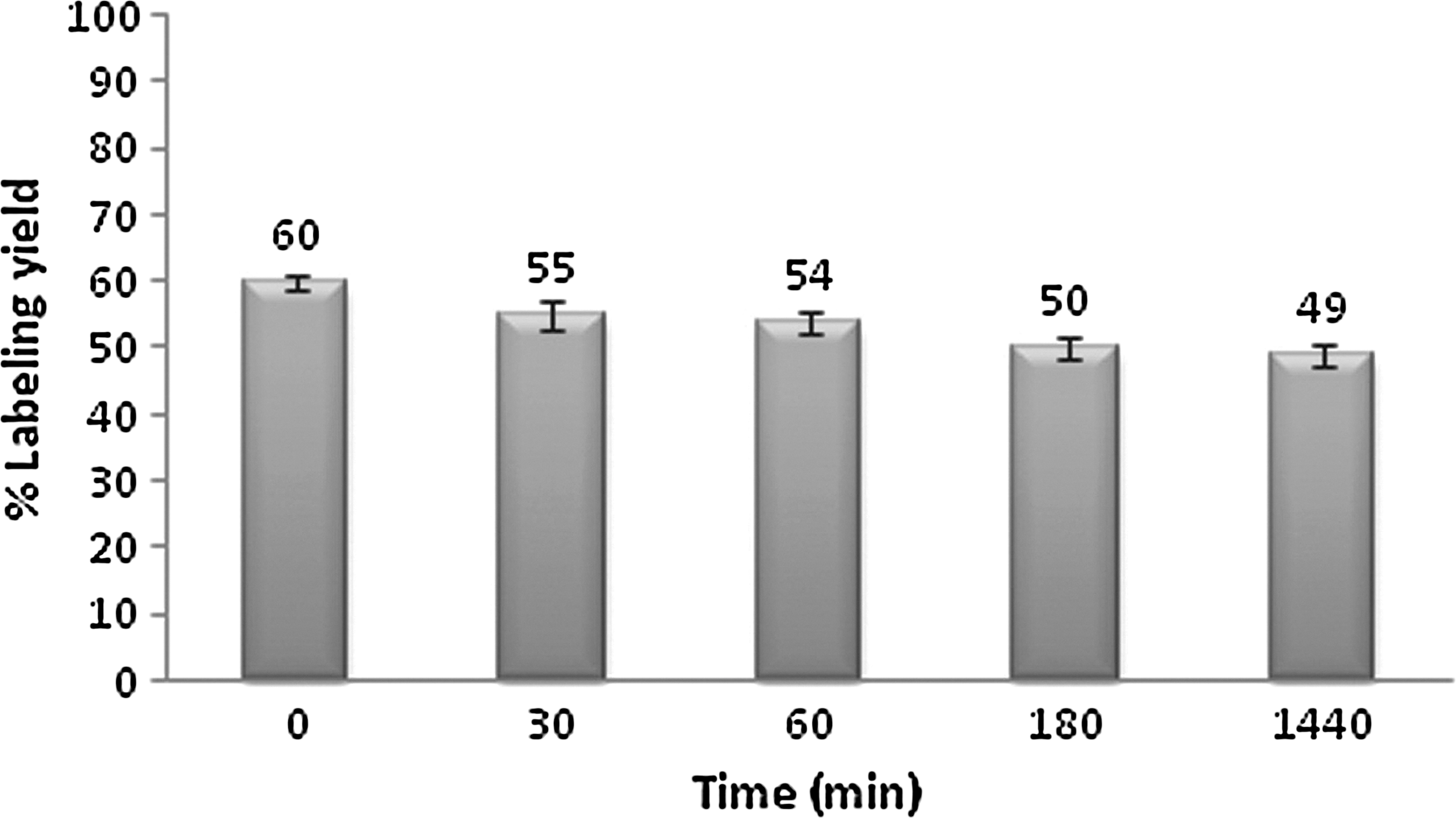
Stability of 131I-BLMG in serum.
Results of scintigraphic studies
Figure 5 shows the dynamic image corresponding to 30 minutes after the administration of 131I-BLMG. Figure 6 indicates the static scintigram corresponding to 30 minutes after the administration of 131I-BLMG. As seen on this scintigram, 131I-BLMG was eliminated via kidneys and accumulated in the stomach and the bladder within 30 minutes, and some thyroid uptake was observed. This also indicates a radiolabeling yield of about 60% for this compound. After 4 hours, the activity was completely cleared from the bladder. On the other hand, it was also observed that 131I radioactivity remained for a sufficiently long time in the metabolism and was not rapidly cleared.
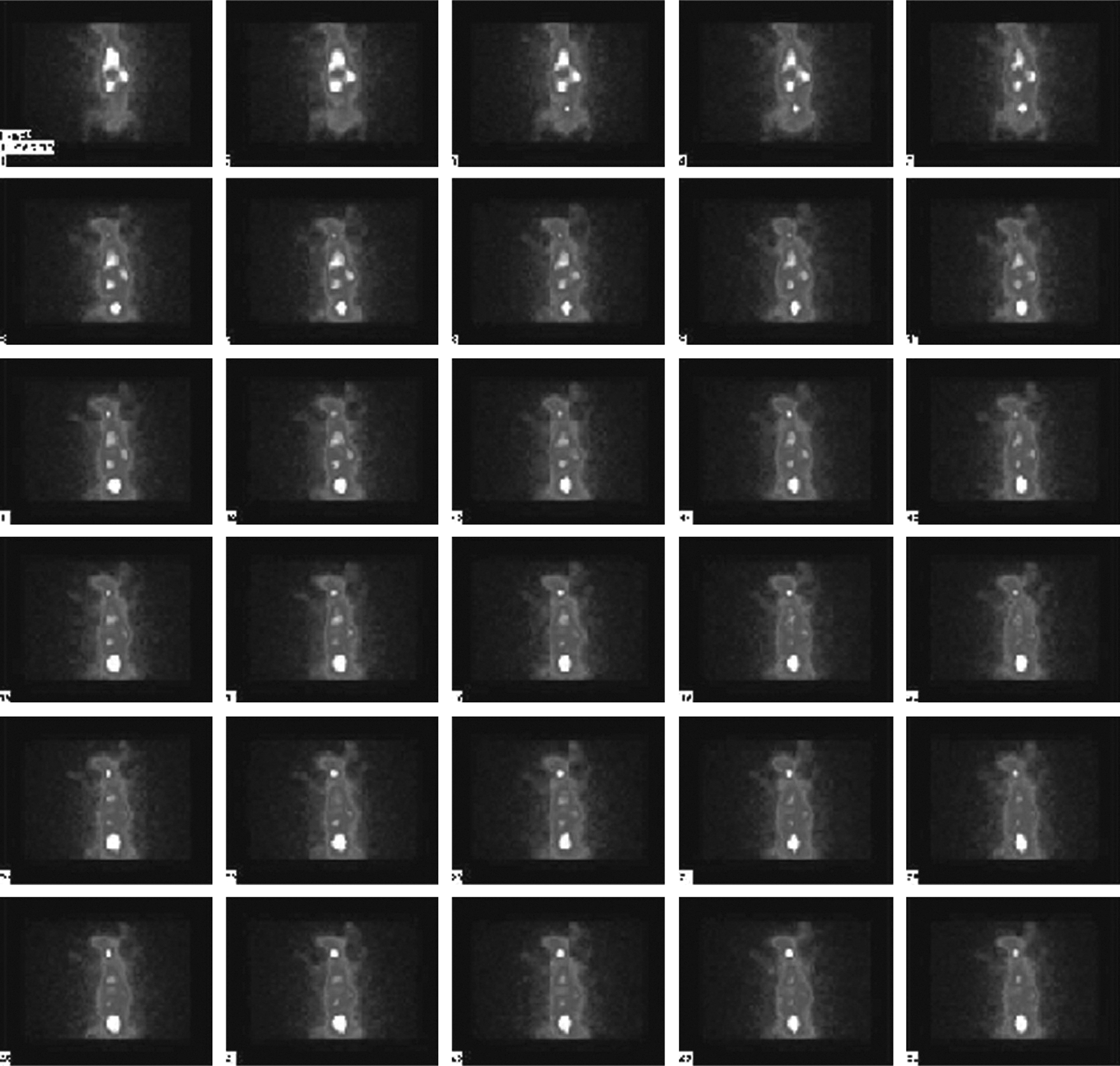
Dynamic scintigrams of 131I-BLMG, which was administered to a rabbit via the ear vein in 30 minutes. (The mixture of xylazine and ketamine anesthesia was used in the scintigraphy studies. Dynamic scintigrams were obtained over the first half hour with frames of 1 minute following the administration of the labeled compound.)
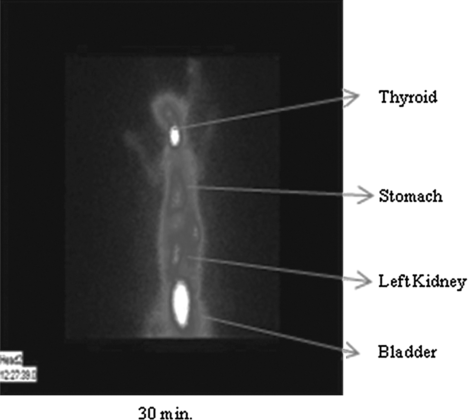
Static scintigram of 131I-BLMG, which was administered to a rabbit via the ear vein in 30 minutes. (The mixture of xylazine and ketamine anesthesia was used in the scintigraphy studies. Static image was obtained from posterior projection following the administration of the 131I-BLMG.)
Biodistribution studies
131I-BLMG at 60% radiochemical purity [specific activity: 3.7 MBq (100 μCi)/μg radiolabeled compound per rat] was injected to rats (for each time intervals, n=3). Figure 7 represents the biodistribution results of 131I-BLMG obtained at 30, 120, and 240 minutes after its administration to the rats. The %ID/g values of the radiolabeled BLMG in the stomach, bladder, prostate, and spinal cord were 1.16, 3.32, 0.39, and 0.43 at 120 minutes, respectively. It is obvious that the activity in none of the followed organs at none of the measured time points exceeded 3.5% of the injected activity. Very low accumulation in the liver and the spleen is in particular favorable, since accumulation in the liver and the spleen is often considered as a sign of poor biocompatibility. No significant deposition of the released activity in the thyroid is important, but indirect demonstration of the 131I-BLMG stability in vivo is. The uptake in these organs decreased with time. Significant amount of radioactivity was also seen in the prostate, the spinal cord, and the large intestine. Radioactivity in these organs was not cleared after 240 minutes. Biodistribution results agreed well with that of scintigraphic results obtained with this compound. In this study, a significantly positive correlation was shown in lung–kidney (r=0.805, p<0.02), lung–spinal cord (r=0.920, p<0.04), kidney–spleen (r=0.867, p<0.03), thyroid–blood (r=0.978, p<0.02), stomach–prostate (r=0.842, p<0.04), blood–kidney (r=0.940, p<0.02), and prostate–stomach (r=0.891, p<0.02) in rat's organs.
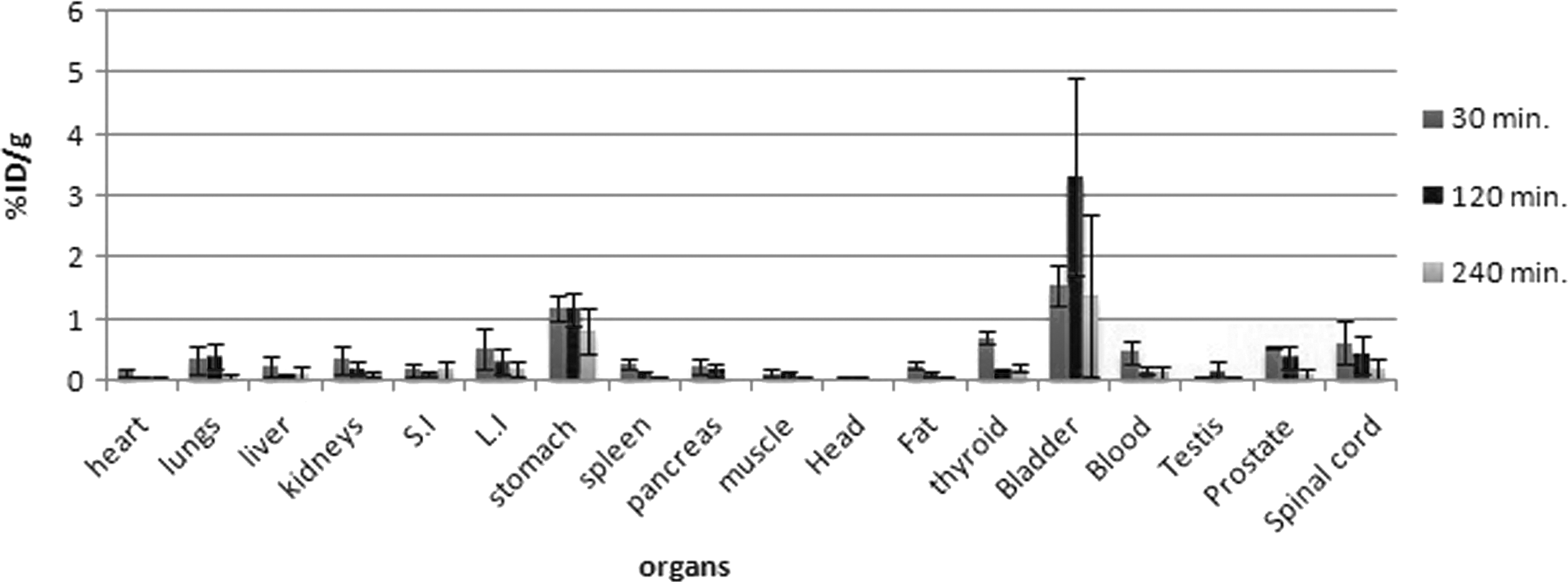
Activity (%ID/g) of 131I-BLMG in major organs of the rats in the given time intervals (n=3); error bars mean standard deviation; %ID/g=percent of the injected dose per gram tissue. All the presented data are decay corrected. S.I., small intestine; L.I., large intestine.
In vivo targeting activities of 131I-BLMG and 131I-BLM
Efficiencies of 131I-BLMG and 131I-BLM to target human colon carcinoma xenographies (Caco-2 cell line xenografts) were explored in biodistribution studies. At various times after 131I-BLMG and 131I-BLM injection, blood, the tumor, and the normal organs were analyzed to determine the amount of each radionuclide retained per gram of tissue. Biodistribution results (given as %ID/g vs. organ, organ-to-muscle ratio vs. organs) of 131I-BLM and 131I-BLMG in Balb/C mice tissues are given in Figures 8 –11. The high values of organ-to-muscle ratio for 131I-BLM were observed within 30 minutes after the administration of 131I-BLM in the tumor, the bladder, the breast, the spleen, and the lung. Some amount of the radioactivity was seen in the stomach. Stomach-to-muscle ratios were 1.17, 0.51, and 1.19 at 30, 120, and 240 minutes, respectively. In addition to this, the bladder-to-muscle ratio was highest at 30 minutes. The liver-to-muscle ratios of 131I-BLM were 0.64, 0.26, and 0.66 in the time intervals. Activity uptake in the thyroid and, evenmore, in the stomach is a (well-known) strong indication for the presence of radioiodide. Therefore, it is to be assumed that elimination of radioactivity is mainly due to the clearance of radioiodide beside the metabolism of 131I-BLM. Radioactivity in this organ was not cleared after 240 minutes. After intravenous (i.v.) administration of 131I-BLMG, radioactivity was widely distributed into most tissues. Blood radioactivity kinetics displayed a maximum with levels of around 1.3%ID/g at 240 minutes postinjection (p.i.). After administration of 131I-BLMG, radioactivity was widely distributed into most tissues. All tissues showed high uptake rapidly achieved 30 minutes p.i. The highest concentrations were found in the lungs, the spinal cord, and the stomach. The highest values of 131I-BLMG in the organ-to-muscle ratio were observed in the bladder, the stomach, the spinal cord, the lung, and the tumor within 30 minutes after the administration of 131I-BLMG. The lowest radioactivity levels were observed in the head and the muscles. At 240 minutes p.i., most of the radioactivity has been eliminated from the tissues, except for the stomach, the liver, and the spinal cord. In contrast, tumor uptake did not vary significantly over the time frame studied, and remained at around 2–3%ID/g globally. Moreover, radioactivity accumulated in the thyroid and the stomach as generally observed with molecules directly radiolabeled with iodine. Stomach-to-muscle ratios of 131I-BLMG were obtained as 3.32, 11.22, and 19.12 at 30, 120, and 240 minutes, respectively. The highest value of the bladder-to-muscle ratio was observed at 30 minutes as ∼24. The ratio in the tumor decreased with time.
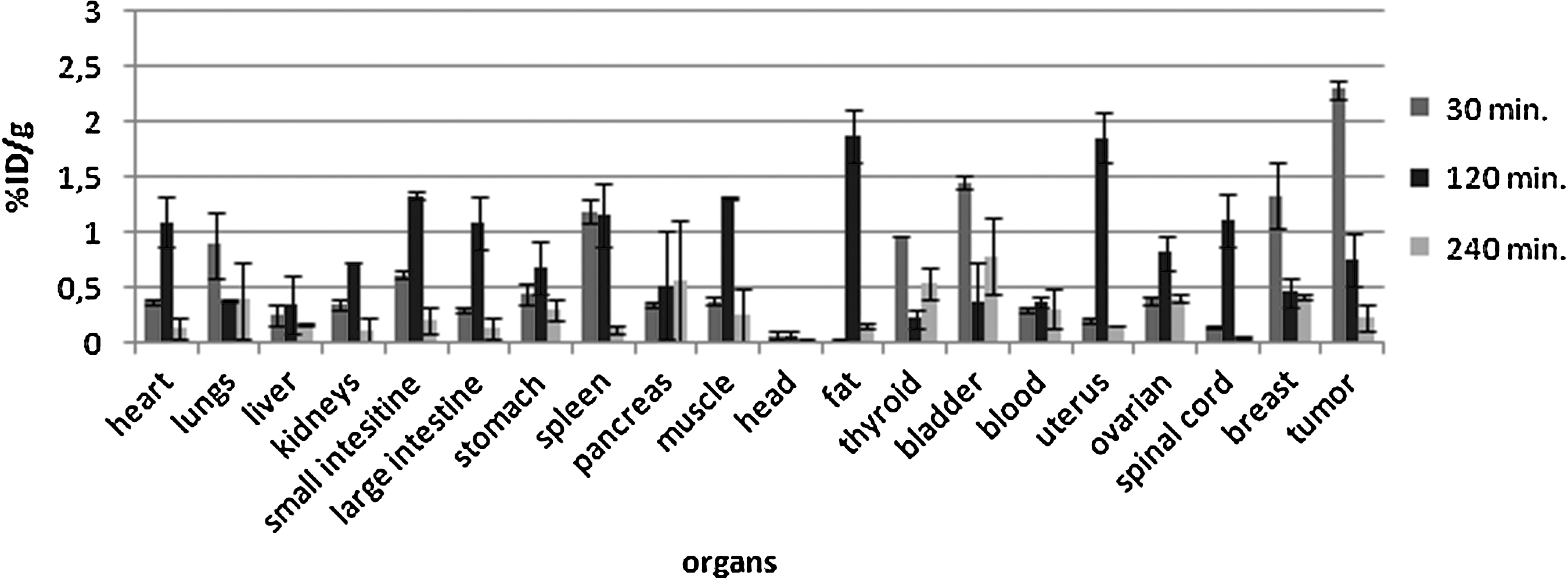
Activity (%ID/g) of 131I-BLM in major organs of the Balb/C mice in the given time intervals (n=3); error bars mean standard deviation; %ID=percent of the injected dose per gram tissue. All the presented data are decay corrected.
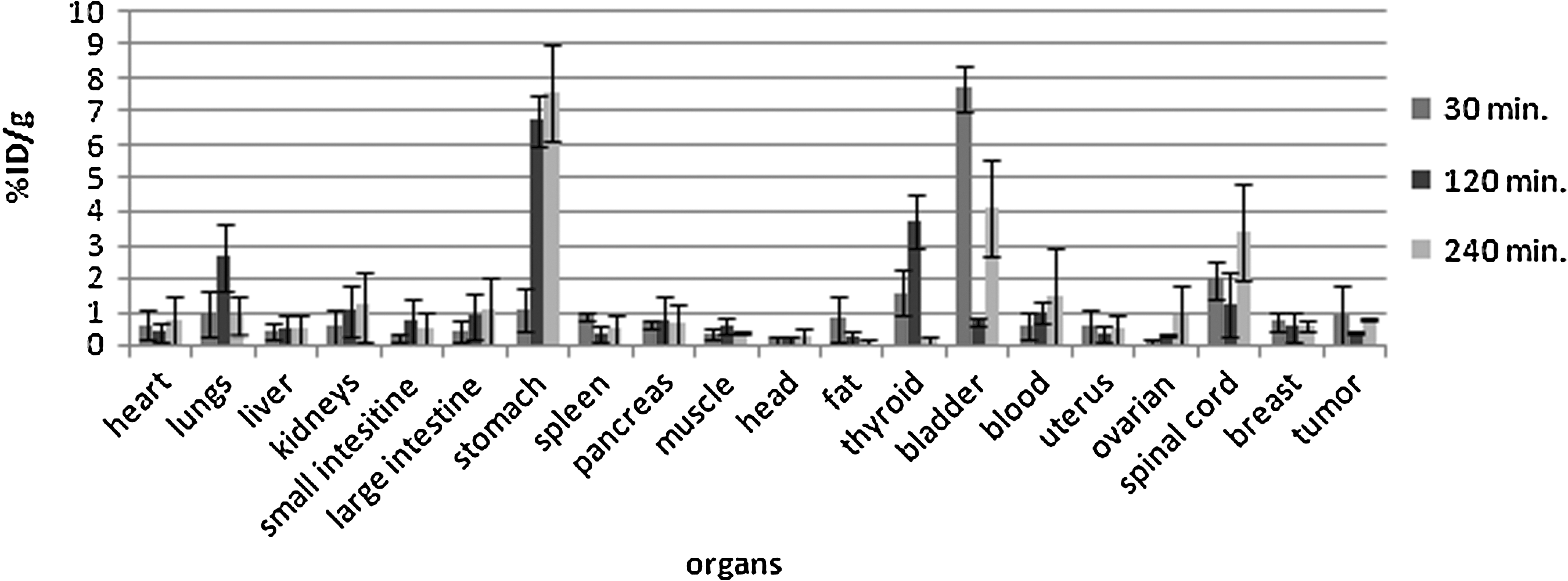
Activity (%ID/g) of 131I-BLMG in major organs of the Balb/C mice in the given time intervals (n=3); error bars mean standard deviation; %ID=percent of the injected dose. All the presented data are decay corrected.
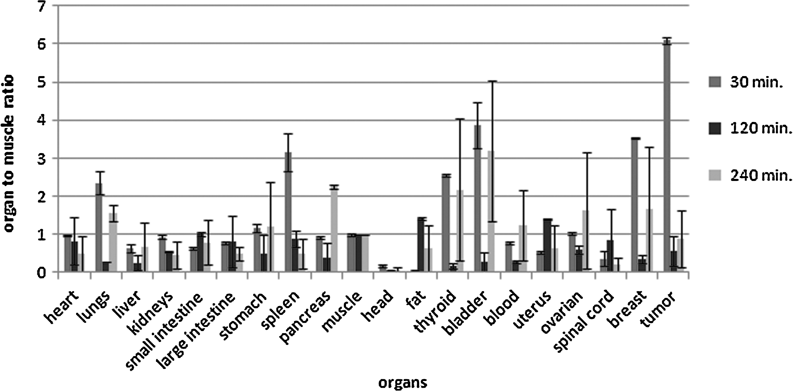
Organ-to-muscle ratio of 131I-BLM in the Balb/C mice in the given time intervals (n=3); error bars mean standard deviation; %ID=percent of the injected dose per gram tissue. All the presented data are decay corrected.
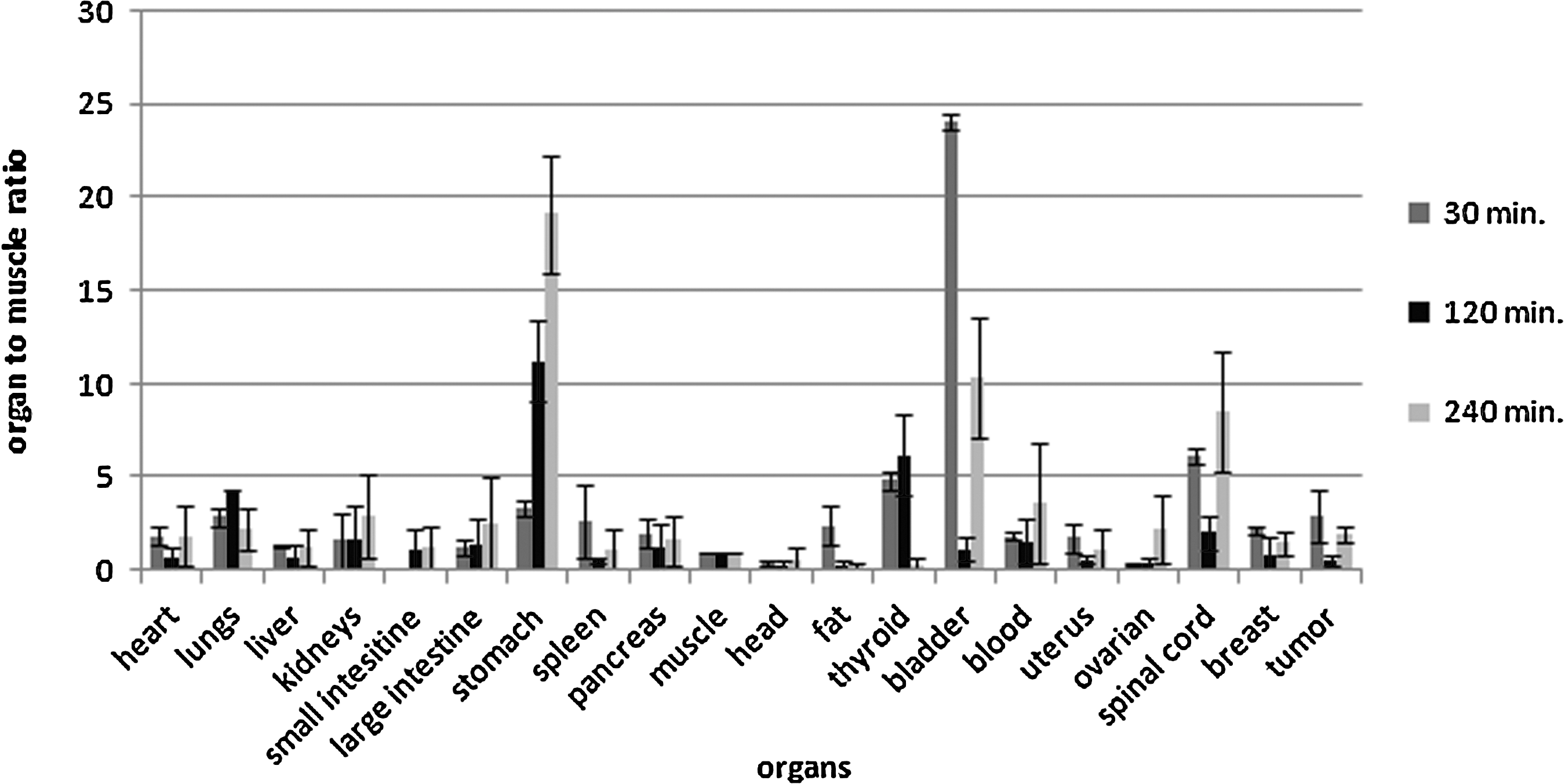
Organ-to-muscle ratio of 131I-BLMG in the Balb/C mice in the given time intervals (n=3); error bars mean standard deviation; %ID=percent of the injected dose per gram tissue. All the presented data are decay corrected.
There were also significant relations between breast and heart (p<0.01); breast and large intestine (p<0.05); blood and large intestine (p<0.05); blood and kidney (p<0.05); spinal cord and blood (p<0.01); muscle and small intestine (p<0.01); muscle and fat (p<0.05); breast and tumor (p<0.01); testis and large intestine (p<0.01); testis and blood (p<0.05); and testis and spinal cord (p<0.05).
Figure 12 represents the tumor-to-large intestine ratio of 131I-BLMG, and 131I-BLM was obtained 240 minutes after their administration to mice. High levels of tumor-to-large intestine ratios of 131I-BLMG and 131I-BLM were found as 7.81 and 2.29 at 30 minutes, respectively. These results showed that 131I cleared away from the large intestine quickly, but accumulated gradually in the tumor.
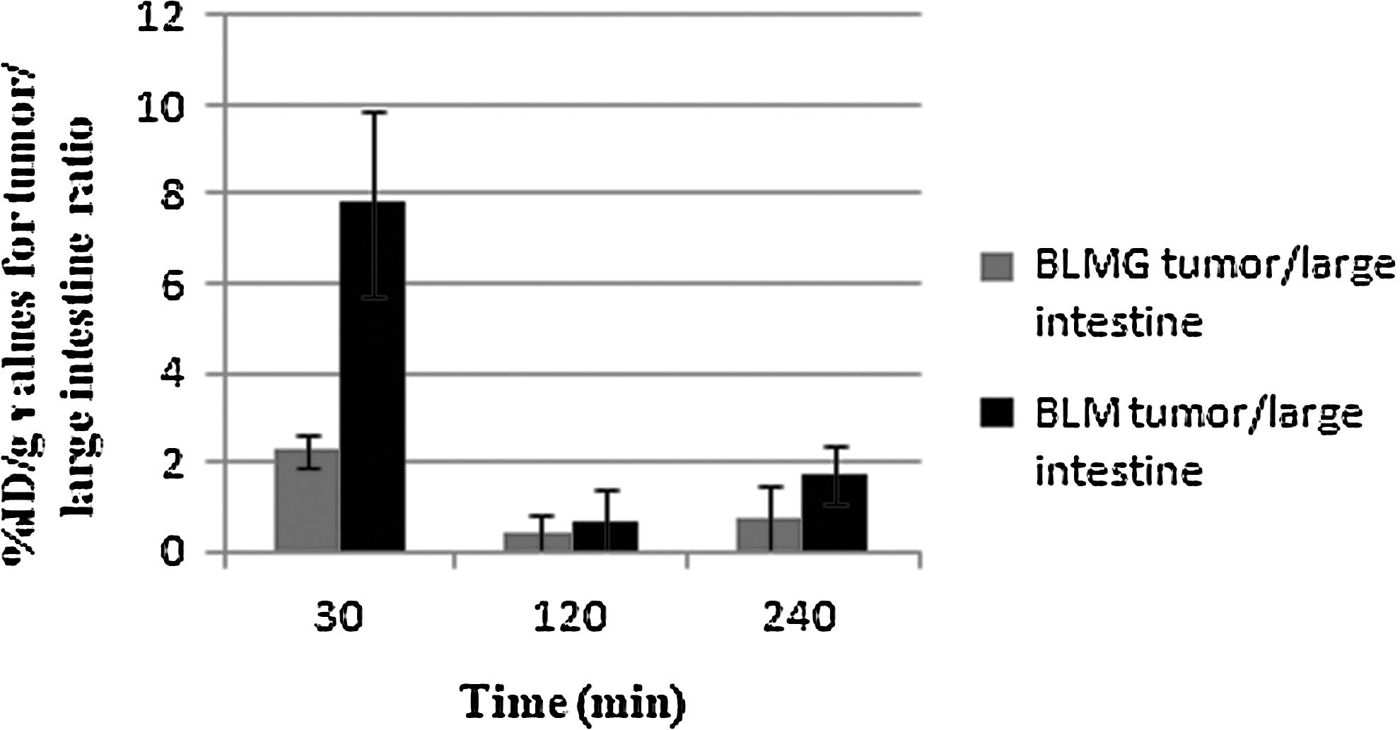
Tumor to large intestine ratio of 131I-BLM and 131I-BLMG.
Discussion
In a recent study, BLMs were separately labeled with 131I, and radiopharmaceutical potentials were investigated using animal models by Avcıbaşı et al. 43 In this respect, they have reported that these labeled compounds showed high uptake in the stomach, the bladder, the prostate, the testicle, and the spinal cord in rats, and the scintigraphies of radiolabeled isomers (131I-A2 and 131I-B2) were similarly found with that of 131I-BLM.
Pharmacokinetics of BLM was compared with those of 99mTc-, 111In-, and 57Co-BLM, and 67Ga citrate in mice bearing a transplanted KHJJ tumor by Krohn et al. 44 It was indicated that the in vivo kinetics and stability of 123I- and 57Co-BLM were similar: both were acceptable, although not equivalent, tag for BLM and, along with 67Co citrate, both had biologic properties suitable for tumor detection. Both 99mTc- and 111In-BLM dissociated rapidly in vivo and hence did not represent legitimate tags for BLM. Tumor-to-blood and tumor-to-liver ratios were higher for 123I-BLM than for 67Ga- or 57Co-BLM. Antunes et al. have described their first PET tracer for extracellular β-glucuronidase (β-GUS), fluoroethylamine glucuronic acid labeled with 18F ([18F]-FEAnGA), which consists of an [18F]-fluoroethylamine ([18F]-FEA) group bound to a glucuronic acid via a self-immolative nitrophenyl spacer. [18F]-FEAnGA was synthesized by alkylation of its imidazole carbamate precursor with [18F]-FEA, followed by deprotection of the sugar moiety with NaOH in the 10–20% overall radiochemical yield. At the end of this study, they carried out a micro-PET study in mice bearing tumor. In a preliminary micro-PET study, in mice bearing both CT26 and CT26m_GUS tumors, [18F]-FEAnGA exhibited a 2-fold higher retention of radioactivity in the tumor expressing β-GUS than in the control tumor. [18F]-FEA did not show any difference in tracer uptake between tumors. 38
In the presence of BLMG, electrophilic aromatic substitution reactions might probably take place in the 2 position of the imidazole ring on the compound. This is supported by the given m/z values of fragment 587, 1197, 1465, 1851, 1724, 1699, and 1564. The stability of radioiodinated compounds was long enough to be able to complete the scintigraphic studies. For these reasons, the 131I-BLMG was directly injected to the experimental animals without needing any separation or purification procedures.
It is well known that many aromatic amines, such as β-naphthylamine and 4-aminobiphenyl, are known to induce tumors, most notably in the urinary bladder. 45 N-hydroxylation of arylamines has been demonstrated to be involved in producing toxicity from these compounds. 46,47 Another major metabolic pathway for aromatic amines, which may compete with N-hydroxylation and thereby influence carcinogenic potential, is N-glucuronidation. 48,49 Glucuronidation of xenobioics is generally considered to be a significant step in their detoxification and elimination from the body. However, N-glucuronides are labile and may be easily hydrolyzed to the parent amine derivative under weakly acidic conditions, which exist generally in the urinary bladder. 50 As is expected, the scintigrapic image of the glucuronide derivative of 131I-BLM (131I-BLMG) supported this hypothesis. Therefore, activity in the bladder was probably due to accumulation of the 131I-BLMG. In addition to this, high activity in the urogenital zone was not only due to uptake of the 131I-BLMG in the bladder but also due to the accumulation of a radiolabeled glucuronide derivative of BLM in the prostate and the testis.
In case of administration of phenolphthalein (PPH) with a regular dose, it is metabolized as phenolphthalein-O-glucuronide (PPHOG) in the intestinal lumen and in the liver and excreted largely as this glucuronide conjugate. 51 The intestine contains significant amounts of the enzyme β-glucuronidase as a result of enzymatical hydrolyses of the glucuronide conjugate resulting in the formation of free-131I-PPH. This may then be reabsorbed and transported to the liver and undergo reconjugation and re-excretion. This behavior is termed enterohepatic recirculation and makes a significant contribution, prolonging the half-life of the drug in the body, with the obvious result of potential of the pharmacological action of the 131I-PPH. In the biodistribution results obtained for BLMG, it was shown that 131I-BLMG was significantly localized in the large intestine within 30 minutes as is shown in Figure 9. Biber et al. observed similar results with the glucuronide derivative of 99mTc-labeled β-estradiol (1,3,5,[10]-estratriene-3,17β-diol) attached to diethylenetriamine pentaacetic acid. 39,52 Results supported that intestines express the enzyme β-glucuronidase, which hydrolyzes the glucuronide bond similar to radioiodinated PPHG. 11
As seen in Figures 8 and 9, 131I-BLM exhibited about 2-fold higher retention of radioactivity than that of 131I-BLMG in the tumor. The liver-to-muscle ratios of BLMG were obtained as high values than those of BLM. This was probably because of the significant amounts of β-glucuronidase activity in the liver.
A specific enzyme detoxifying BLM, BLM hydrolase (BLMH), was recently put forward as a plausible candidate for associations with mutagen sensitivity (Caporaso, 1999). 53 BLMH, a neutral cysteine protease with exopeptidase activity that detoxifies BLM through deamination of the beta-aminoalanine moiety, 54 has been found in most human tissues, including peripheral blood leukocytes. In fact, resistance to BLM chemotherapy, observed in some cancers, has been postulated to result from overactive BLMH. BLM detoxification may also involve N-acetyltransferases (NATs). In Streptomyces verticillus, the BLM-producing antinomycete bacterium, resistance to BLM is conferred by an NAT enzyme. 55
With respect to the explanations given above about some glucuronide conjugate, Biber et al. synthesized a derivative of estradiol glucuronide that can be labeled with 99mTc and investigated its radiopharmaceutical potential using imaging and biodistribution studies. In conclusion, all these studies indicated that 99mTc-estradiol–glucuronide conjugates have not showed estrogen receptors specificity. Enzymatic mechanism seemed more effective than receptor specificity in tumor uptake. It was suggested that radionuclide-labeled estrogen–glucuronide conjugates may be a new series of radiopharmaceuticals for the enzyme β-glucuronidase-rich tissues and tumors. 39 Koçan et al. investigated the radiopharmaceutical potential of 99mTc-BLM and 99mTc-BLMG. They found that maximum uptakes of 99mTc-BLM and 99mTc-BLMG metabolized as N-glucuronide were observed within 2 hours in the liver, the bladder, and the spinal cord for 99mTc-BLM and the lung, the liver, the kidney, the large intestine, and the spinal cord for 99mTc-BLMG, respectively. 56
In conclusion, 131I-BLMG, which has diagnostic and therapeutic application potentials in nuclear medicine, was first synthesized and radioiodinated using the iodogen method and investigated to evaluate its biodistribution in the human colon cancer xenograph–bearing Balb/C mice. 131I-BLMG was obtained in high radiochemical purity and high yield by direct electrophilic iodination of BLMG. Radiolabeling of BLMG with 131I means that it can also be radioiodinated with other radioiodine isotopes such as 123I, 124I, and 125I under similar conditions. 131I-BLMG has shown specifity in the prostate, the large intestine, and the spinal cord in the biodistribution studies in male rats. Tumor-bearing mice model studies demonstrated that the highest uptake of 131I-BLMG was obtained in the tumor in the first 30 minutes. Radiolabeled BLMG proved a useful tool for assessing the in vivo behavior of the drug in mice bearing subcutaneous Caco-2 human colorectal adenocarcinoma, consequently validating its potential for the treatment of colorectal cancers.
Footnotes
Acknowledgments
The authors thank Celal Bayar University Research Fund (Contract no. 2007 FEF 007) for financial support. The authors also thank Dr. İlker Medine; Research Assistant Feray Koçan; M.Sc. student Gökçen Topal; and Ph.D. student Yasemin Parlak for their technical assistance during the animal experiments and scintigraphic studies.
Disclosure Statement
There is no any conflict of interest among the authors in this study.