
Abstract
The absorption and fluorescence spectra of the 7
Introduction
Coumarin analogs are the most important compound for photophysical studies during the last few decades, as they are highly fluorescent molecules that have many applications. Substituents play a very important role to deciding photophysical properties of coumarin as their nature and position changes. Coumarins substituted at position fourth and seventh with an electron-donating group are known to exhibit fluorescence. 1 As 7-aminocoumarins are highly fluorescent, they have been used as optical brighteners and fluorescent probes. Substituted 7-aminocoumarins also form an important class of laser dyes for the blue–green region. The photophysical properties of these compounds depend on the nature and the position of a substituent group in the parent molecule and also vary with the surrounding medium. Coumarins are used as nonlinear optical chromophores and as an excellent probe to study solvation dynamics in homogeneous solutions as well as organized media. 2 –8 In the recent past, numerous coumarin heteroderivatives have been synthesized, and the possibility of their applications as laser dyes, organic scintillators, and triplet sensitizers has been explored. 9 –13 In a series of earlier work, the effect of solvents, substituents, and temperature on the various photophysical properties of coumarin compounds has been reported. 14 –19 It is found that the nature of solvents and substituents brings about changes in the values of fluorescence wavelength maxima, quantum yield, lifetime, polarization, and excited-state dipole moment of the coumarin. 18 A systematic study of fluorescence quenching of 4- and 7-substituted coumarins by halide ions in an aqueous medium has also been conducted previously. 3,19
The importance of coumarins also becomes so important, because during the last few years, there has been a remarkable growth in the use of fluorescence in biological sciences, especially in biochemistry and biophysics along with environmental monitoring, clinical chemistry, DNA sequencing, and genetic analysis by fluorescence in situ hybridization. In molecular biology, fluorescence is used for cell identification and sorting in flow cytometry, and in cellular imaging to reveal the localization and movement of intracellular substances by means of fluorescence microscopy. Because of the high sensitivity of fluorescence detection, there is continuing development of medical tests based on the phenomenon of fluorescence. These tests include the widely used enzyme-linked immunoassays and fluorescence polarization immunoassays.
Most drugs administered nowadays are distributed through the circulatory system by means of their binding to some plasma protein. Human serum albumin (HSA) is the serum protein that shows the highest nonspecific binding capacity, and is the principal carrier of drugs and endogen substances in blood. Serum albumin, the most abundant protein in the circulatory system, has been one of the most extensively studied of all proteins. 20 –22 It is synthesized in the liver, exported as a nonglycosylated protein, and is present in the blood at around 40 mg/mL. 23 It is the major transport protein for unesterified fatty acids, but is also capable of binding an extraordinarily diverse range of metabolites, drugs, and organic compounds. The remarkable binding properties of albumin account for the central role, that is, it can play in both the efficacy and rate of delivery of drugs.
Bovine serum albumin (BSA) was selected as a protein model because of its medical importance, stability, and structural homology with human serum albumin. Thus, it is important to study the interaction of coumarin analogs with BSA experimental.
Materials
All the chemicals, including BSA, were purchased from Sigma Chemical Company. The water used for preparation of solution was of 18 Ω MQ grade derived from the Millipore water system. For fluorescence and absorption measurements, the BSA solution of pH 7.4 was prepared in 0.1 M phosphate buffer (mixture of sodium dihydrogen phosphate dehydrate and anhydrous disodium hydrogen phosphate, both were of analytical grade and from E. Mark) containing 0.15 M NaCl (analytical grade from Sigma). Coumarins stock solutions were prepared in MQ water, and then the working solution was made by further dilution with 0.1 M phosphate buffer containing 0.15 M NaCl.
The stock solutions of BSA (2.0×10⁻⁵ M) were prepared in 10 mL of 0.1 M sodium phosphate buffer (pH 7.4) containing 0.1 M NaCl, whereas the stock solutions of 7-diethyl amino-4-methyl coumarin (DAMC) were prepared in an ethanol–water (1:9 v/v) solution. The concentration of the BSA was determined spectrophotometrically using an extinction coefficient 43,824 M−1 cm−1 at 280 nm. 10,11 The pH of solution was adjusted to the desired value with 0.1 M HCl or NaOH.
Spectral measurements
The absorption spectral measurements were recorded on a Perkin Elmer spectrophotometer. The sample cuvettes used were a pair quartz cells of 1.00-cm path length. All scanning parameters were optimized to obtain the best spectra, and in general, the parameters were scan range 200–700 nm, wavelength step 0.5, and all measurements were carried out at room temperature (23±1°C).
Fluorescence measurements for studying quenching mechanism
Spectrofluorimeter model FS1120 of Edinburgh Instruments equipped with a xenon arc lamp was use for fluorescence measurements. The temperature of the sample holder was regulated with a peltier-cooled thermostat. Quartz cuvettes of 3-mL capacity and path length 1 cm were used for all measurements.
Fluorescence excited-state lifetime measurements
The fluorescence lifetime measurements were performed using a customized integrated steady-state spectrofluorimeter. The excitation source was hydrogen gas filled nanosecond flash lamp (model nF900) filled with low H2 gas pressure of 0.4 bar operating at a frequency 40 KHz. The silt width for both excitation and emission monochromators were kept fully open. The intensity decay curves were obtained at emission maximum and fitted as sum of exponentials as
where, τi and A i are representing the fluorescence lifetime and pre-exponential factor for ith decay component. The detailed procedures of measurement and analysis of decay parameters are discussed elsewhere. The excited-state lifetime values were calculated by deconvoluting the intensity decay profiles. More precisely multiple-exponential fittings are applied along with the instrument response function (IPR) of the excitation wavelength in the deconvolution process. The IPR was recorded with Ludox as a scattering medium. The analysis of the fitted data was tested using statistical parameters provided in the analysis software.
A549 human lung cancer cell proliferation assay
Cell proliferation was measured by the MTT method. Briefly, A549 cells were collected and seeded in 96-well plates at a density of 5×10³ cells/well. After incubation of six different concentrations (0.06, 0.23, 0.94, 3.75, 15, and 60 μg/mL) for 48 hours, the medium was removed and replaced with a fresh medium (180 μL/well). Twenty microliters of MTT solution (5 mg/mL) was added to each well, and the plates were incubated for an additional 4 hours at 37°C. The medium was aspirated off, and dimethyl sulfoxide (150 μL) was added to each well. The absorbance was read at 570 nm using a microplate reader. Cell viability was studied as a percentage of the untreated cells.
A549 cellular apoptosis assay
A549 cells were seeded in 24-well plates at a density of 5×10³ cells/well, and incubated for four different concentrations (0, 1, 5, and 10 μg/mL) for 48 hours. The cellular monolayer was fixed and stained with a DNA fluorochrome for 30 minutes. After washing with phosphate-buffered saline, the morphological features of apoptosis (including cellular nucleus shrinkage, chromatin condensation, intense fluorescence, and nuclear fragmentation) were monitored by fluorescence microscopy.
Result
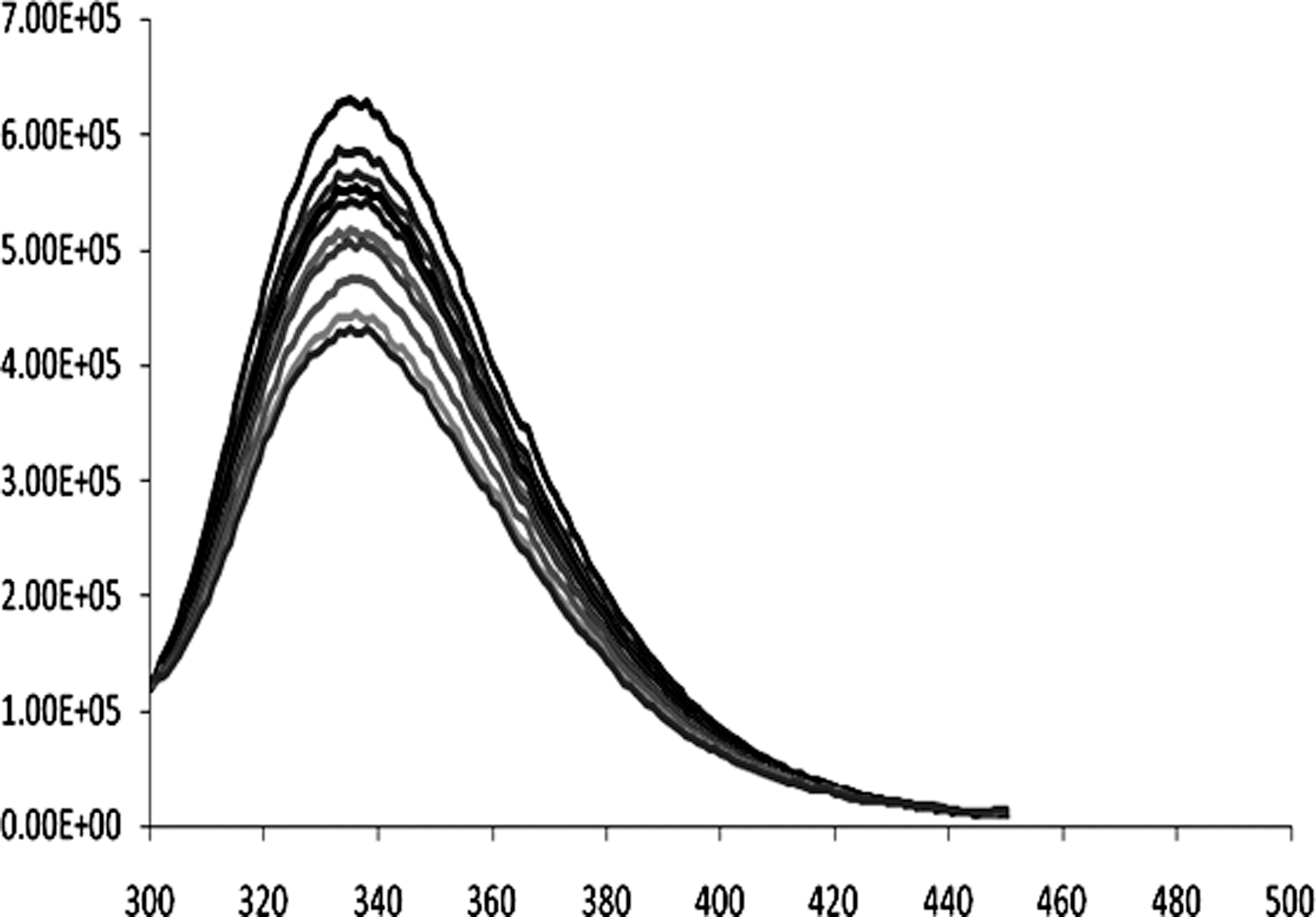
Absorption spectral measurements on BSA in the presence of small molecules provide useful information related to the nature of interaction between a ligand and BSA. In the present study, the BSA solution (10 μM) was titrated against DAMC in 0.1 M phosphate buffer, pH.7.4 (Fig. 1). The absorption spectra of the coumarin analog DAMC in the ethanol–water (1:9 v/v) solution of varying pH are shown in Figure 1. The absorption wavelength showed obvious pH dependence, and the optical density decreases with increasing pH. Formation of new band at 378 nm was observed for the basic form. The isobestic points at 345 nm are due to the protonation–deprotonation equilibriums.

Absorption spectra of DAMC in ethanol–water (1:9 v/v) solution at different pH values. DAMC, 7
The fluorescence spectra of neutral DAMC excited at 335 nm (maximum absorption) give very weak fluorescence intensity with maximum at 450 nm. Figure 2 shows the excitation spectra of DAMC monitoring at 450 nm while the fluorescence spectra of BSA in the presence of different concentrations of DAMC are shown in Figure 3 which shows the quenching nature of compound on BSA.
Fluorescence spectra of DAMC in ethanol–water (1:9 v/v) solution of varying pH.
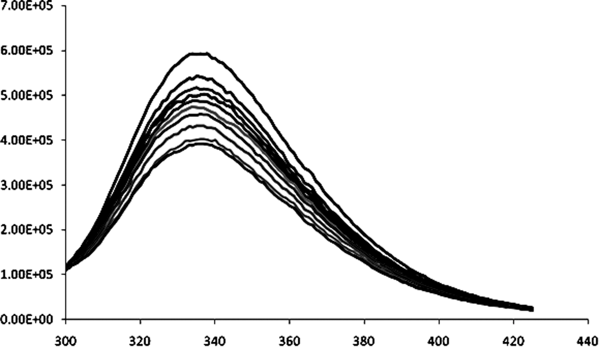
Effect of DAMC on the fluorescence spectra of BSA (wavelength 295 nm, pH 7.4, T0, 298 K). [BSA]=1.2×10⁻⁶ M; [DAMC]=10−6 M×(0, 2.0, 4.0, 7.5, 9.0, 15, 20, 30, 40, and 50]. BSA, bovine serum albumin.
The modified Stern-Volmer plot based on Equation (2) facilitates better description of the quenching data when both dynamic quenching and static quenching are present simultaneously.
where Kq is the collisional quenching constant or Stern-Volmer quenching constant, and V is the static quenching constant. The value of V was obtained from Equation (2) by plotting (F0/FeV [Q])-1 versus [Q] for varying V until a linear plot was obtained. The value of Kq was obtained from the slope of (F0/FeV [Q])-1 versus [Q] (Fig. 4). The values of V and Kq so obtained are found to be 8.2×10⁴ M−1 and 17.3025×10⁴ M−1, respectively.
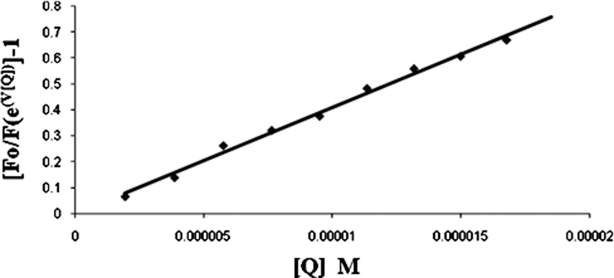
Modified Stern-Volmer Plot [F o/F (eV [Q])]-1 versus [Q] for the binding of BSA with DAMC.
Fluorescence bimolecular quenching rate constant k
q
measurement was evaluated using the equation,
where, τ0 is the lifetime of BSA in the absence of quencher and k
q
is the rate constant for the quenching. The τ0 value for BSA is reported in many studies and a general τ0 value was found to be in 10−9 s and hence the value of k
q
was observed to be of the order of 1013 M−1s−1. The value of k
q
depends on the probability of a collision between fluorophore (in this case it is tryptophan) and quencher and this probability depends on their rate of diffusion (D), size and concentration.
where, D is the sum of the diffusion coefficients of quencher and fluorophore σ is the sum of molecular radii and N a is the Avogadro's number. Since the upper limit of k q expected for diffusion controlled bimolecular process is 1010 M−1s−1. The high magnitude of K q in the present study (1013 M−1 s−1) can be attributed to a specific long-range interaction between drug molecules and tryptophan residues on protein and that the quenching is initiated predominantly by static collision due to complex formation. The biological cell arrest of DAMC is express in Figure 5, which shows it as prominent anti-tumor agent in induced apoptosis in A549 human lung cancer cells.
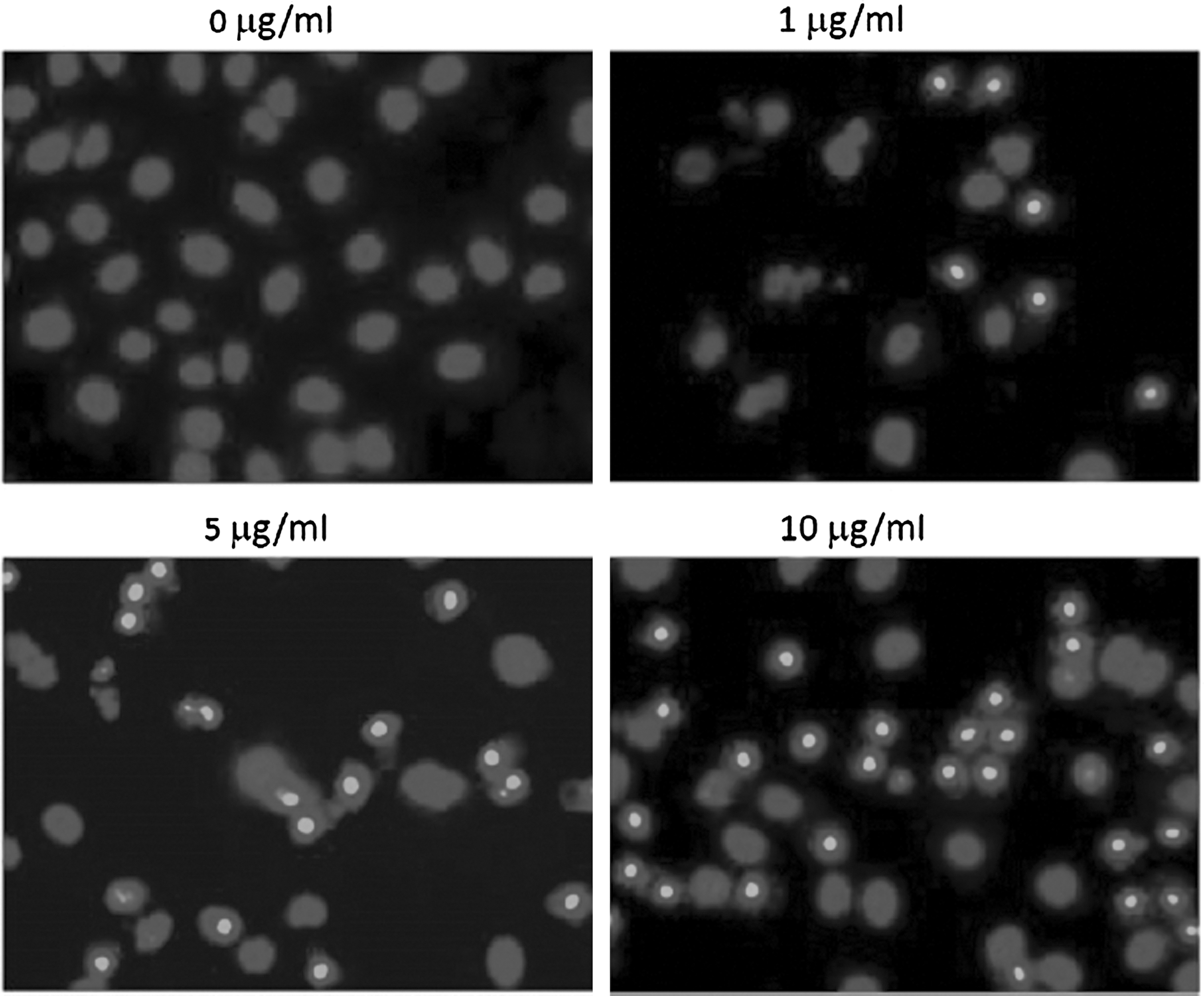
DAMC-induced apoptosis in A549 human lung cancer cells. Condensed and fragmented nuclei with bright staining were considered to be apoptotic cells.
Discussion
Fluorescence spectroscopy is a powerful technique for studying the interaction of drugs with protein. Fluorescence quenching can be induced by a variety of molecular interactions of a fluorophore with a quencher molecule, including ground-state complex formation, energy transfer, molecular rearrangement, excited-state reactions, and collision quenching. The mechanisms of fluorescence quenching are usually classified as either dynamic quenching or static quenching. BSA has three fluorophores, which are tryptophan, tyrosine, and phenylalanine. The fluorescence of tyrosine is almost quenched due to the effect of the nearest amino group, a carboxyl group, or a tryptophan residue. Phenylalanine has a very low fluorescence quantum yield. Therefore, the intrinsic fluorescence of BSA is mainly due to the tryptophan residue. To avoid any contribution from the tyrosine to the fluorescence spectra, the excitation wavelength was selected at 295 nm, in which the absorption is only due to tryptophan. The intrinsic fluorescence of proteins is the high sensitivity of tryptophan to its local environment. Changes in emission spectra of tryptophan are common in response to protein conformational transitions, subunit association, substrate binding, or denaturation. Hence, the intrinsic fluorescence of proteins can provide considerable information about their structure and dynamics, and is often considered in the study of protein folding and association reactions. BSA undergoes a number of pH-dependent conformational transitions. This means that the pH values of the environment have a pronounced effect on the secondary structure of the proteins. The normal form of BSA exists at neutral pH (7.0–8.0). Above pH 10.5, protein gets cleaved. The peptidic residues get positively charged at low pH values that counteract internal stabilization forces of the protein. This leads to the unfolded state of BSA. Therefore, the interaction of BSA with DAMC was investigated at neutral pH. The fluorescence spectra of BSA in the presence of different concentrations of DAMC are shown in Figure 3. The fluorescence intensity of BSA gradually decreased with the increase of DAMC concentration.
The biological studies were performed for DAMC-induced apoptosis in A549 human lung cancer cells. Condensed and fragmented nuclei with bright staining were visualized by a fluorescence microscope, which confirms the potential of this molecule as a potent optical imaging agent. In addition, for further biological application point of view, we then employed the MTT assay to investigate cytotoxicity of DAMC to the cell lines. The cell viability declines by 50%–70% upon DAMC treatment; therefore, it should be noteworthy that DAMC is effective for enhancing cell viability. These results are also found as similar to other known coumarin fluorescence sensors.
In the present study, fluorescence spectroscopic measurements were undertaken to gain insight into the detailed nature of binding of DAMC with BSA as indicated in UV-Vis absorption measurements. Fluorescence spectra were recorded in the range 300–550 nm upon excitation at 295 nm.
Fluorescence decay profiles of DAMC were obtained both for free and in the presence of BSA, and when they were deconvulated for analysis with the instrument, IRF revealed double exponentially decaying constants having short (τ1 ) and long (τ 2) decay components. The numerical values of τ 1 and τ 2 and their relative distribution varied in the presence of BSA. For example, in the absence of BSA, the values of τ1 and τ2 are 0.3 and 3.6 ns, respectively.
The fluorescence quenching data have been further analyzed by the Stern-Volmer equation:
where F0 and F are the fluorescence intensities of BSA at 336 nm in the absence and presence of the quencher (DAMC), respectively. Ksv is the Stern-Volmer quenching constant, and [Q] is the concentration of the quencher DAMC. The linearity or deviation from the linearity of the Stern-Volmer plot F0/F versus [Q] for BSA as shown in various studies revealed the type of quenching viz. static or dynamic. The Stern-Volmer plot (Fig. 4) in the present study has an upward curvature for the complete concentration range of DAMC; it therefore indicates the presence of both static and dynamic quenching by the same fluorophore.
The upward curvature in the Stern-Volmer plot indicates that both tryptophan residues of BSA are exposed. After addition of DAMC to 25 μM BSA solutions, around 70% of the fluorescence intensity was quenched. The maximum quenching was obtained by extrapolating a plot of (F0−F)/FO versus 1/[Q], corresponding to an infinite concentration of DAMC. It was observed that at infinite concentration of DAMC, fluorescence quenching was ∼90%. This again shows that both the tryptophan residues of BSA are accessible to DAMC.
Conclusion
The interaction between DAMC and BSA was studied by fluorescence spectroscopic methods. The results showed that DAMC has a strong ability to quench the BSA fluorescence mainly through a static quenching process. The binding constant K and the number of binding site n were calculated according to the fluorescence quenching results. The biological studies confirm that this molecule has a very good potential for optical imaging. These findings are likely to provide useful insight for interaction of optical agents with serum albumin protein, and thereby lead to design new ligands with improved pharmacokinetic parameters.
Footnotes
Disclosure Statement
There is no conflict of interest exists for any of the authors.