




Abstract
Aims:
The purpose of the present work was to formulate and evaluate cationic nano-liposomes as novel nonviral gene delivery for colon cancer treatment.
Methods:
Recombinant pEGFP-c1-Fms-like tyrosine kinase receptor 3 ligand (FL) plasmids containing human FL gene and green fluorescent protein (GFP) reporter genes were constructed. FL and GFP Gene-carrying cationic nano-liposomes were prepared based on the electrostatic adherence principle and then transfected into Lovo cells. The morphology, particle size, and zeta potential of gene-carrying cationic nano-liposomes were observed using an electron microscope. GFP expression was observed by fluorescence microscopy to assay the transfection efficiency. The cytotoxicity of FL/nano-liposomes was evaluated by the MTT method.
Results:
Recombinant plasmids pEGFP-c1-FL are successfully constructed using gene cloning methods and confirmed by restriction enzyme digestion and sequencing. The cationic nano-liposomes carrying pEGFP-cl-FL were observed by an electron micrograph and showed uniform spherical or elliptical shapes and many pores. The fluorescence microscopy images of gene-carrying cationic nano-liposomes showed good expression of GFP in pEGFP and pEGFP-cl-FL groups. The MTT assay of cell death indicated a significantly higher level of cell death between the FL group and the control group at 24, 48, and 96 hours after transplantation.
Conclusion:
Cationic nano-liposomes show safe and high-performance transfection as gene carriers. Gene therapy has significant implications for colon cancer treatment in future.
Introduction
Up to date, the traditional treatments for colorectal cancer such as surgery and chemotherapy not only cannot completely eliminate all the cancer cells in vivo but also have serious side effects. At the same time, gene therapy is a rapidly advancing field with enormous potential to treat vital malignant diseases and brings hope to human beings to overcome the threat of cancer.
The development of potent gene transfer systems that can deliver foreign genes efficiently and safely into target cells is of crucial importance for successful gene therapy. Although having better efficiency of transfection, genetic carrier of retrovirus or adenovirus had many shortcomings, such as complications of preparation, immunogenicity, grave hidden danger of safety, and so on. The nonviral vector, possessing significantly low safety risks and convenient preparation in large quantities easily and inexpensively, has been suggested as an alternative to viral vector. 1 Therefore, the development of a safe and effective nonviral vector system is an urgent matter.
As a new kind of nonviral vector, the cationic nano-liposome is becoming one of the most important gene carriers in the biomedicine. Research conducted on cationic nano-liposomes has significant and subsistent meaning for tumorous gene therapy and the living quality of cancer patients due to its safe and high-performance transfection. The regulatory Food and Drug Administration (FDA) approval of nano-liposomes for diseases such as breast cancer and ovarian cancer has fueled research in the development of targeted stealth liposomal systems. 2 Recently, developments in liposome technology are leading to a burgeoning number of promising experimental and clinical applications. 3 The nano-liposome has been investigated as a nonviral vehicle for reaching targeted cells when performing gene therapy. 4 Therefore, the cationic nano-liposome might be considered a promising and suitable candidate for gene delivery application.
Currently, significant progress has also been made with regard to colon cancer genes. The antitumor effects of Fms-like tyrosine kinase receptor 3 ligand (FL) are becoming hot research topics. FL is a hemopoietic growth factor that is produced by bone marrow stromal cells and lymphocytes. The expression of FL has been demonstrated in a number of human leukemia cell lines of both myeloid and lymphoid lineage. 5 FL is important in the proliferation and differentiation of hematopoietic progenitor cells, and its receptor is mainly expressed in hemopoietic stem and progenitor cells. 6,7 It can enhance the proliferation of dendrtic cells (DCs) and promote the downstream T-effective cell immunity reaction, thus displaying an important role of antitumor immunity. Maliszewski reported that immunization of FL-treated mice with a protein antigen leads to increased production of antibodies specific for that protein. 8
Therefore, to explore the role of cationic nano-liposome and FL gene in the treatment of colorectal cancer, the FL gene was delivered into colon cancer Lovo cells through the cationic nano-liposome carrier, and their effects on the apoptosis of cancer cells were analyzed in this study.
Materials and Methods
Materials
The Lovo cell line was initiated in 1971 from a fragment of a metastatic tumor nodule in the left supraclavicular region of a 56-year-old Caucasian male patient with a histologically proven diagnosis of adenocarcinoma of the colon and kindly provided by the laboratory of the department of General Surgery, Affiliated Union Hospital of TongJi Medical College, Huazhong University of Science and Technology. 9
The instruments used in this study include the following: a 5414D-refrigerated centrifuge, a DYY-III-6B electrophoresis apparatus, a vertical −80°C freezer, a TD5 low-speed desktop centrifuge, an AE200 electronic balance, a JEM-1200EX transmission electron microscope, a Zetasizer 3000-particle analyzer, a fluorescence microscope, an FACS flow cytometer, a T4 DNA ligase, a pEGFP-C1 vector, a pUC57 vector, Lipofectamine 2000. All the other chemicals and reagents used were of an analytical purity grade or higher and were commercially obtained.
Methods
Recombinant pEGFP-c1-FL plasmids construction
The full-length FL gene was synthesized by a DNA synthesizer according to the cDNA sequences of human FL in GeneBank (510 bp, 169 amino acids). The synthesized FL genes were digested by restriction enzyme SalI and BamHI and bound with a puc57 vector to obtain a cloning vector of puc57-FL plasmids. Then, the target genes were subcloned again from puc57-FL into pEGFP-c1 to obtain the eukaryotic expression vector of recombinant pEGFP-c1-FL plasmids. Agarose gel electrophoresis was used to identify the recombinant pEGFP-c1-FL plasmids.
Gene-carrying cationic nano-liposomes preparation based on electrostatic adherence principle
First, 0.2 mL of Lipofectamine 2000 was taken into a 1.5 mL EP pipe and mixed with 1.2 mL serum-free RPMI1640 medium for 5 minutes. Appropriate amounts of recombinant pEGFP-c1-FL plasmids were also mixed with 1 mL serum-free RPMI1640 medium for 5 minutes. Then, the two mixtures just referred to were combined and incubated for 30 minutes at a temperature of 25°C to obtain FL-carrying cationic nano-liposomes (FL/nano-liposomes).
Morphology, particle size, and zeta potential of gene-carrying cationic nano-liposomes
The morphology of nano-liposomes and FL/nano-liposomes was examined under a transmission electron microscope. The average particle size, size distribution, and zeta potential of the nanoparticles were measured by photon correlation spectroscopy (PCS) using Zetasizer 3000. The average particle size was expressed in volume mean diameter, and the reported value was represented as mean±standard deviation (SD) (n=3).
In Vitro Transfection Assays of FL/nano-liposomes
The transfection activity of FL/nano-liposomes was evaluated in Lovo cell lines, encoding enhanced green fluorescent protein (GFP) as the reporter gene in the transfection studies. Cells were inoculated on a 12-well plate at a density of 1×10⁵ cells/well 24 hours before the transfection and then cultured using a complete medium plus rmGM-CSF/rmIL4 under normal conditions. When the cells reached 70%–80% confluence, appropriate amounts of serum-free medium diluted gene-carrying cationic nano-liposome FL/nano-liposomes (1 μg/8 μL) were added to each well. Cells were then cultured at 37°C at 5% CO2 in an incubator for 24–72 hours to allow the expression of transfected genes. Nano-liposomes/pEGFP was used as a positive control, and 1 μg of the plasmid pEGFP-c1-FL was used as a negative control. The detection of the expression of EGFP was carried out using an inverted fluorescent microscope with an attachment for fluorescent observation, and the picture was captured using a 400× objective.
Cell viability test of FL/nano-liposomes
The cytotoxicity of FL/nano-liposomes was evaluated by the MTT method in the Lovo cell line. Briefly, the cells were seeded at a density of 1×10⁴ cells per well in 0.2 mL of RPMI 1640 culture medium supplemented with 10% fetal bovine serum and antibiotics in a 5% CO2 incubator at 37°C overnight. After that, the culture medium was replaced by a 200 μL fresh serum-free RPMI 1640 medium with different concentrations of the nanoparticles and Lipofectamine 2000 in comparison. After incubation for 24 hours, 20 μL of MTT stock solution in PBS (5 mg/mL, pH 7.4) was added into each well at a final concentration of 0.5 mg/mL MTT. The medium was removed and DMSO was then added, and the absorbance was measured using the ELISA instrument. The cell viability (%) was calculated and compared with the untreated control (100%) according to the following equation: cell death rate=(l-average D value in experimental group/average D value in control group)×100%.
Statistical analysis
Statistical analyses were performed using SPSS software. Continuous variables were expressed as mean±SD, and categorical variables were shown as percentages. Continuous variables were analyzed using the Student's test, whereas categorical variables were analyzed using the Pearson's chi-square test. p-values<0.05 were considered statistically significant.
Results
Construction and identification of recombinant plasmids pEGFP-c1-FL
Recombinant plasmids pEGFP-c1-FL was digested by two enzymes and identified by agarose gel electrophoresis, which clearly showed the FL gene fragments in matched sizes according to the DNA marker (Fig. 1A). Furthermore, the plasmids were sequenced, and the results confirmed that the nucleotide sequences were the same as designed (Fig. 1B).
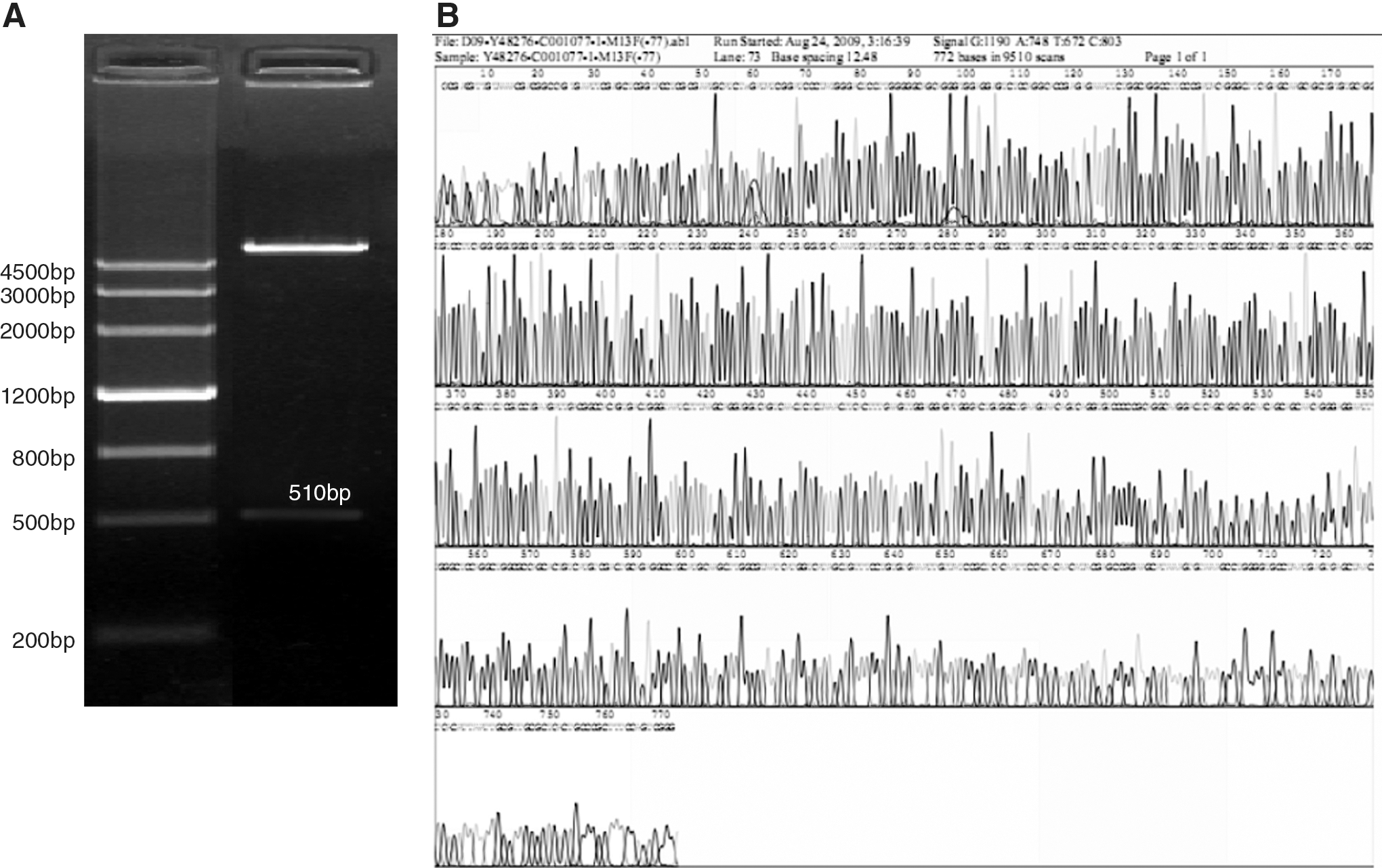
The agarose gel electrophoresis that produced recombinant plasmid pEGFP-c1-FL was digested by two enzymes of SalI and BamHI
Morphology, particle size, and zeta potential of FL/nano-liposomes
The cationic nano-liposomes carrying pEGFP were observed by an electrom micrograph and showed uniform spherical or elliptical shapes. Many pores were also found in pEGFP-cl-FL cationic nano-liposomes (Fig. 2A, B).
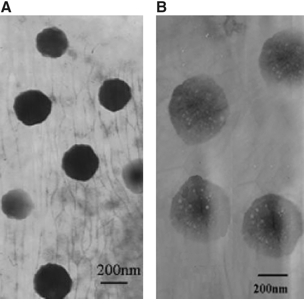
The electron microscope graph of two group cationic nano-liposomes, the cationic nano-liposomes carrying pEGFP
The average particle size, size distribution, and zeta potential of the nanoparticles were measured by PCS using Zetasizer 3000. The average particle size was expressed in volume mean diameter, and the reported value was represented as mean±SD (n=3). The electron microscopy parameters for each cationic nano-liposome are listed in Table 1.
and #represent p<0.05.
The average particle size and zeta potential of the nanoparticles were measured by photon correlation spectroscopy (Zetasizer 3000) as mean±SD (n=3).
FL, Fms-like tyrosine kinase receptor 3 ligand.
In Vitro transfection assays of FL/nano-liposomes
Nano-liposomes/pEGFP was used as a positive control, and 1 μg of plasmids pEGFP-c1-FL was used as a negative control. Fluorescence microscopy images of naked DNA transfections showed that the naked DNAs expressed little fluorescent protein, indicating that they did not enter cells, and no gene expression occurred. However, the fluorescence microscopy images of gene-carrying cationic nano-liposomes showed good expression of GFP in pEGFP and pEGFP-cl-FL groups. In addition, the flow cytometrys showed the protein expression level augmented as the culture time increased, with the highest level at 72 hours (Fig. 3A, B). The detection of expression of EGFP was carried out using an inverted fluorescent microscope with an attachment for fluorescent observation, and the picture was captured using a 200×objective.
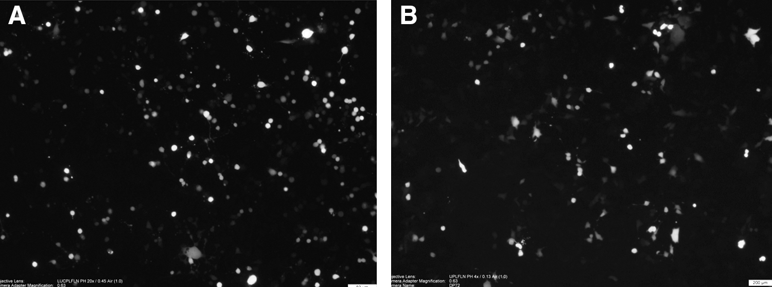
Fluorescence microscopy shows that gene-carrying cationic nano-liposomes exhibit good expression of green fluorescent protein in both the pEGFP group
Cell viability test of FL/nano-liposomes (MTT assay of cell death)
Tumor cell death in different groups after 24, 48, and 96 hours of transplantation is shown in Table 2. Statistical analysis indicated a significantly higher level of cell death between the FL group and the control group at 24, 48, and 96 hours after transplantation, with a lower level of cell death in the control group (Table 2).
The cell death rates after gene transplant were measured using the MTT assay. The FL group has significantly higher levels of cell death at 24, 48, and 96 hours after transplant than those of the control group (p<0.05).
Discussion
As the second highest incidence of tumor in the world, colon cancer accounts for 677,000 deaths per year. 10,11 Nowadays, surgery is still the most important treatment for colon cancer. After curative resection, chemotherapy and radiotherapy are always used to prevent tumor recurrence. However, severe side effects associated with these chemotherapeutic strategies decrease the patient's quality of life and can even be fatal. Hence, treatment strategies with minimal or no toxicity are vital and needed to prevent the side effects just mentioned. 12
Recently, gene therapy is becoming a new important treatment for malignant diseases and brings new hope for overcoming the threat of cancer. However, due to many factors, gene therapy did not accomplish ideal results over the years. The problems of how to improve gene transfection efficiency and safely remain to be solved. Various gene delivery carrier systems such as nanoparticles, liposomes, polymeric micelles, and parentral emulsions have been evaluated for cancer gene therapy. 13 –15 Among these systems, nanoparticle-mediated delivery provides a number of advantages, including small particle size, increased gene efficacy, lowered toxicity, enhanced gene stability, and an ability to achieve steady-state therapeutic levels over an extended time frame. 16,17
In addition, gene research on colon cancer has made significant progress in recent years, and the antitumor effects of FL are among the hot research topics. 18 As an early-stage hematopoietic growth factor, FL regulates the proliferation and differentiation of hematopoietic progenitor cells and cooperates with various cytokines to enhance hematopoietic function. 19 It can especially enhance the proliferation of DCs and promote the downstream T-effective cell immunity reaction, thus displaying an important role of antitumor immunity. 20,21
In this study, the recombinant plasmid EGFP-cl-FL, which contains the human FL gene and the GFP reporter gene, was successfully constructed. They were successfully validated by restriction enzyme digestion and sequencing, confirming that the nucleoside acid sequences are identical as designed. Since the recombinant plasmid contains the GFP reporter gene, it is convenient to examine the gene expression in the following experiments.
Human Lovo cells were used as a colon cancer in vitro model and successfully transfected the FL gene and the GFP gene through gene-carrying cationic nano-liposomes in this study. The target genes were highly expressed in the Lovo cells, and their protein expression augmented as time increased, thus confirming the feasibility of using cationic nano-liposomes as a gene vehicle.
The cell death, as shown by MTT assay, revealed a higher level of cell death and a lower level of proliferation in tumor cells in the FL group. The parameters for FL groups were between the two groups just mentioned. These data show that the FL genes have a better therapeutic outcome than the control group with regard to cancer treatment.
In summary, in vivo tumor inhibition efficacy of the cationic nano-liposomes formulation was evaluated in a human colon cancer xenograft mouse model. Although the exact antitumor mechanism and the side effects still require further quantitative research, this study qualitatively investigated the gene therapy of the tumor, with the hope of finding a strategy to increase gene therapy efficiency and improve the survival rate and the life quality of colon cancer patients.
Footnotes
Acknowledgment
The authors want to thank Professor Na Zhang and Dr. Dong-hua Liu, the School of Pharmaceutical Science, Shandong University, for the support of preparation of gene-carrying cationic nano-liposomes.
Disclosure Statement
The authors have declared that no competing interests exist.