
Abstract
Bevacizumab is a humanized monoclonal antibody that inhibits vascular endothelial growth factor A and is used for the treatment of several cancers. We labeled this monoclonal antibody with Iodine-131 (131I) and performed in vitro quality control and tumor cell growth inhibition tests. Bevacizumab was labeled with 131I using chloramine T. Radiochemical purity and stability in phosphate-buffered saline and human blood serum were determined using thin-layer chromatography and radio-sodium dodecyl sulfate-polyacrylamide gel electrophoresis, respectively, performed at different times. Cell-specific binding, internalization, and toxicity of the radiolabeled antibody were tested using the SKOV-3 ovarian cancer cell line. The biodistribution of 131I-bevacizumab was investigated using male mice. The radiochemical purity of the complex was 99%±0.7%. Its stability in phosphate-buffered saline and human blood serum at 48 hours postpreparation was 78%±1.2% and 93%±0.6%, respectively. 131I-bevacizumab was significantly bound to SKOV-3. The internalization of 131I-bevacizumab was time dependent, and it was cleared from the blood after 24 hours. Significant reductions in SKOV-3 cell viability were achieved with 131I-bevacizumab at a concentration of 500 nM. A low accumulation of 131I-bevacizumab was observed in the stomach and salivary glands after 24 hours and 48 hours. These findings indicate that the new radiolabeled antibody should be further evaluated in animals and, possibly, in humans as a new radiopharmaceutical agent for use in radioimmunotherapy for ovarian cancer.
Introduction
While several strategies have been used for cancer therapy, such as external irradiation, these do not specifically target tumor cells and, thus, are often associated with indiscriminate toxicity in normal cells. One potential means of reducing toxicity is to deliver specific toxic ionizing radiation to malignant cells via molecular targeting molecules. In radioimmunotherapy (RIT), an antibody with specificity for a tumor-associated antigen is used to deliver a lethal dose of radiation to the tumor cells, avoiding harming normal cells. 1 There are two proposed advantages for RIT over the use of monoclonal antibodies (MoAbs) conjugated with either drugs or toxins. First, tumor cells not expressing the target antigen can still be sterilized by the so-called crossfire phenomenon, that is, radiation energy emitted by the radionuclides bound to antibodies targeting adjacent tumor cells. 2 Second, radionuclides are not subject to multidrug resistance. 3,4 Iodine-131 (131I) is one of the most important and effective radioisotopes that is used for radionuclide therapy in cancer. 131I is a beta minus-emitting radionuclide with a physical half-life of 8.04 days, a principal gamma ray of 364 keV (81% abundance), and beta particles with a maximum energy of 0.61 MeV and an average energy of 0.192 MeV. 5 Its medium-energy beta emissions can kill cells, and it has been proved to be useful against thyroid cancer. 6,7 Iodine 131I-tositumomab (Bexxar) was approved for the treatment of refractory non-Hodgkin's lymphoma and brought about clear improvement in treatment response compared with nonradiolabeled antibodies for solid tumors. 8,9
Bevacizumab is a humanized MoAb that inhibits vascular endothelial growth factor A (VEGF-A). 10 VEGF-A is a protein that stimulates angiogenesis in various diseases, especially cancer and diabetes retinopathy. 11 Bevacizumab is currently approved by the U.S. Food and Drug Administration (FDA) for metastatic cancers. It received its first approval for combination use with standard chemotherapy for metastatic colon cancer, non-small-cell lung cancer, and metastatic breast cancer. 12 We recently showed that bevacizumab labeled with 111In has good specific binding to the SKOV-3 ovarian cancer cell line, and is a promising radiotracer for tumor imaging. 13 Recently, bevacizumab was labeled with 64Cu for use as a positron emission tomography tracer. The tumor accumulation of 64Cu-bevacizumab was found to be correlated with VEGF expression, and it was clearly accumulated in mice bearing human colorectal cancer xenografts. 14 These findings show that bevacizumab is a MoAb that is significantly accumulated in tumor cells, thus having the potential to be a molecule for the specifically targeted delivery of radionuclides to cancerous cells. It is clear that radiolabeling bevacizumab with 131I to form a beta-emitting radionuclide is potentially suitable for tumor therapy. In the present study, we describe the preparation of 131I-bevacizumab, its stability and specific binding to tumor cells, and its effect on the viability of SKOV-3 cells.
Materials and Methods
Materials
Bevacizumab was obtained from Roche. 3-(4,5-Dimethylthiazol-2-yl)-2,5-diphenyltetrazolium bromide (MTT), chloramine T, sodium metabisulfite, and phosphate-buffered saline (PBS; pH 7.4) were purchased from Sigma. Sodium 131I was purchased from Izotop. NuPAGE® 4%–12% Bis-Tris gels (Invitrogen) were used for the analysis of the radiolabeled antibody and the in vitro stability studies. The radioactivity along the instant thin-layer chromatography (ITLC) strip was determined using a Lablogic mini-scan TLC scanner and analyzed with Lura image analysis software (Sheffield). Radioactivity in the samples was measured using a gamma counter and an NaI(Tl) detector gamma detector (Delshid).
Animals
Male NMRI (Naval Medical Research Institute) mice weighing 25±3 g were purchased from the Pasteur Institute (Amol). The mice were housed under suitable conditions at the university animal laboratory, fed with standard mouse pellets, and given water ad libitum. All animals were kept under controlled light (light:dark cycle 12:12 hours) and temperature (22°C±1°C) conditions. Animal experiments were approved by the Research Committee of the Mazandaran University of Medical Sciences, Sari, Iran.
Cell line
The SKOV-3 human ovarian cancer cell line was purchased from the Iranian Pasteur Institute (Tehran). The cells were grown in RPMI 1640 medium (Biosera) supplemented with 10% fetal bovine serum (FBS), L-glutamine (2 mM), and PEST (penicillin 100 IU/mL and streptomycin 100 μg/mL), all of which were obtained from Gibco
Radiolabeling
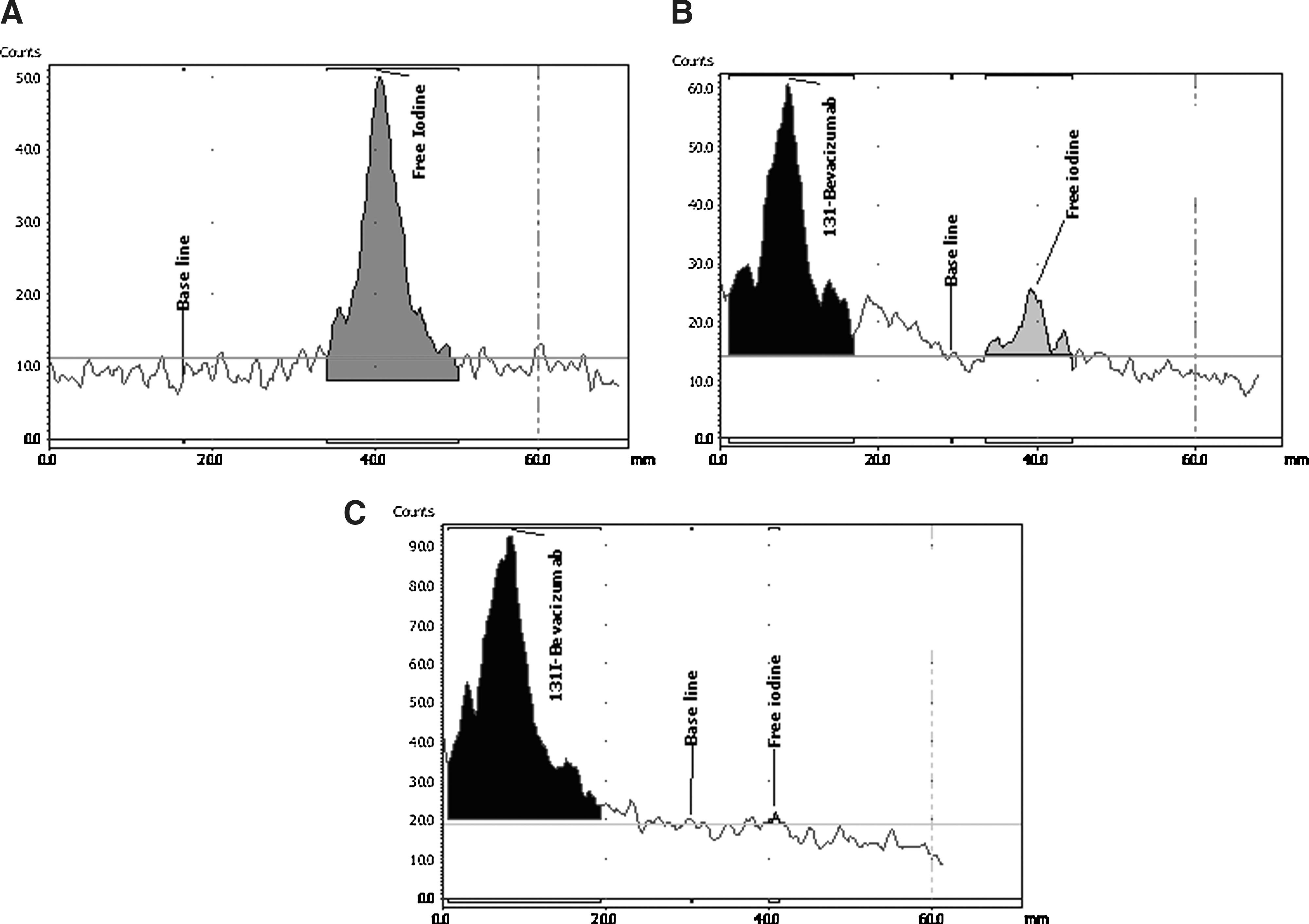
Bevacizumab was labeled with 131I using chloramine T. Briefly, 10 μL chloramine T (40 μg in phosphate-buffered saline) and 11–22 MBq of sodium 131I were added to 25 μL of a fresh antibody solution containing PBS (100 μg). Iodination was carried out by vortexing the mixture for 2 minutes, and then adding 20 μL of sodium metabisulfite in phosphate buffer. Quality control was performed using ITLC (Biodex) with acetone 70% as the mobile phase. In this system, free iodine-131 is moving with an Rf=0.9, while 131I-bevacizumab is the origin.
Stability control
Stability of the labeled antibody in the presence of PBS solution and human serum was assessed for approximately 48 hours at 37°C. Briefly, 10 μL of radiolabeled antibody and 90 μL of PBS or human serum were mixed and incubated at 37°C, and an aliquot was sampled for ITLC at each time point.
Serum and PBS stability studies were performed with sodium dodecyl sulfate polyacrylamide gel electrophoresis (SDS-PAGE) to determine protein binding of the 131I-antibody. At 48 hours, a serum or PBS sample containing 131I-bevacizumab (10 μL) was mixed with 30 μL sample buffer and incubated for 10 minutes at 70°C. The samples were incubated and analyzed using radio-SDS-PAGE in 2-(N-morpholino)ethanesulfonic acid (MES) buffer (200 V constant for 40 minutes, BioRad). A sample of 131I was used as a reference standard for low-molecular-weight compounds. Both serum and PBS samples were run in parallel with the reference sample on the same gel.
Cell-binding specificity test
SKOV-3 cells were used to assess cell-binding specificity. 13 Ten petri dishes (3.5 cm diameter) containing a cell monolayer (1×10⁶ cells/dish) were used. Cells in five dishes were presaturated with 300-fold excess unlabeled bevacizumab 5 minutes before labeled 131I-bevacizumab (12 nM) was added. The cells were then incubated with the radiolabeled antibody in a humidified incubator (5% CO2, 37°C) for 1 hour. The medium was subsequently collected, and the dishes were washed once with cold serum-free medium before 0.5 mL trypsin-EDTA solution was added to each dish, and the dishes were incubated for 10 minutes. The detached cells were diluted with a complete medium containing FBS, glutamine, and PEST, re-suspended, and transferred to fraction tubes. The radioactivity of the cells and medium was measured using an automated gamma counter, and the percent cell-bound radioactivity was calculated.
Cell processing and retention
SKOV-3 cells (1×10⁶ cells per dish) were incubated with 131I-bevacizumab at 4°C. After 1 hour of incubation, the medium with the labeled compound was removed, and the cells were washed thrice with ice-cold, serum-free medium. One milliliter of complete medium was then added to each dish, and the cells were incubated at 37°C in an atmosphere containing 5% CO2. At designated time points (0, 1, 2, 4, 24, and 48 hours), a group of three dishes was removed from the incubator, the medium was collected, and the cells were washed thrice with ice-cold, serum-free medium. Thereafter, the cells were treated with 0.5 mL 0.2 M glycine buffer (pH 2) containing 4 M urea for 5 minutes on ice. The acidic solution was then collected, and the cells were washed with 0.5 mL glycine buffer. The acidic fractions were then pooled. The cells were subsequently incubated with 0.5 mL 1M NaOH at 37°C for 10 minutes, after which the cell debris was collected, and the dishes were washed with 0.5 mL NaOH solution. Finally, the alkaline solutions were pooled. Radioactivity in the acidic solution was represented by a membrane-bound tracer compound and in the alkaline fraction, by an internalized tracer compound.
MTT growth inhibition assay
The MTT colorimetric assay is used for measuring the cell toxicity of biomaterials. The test is based on the ability of mitochondrial enzymes to reduce MTT (pale yellow) to formazan crystals (dark blue). Due to their impermeability relative to the cell membrane, formazan crystals accumulate in cells. Since a reduction in MTT only occurs in metabolically active cells, the level of accumulation can be considered a measure of cell viability. 15 SKOV-3 cell samples (6.0×10⁴ cells/well) were seeded for 24 hours. 131I-bevacizumab in concentrations of 0–500 nM was added to the samples, and the cells were incubated for 1 hour at 37°C. The medium was aspirated and washed thrice with fresh medium, and the cells were rested for 48 hours. Subsequently, 20 μL MTT was added to each well, and the cells were incubated for 3.5 hours. A mixture of acid isopropanol was used to solubilize the formazan compounds formed during incubation. The absorption of the samples under visible light was measured with an ELISA microreader (Biotek). The same procedure was repeated for bevacizumab, 131I, and bevacizumab+131I at different concentrations.
Animal studies
Biodistribution studies were performed using male NMRI mice. All mice were acclimatized for 1 week at the Mazandaran University of Medical Sciences Laboratory animal facility before any experimental procedure was performed. Twelve mice were randomized into groups of 3. Each mouse was intravenously injected with 1 μg 131I-bevacizumab in the 100 μL PBS. One group of mice was sacrificed at each predetermined time point (1, 24, and 48 hours after an intravenous injection) by an intraperitoneal injection of an anesthetic (ketamine/xylazine solution; Sigma) with subsequent exsanguination by heart puncture using a 1 mL syringe prewashed with diluted heparin. The blood, lungs, liver, spleen, stomach, kidneys, salivary glands, muscles, intestines, femurs, heart, and the remaining carcass were collected. Organs and tissue samples were weighed, and their radioactivity was measured. The tissue uptake values were calculated as a percent of injected activity per gram tissue (%ID/g), except for the intestines and the carcass, which were calculated as %ID per sample.
Statistical analysis
The data are presented as means±SD. One-way analysis of variance and Tukey's HSD test were used for multiple comparisons.
Results
Radiolabeling and in vitro stability
Bevacizumab was efficiently labeled with 131I with a labeling yield of 99%±0.7% (n=10), rendering a highly purified radiolabeled antibody without requiring further purification. The results from the stability test are presented in Figure 1. 131I-bevacizumab was more stable in serum than PBS and had 93%±0.6% stability in serum after 48 hours of incubation at 37°C; in PBS, its stability was reduced to 78.65%±1.2%. SDS-PAGE (Fig. 2) revealed that 131I-bevacizumab was highly stable after 48 hours of incubation with serum. SDS-PAGE analysis also showed no aggregation or binding to serum proteins, and no low-molecular-weight compounds arising from the release of radionuclides into the serum sample containing 131I-bevacizumab were detected; however, free 131I had a low stability value of 25% after 131I-bevacizumab was incubated in PBS.

The stability of 131I-bevacizumab in phosphate buffer and human blood serum at different times after labeling (n=3).
SDS-PAGE analysis of the stability of 131I-bevacizumab in PBS
Binding specificity and cell retention
131I-bevacizumab showed specific binding to VEGF. Since the uptake by SKOV-3 cells was significantly blocked by presaturation with a 300-fold molar excess of unlabeled antibody, cell-associated radioactivity was decreased to 55% (p<0.001) (Fig. 3).
SKOV-3 cells were loaded with 131I-bevacizumab at 4°C for 1 hour. Then, after removing unbound 131I-bevacizumab, the cells were incubated at 37°C for different times to allow for internalization. The fraction of 131I-bevacizumab that remained associated with the cells after treatment with a low-pH buffer increased with longer incubation periods at 37°C from 29% (t=0 minutes) to 54% and 70% at 24 and 48 hours, respectively (Fig. 4). The radioactivity of the medium was increased in a time-dependent manner; free 131I was released from the inner cells into the medium after internalization.
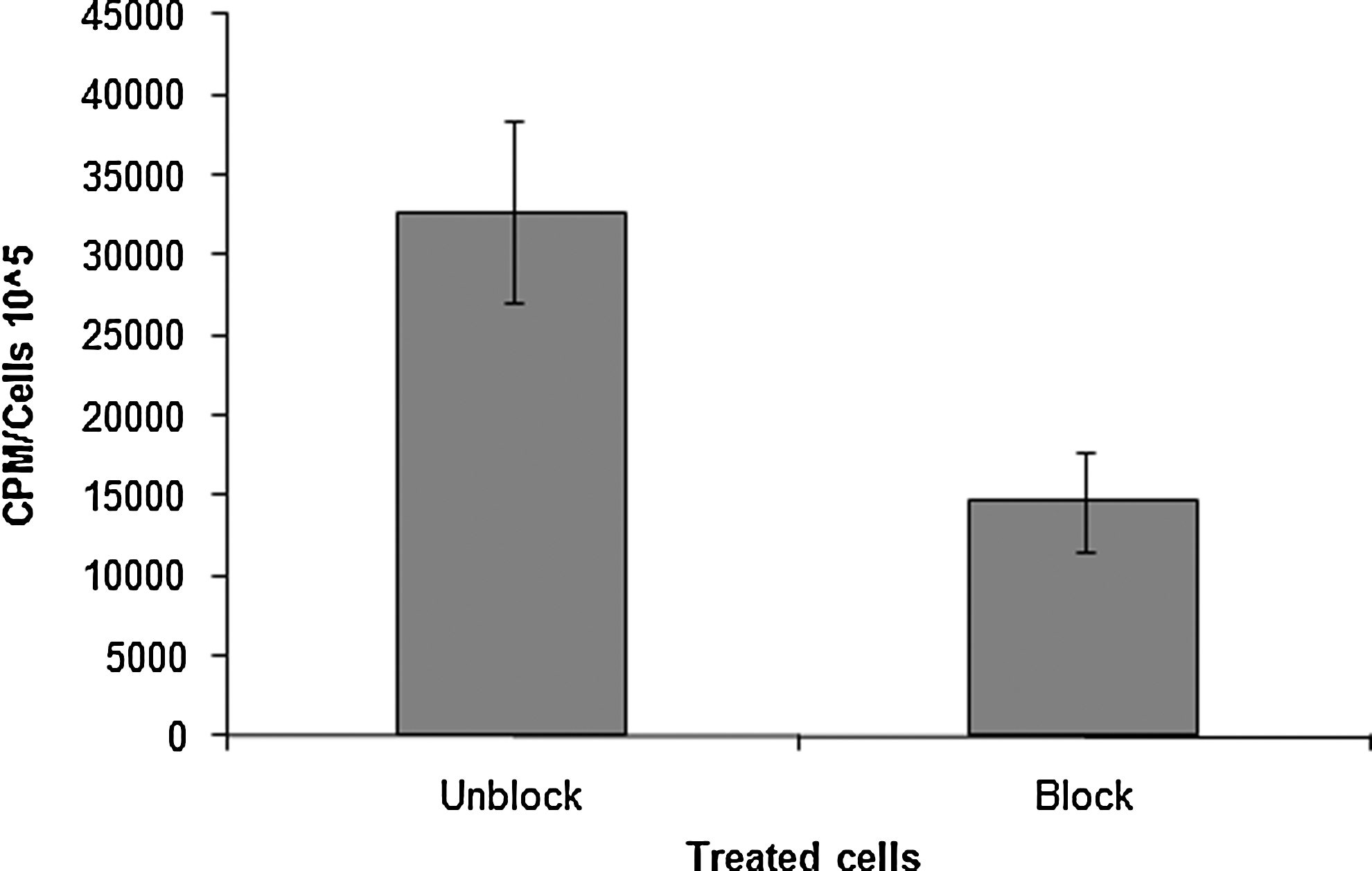
Specificity of the binding of 131I-bevacizumab to VEGF-expressing cells in vitro. The specificity test was performed using SKOV-3 ovarian cancer cell lines. For the presaturation of VEGF, a 300-fold molar excess of nonradioactive bevacizumab was added. Data are presented as mean values from five cell dishes with standard deviations. Presaturation of VEGF caused a significant decrease of the binding, which demonstrated specific binding to VEGF (p<0.001) (n=5). VEGF, vascular endothelial growth factor.
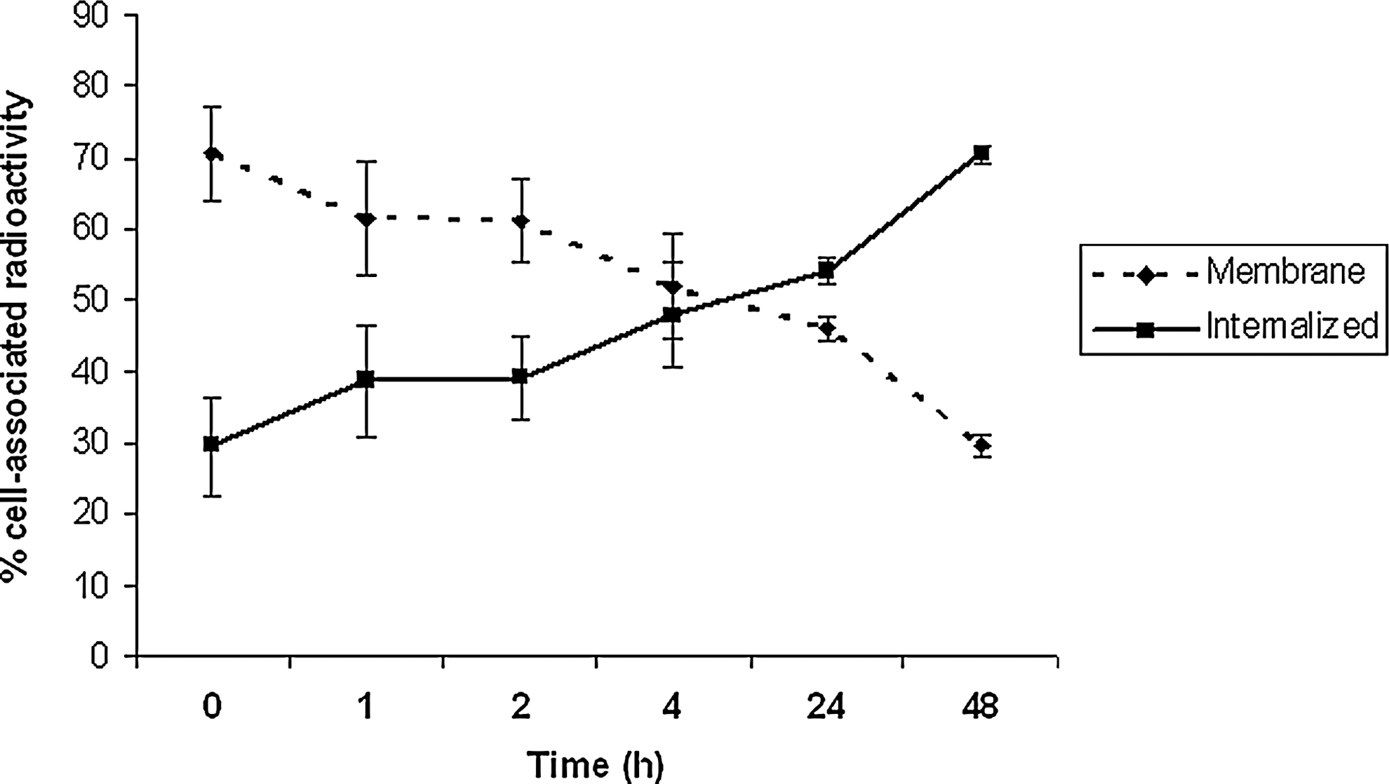
Cellular processing of 131I-bevacizumab by SKOV-3, 131I-bevacizumab was internalized by cells at different times after incubation at 37°C (n=3).
Cytotoxicity (MTT colorimetric assay)
The cytotoxicity of different concentrations of 131I-bevacizumab, bevacizumab, and free 131I in SKOV-3 cells is shown in Figure 5. The MTT absorbance of cells treated with 131I-bevacizumab was reduced at a concentration of 500 nM compared with controls, and the cell toxicity associated with 500 nM of 131I-bevacizumab was 25% (p<0.05). There was no statistically significant difference relative to the control for any of the other groups. Bevacizumab and 131I alone were not toxic according to the MTT test.

The results of MTT colorimetric assay after 48 hours of treatment of SKOV-3 with different concentrations (50, 100, and 500 nM) of 131I-bevacizumab; p<0.05 131I-benacizumab at 500 nM (IA500) as compared with control (n=3).
Biodistribution
The pharmacokinetic properties of 131I-bevacizumab were studied in NMRI mice at 1, 24, and 48 hours after an intravenous injection, and were expressed as a percentage of injected dose per gram of tissue (%ID/g) (Fig. 6). 131I-bevacizumab was cleared from the blood after 24 hours. The low accumulation of 131I-bevacizumab in the stomach and salivary glands after 24 and 48 hours indicates the in vivo stability and low dehalogenation of the radiolabeled antibody.

Biodistribution of 131I-bevacizumab in NMRI mice after an intravenous injection at 1, 24, and 48 hours after the injection (n=4).
Discussion
Radiolabeled MoAbs are effective for delivering ionizing radiation that kills tumor cells. This study described the preparation and use of a 131I-labeled anti-VEGF MoAb (bevacizumab) against human ovarian carcinoma tumor cells (SKOV-3). Bevacizumab was successfully radiolabeled with 131I using chloramine T, a simple and rapid method that yielded a high purity tracer compound. 131I-bevacizumab was stable in serum.
Angiogenesis plays an important role in the development of aberrant blood vessels that are required for malignant growth, invasion, and progression. Tumors can express VEGF, and some of their isoforms are freely soluble, whereas VEGF remains localized in the extracellular matrix. 16 VEGF is a critical marker of angiogenesis and tumor development. Bevacizumab, the humanized MoAb targeting VEGF, was approved for use in a number of different tumor types after successful clinical trials demonstrated clear benefits in overall survival when it was combined with common cytotoxic chemotherapies. 17,18 Bevacizumab binds to all VEGF isoforms, thereby preventing any interaction with VEGF receptor kinases. 19,20111In-bevacizumab was applied as a specific VEGF tracer in mice with human tumor xenografts. 21 –23 It was also used for the visualization of liver metastasis in patients. 24 In our previous study, three tumor cell lines, SKOV-3, DU145, and LS174T, were investigated for specific binding with 111In-bevacizumab; SKOV-3 showed significant specific binding to 111In-bevacizumab. 13 Tumor targeting with radiolabeled bevacizumab is a possible strategy for anticancer therapies if a beta-emitting radionuclide is used. In this study, 131I-bevacizumab was significantly bound to SKOV-3, which was significantly removed by saturated unlabeled bevacizumab. Our findings indicate that radiolabeled bevacizumab is suitable as a tumor-targeting agent for imaging and treatment. Tumor targeting is a critical parameter for radio- and chemotherapy of cancer, as radiolabeled MoAb should be accumulated in greater amounts by tumor cells compared with normal tissues. 131I has been widely used for radionuclide therapy in cancer. Beta radiations from 131I are used for treatment, whereas gamma radiations from 131I are used for imaging. In this study, MTT tests indicated that 131I-bevacizumab inhibited SKOV-3 cell growth. This effect was dose dependent and was associated with 131I-bevacizumab radioactivity, and this cytotoxicity was not observed at different doses of bevacizumab and/or free 131I. Internalization data showed that 131I-bevacizumab was transported across the cell membrane and accumulated by cells after 1 hour. To maximize the tumor-killing effects of the radiolabeled tracer, it should be accumulated in cells and used to deliver ionizing radiation to critical macromolecules such as DNA. 25,26 This cell-killing effect of 131I-bevacizumab is related to the specific delivery of 131I (a beta emitter) to tumor cells by bevacizumab. Since most solid tumors require the formation of new blood vessels for growth and development through angiogenesis, this process is essential if solid tumors are to grow beyond 2–3 mm3, at which size, diffusion is no longer sufficient to supply the tissue with oxygen and nutrients. 27 Both the present and previous studies have established that radiolabeled bevacizumab is significantly accumulated in tumor cells; 131I-bevacizumab is, thus, a candidate for specific tumor therapy by which tumor blood vessels are destroyed with the beta particles emitted by 131I.
In this study, 131I-bevacizumab was prepared and was confirmed to have high purity and high stability. In vitro experiments showed that this radiolabeled antibody was capable of specific binding to SKOV-3 cells. It was internalized by the cells in a time-dependent manner, which helped it to deliver 131I into cells. MTT assay showed that 131I-bevacizumab inhibited cell growth. These data provide new and beneficial information relating to the potential treatment effects of 131I-bevacizumab in patients.
Footnotes
Acknowledgment
This work was supported by a grant from the Research Council of Mazandaran University of Medical Sciences, Sari, Iran.
Disclosure Statement
No competing financial interests exist.