
Abstract
Silver nanoparticles (SN) of particle size of less than 50 nm were dispersed in an aqueous solution of Pluronic F127 and complexed with the phytoceutical, glyzyrrhizic acid (GLY). Radioprotecting ability of the obtained nanoparticle–glyzyrrhizic acid complex (SN-GLY) was evaluated in an in vivo model using Swiss albino mice. The potential of the complex as an adjuvant during radiotherapy was also analyzed in tumor-bearing mice. The administration of SN-GLY, SN, and GLY protected the hemopoetic and gastrointestinal system against radiation-induced damages as revealed by the total white blood cell count, bone marrow cellularity, endogenous spleen colony formation, levels of cellular antioxidants, and histopathologcal examination of gastrointestinal tract. Oral administration of SN-GLY, SN, and GLY 1 hour before a sublethal dose of radiation exposure reduced the radiation-induced depletion of cellular antioxidants and lipid peroxidation in various tissues of mice. Survival of animals following exposure to a lethal dose of gamma radiation was also improved. It was also found that the oral administration of the complex to tumor-bearing mice before 4 Gy gamma irradiation resulted in a faster tumor regression.
Introduction
Total-body exposure to ionizing radiation in humans and animals results in multiple organ dysfunctions as a consequence of toxicity to the hematopoietic, gastrointestinal, or cerebrovascular systems, depending on the total dose of radiation absorbed. 1,2
These deleterious effects are a product of the cellular reactions resulting from the interaction of several reactive oxygen species, such as superoxide, hydrogen peroxide, hydroxyl radical, generated by radiolysis of water, with cellular macromolecules, such as DNA, lipids, and proteins, manifesting as damage to genomic DNA, peroxidation of membrane lipids, protein oxidation, and altered gene expression causing activation of cytotoxic and cytoprotective cellular signaling pathways. 3 Survival following radiation exposure results from the recovery in mammalian system from the damage to target systems, such as the bone marrow, gastrointestinal tract, and hemostatic systems. 4 The hematopoietic system is the most radiosensitive system, and its damage may lead to the development of hematopoietic syndrome. Death from the hematopoietic syndrome results from infection due to the impairment of the immune system. 5
The gastrointestinal mucosa is an early radiation response tissue, 6 as it depends on a rapid and incessant proliferation of a pool of pluripotent stem cells and uncommitted clonogens localized at the bottom of the crypt of Lieberkuhn. 7 –9
The development of effective radioprotectors and radiorecovery drugs is of great importance in view of their potential application during both planned radiation exposure (e.g., radiotherapy) and unplanned radiation exposure (e.g., in the nuclear industry, natural background radiation emanating from the earth or other sources). 10 Effective radioprotectors are required in the event of a massive radiological accident, a nuclear terrorist attack, or prolonged space travel. 11 –13
Agents that accelerate the regeneration of hemopoetic system and gastrointestinal system can enhance survival of animals following a sublethal or lethal dose of gamma irradiation.
Short-term in vitro tests, such as lipid peroxidation (LPO), assay of free radicals and antioxidant status, cell survival and micronuclei assays, can provide an idea about the radioprotective activity of an agent. However, the gold standard for radioprotective activity is the evaluation of a 30-day survival in rodents, because the 30-day survival after lethal whole-body irradiation clearly indicates the capacity of the agent in test to modulate the recovery and regeneration of the gastrointestinal epithelium and the hemopoietic progenitor cells in the bone marrow, the two most radiosensitive organs that are essential for sustenance of life. 14 The GI syndrome in mice can be assessed by determining survival up to 10 days (measure of GI death) after exposure to comparatively high doses of whole-body radiation, whereas hemopoeitic syndrome can be assessed by monitoring the survival of irradiated animals up to 30 days postirradiation. 15,16
Radiotherapy is a common modality of cancer treatment for patients with solid tumor, but its effectiveness frequently is hampered by the development of radioresistance. 17 To obtain better tumor control with a higher dose, the normal tissues should be protected against radiation injury, where radioprotective compounds can play an important role. 10 In a clinical perspective, radioprotectors that protect against radiation damage to normal cells, but do not provide such protection to tumor cells could be used as adjuvants during radiotherapy.
The present study is focused on analyzing the ability of glycyrrhizic acid–silver nanoparticle complex (SN-GLY) to offer protection against radiation-induced hemopoetic and gastrointestinal system damages, and studies were also carried out to analyze the potential of the complex to offer preferential protection to normal cell in tumor-bearing Swiss albino mice exposed to gamma radiation.
Materials and Methods
Animals
Male Swiss albino mice of 8–10-weeks old, weighing 22–25 g were obtained from the Small Animal Breeding Section (SABS), Mannuthy, Thrissur, Kerala. They were kept under standard conditions of temperature and humidity in the Amala Cancer Research Centre Animal house facility. The animals were provided with standard mouse chow (Sai Durga Feeds and Foods, Bangalore, India) and water ad libitum. All animal experiments in this study were carried out with the prior approval of the Institutional Animal Ethics Committee (IAEC) and were conducted strictly adhering to the guidelines of Committee for the purpose of Control and Supervision of Experiments on Animals (CPCSEA) constituted by the Animal Welfare Division of Government of India.
Chemicals
Nitroblue tetrazolium (NBT), ethylene diamine tetra acetic acid (EDTA) and Riboflavin were from Sisco Research Laboratories Ltd. (Mumbai, India). Reduced glutathione (GSH), 5′-5′dithiobis-(2-nitrobenzoic acid) (DTNB), glycyrrhizic acid, thiobarbituric acid, and bovine serum albumin were from Sigma Chemical Company, Inc. (St Louis, MO). All other chemicals were of analytical grade procured from reputed Indian manufacturers.
Preparation of SN complex of glycyrrhizic acid
SN of less than 50-nm size were obtained in a powder form from Dr. P.K Khanna, CMET (Pune, India). SN (0.1%) were redispersed in 1% Pluronic F-127 under ultrasonication to obtain colloidal silver (SN) having the distinct yellow color characteristic of SN. The pale yellow solution was centrifuged to remove residues. To the supernatant, GLY was added to a final concentration of 10 mM to get the SN-GLY complex.
Exposure to gamma radiation
Irradiation was carried out using a 60Co-Theratron Phoenix teletherapy unit (Atomic energy Ltd., Ottawa, Canada) at a dose rate of 1.88 Gy per minute.
Animals were divided into eight groups for the experiments. The animals were orally administered with GLY, SN, or SN-GLY (equivalent to 50 mg GLY/kg) and exposed to gamma radiation as detailed below, to study gamma radiation-induced alterations and regeneration in various tissues of mice and survival of animals.
Group I-0.2 mL distilled water (oral) + sham irradiation
Group II-0.2 mL distilled water (oral) + 4, 6 or 8 Gy 60Co-γ- rays
Group III-0.15 mL SN (oral) + sham irradiation
Group IV-0.15 mL GLY (oral) + sham irradiation
Group V-0.15 mL SN-GLY (oral) + sham irradiation
Group VI-0.15 mL SN (oral) + 4, 6 or 8 Gy 60Co-γ- rays
Group VII-0.15 mL GLY (oral) + 4, 6 or 8 Gy 60Co-γ- rays
Group VIII-0.15 mL SN-GLY + 4, 6 or 8 Gy 60Co-γ- rays
To study the potential of GLY, SN, or SN-GLY as an adjuvant during radiotherapeutic situations, Swiss albino mice were transplanted with Daltons lymphoma ascites (DLA) cells subcutaneously onto the hind limbs. The animals were divided into eight groups as detailed above once the tumor reached a size of 1.0 cm3 and were exposed to a single dose of whole-body 4 Gy gamma radiation.
Effect of GLY, SN, or SN-GLY on gamma radiation-induced alterations and regeneration in various tissues of mice
Animals in Group I and II were administered orally with 0.15 mL distilled water and SN, GLY, or SN-GLY (equivalent to 50 mg GLY/kg) was given orally to animals in Group III and VI, IV and VII, and V and VII, respectively. The animals in Group II, VI, VII, and VIII were treated with a single dose of 6 Gy whole-body gamma irradiation, 1 hour after SN, GLY, SN-GLY, or distilled water administration.
The animals were exposed to a sublethal dose of 6 Gy whole-body gamma radiation to study the radioresponse of the animals in terms of spleen colony formation, bone marrow cellularity, total white blood cell (WBC) count, tissue antioxidant parameters, etc.
After 24 hours following irradiation, 5 animals from each group were sacrificed by cervical dislocation. The bone marrow cells were collected from both femurs into phosphate-buffered saline (pH 7.4) containing 2% fetal calf serum. The number of cells was determined with a hemocytometer and expressed as total cells (×106)/femur. Blood was collected from each of the animals by heart puncture into heparinized tubes and blood count was performed using automatic hemoanalyzer. Liver, kidney, and brain were quickly excised, and the surface was washed with ice-cold phosphate-buffered saline and kept at −20°C. A part of the intestine was removed from the animals; the intestinal mucosa was scrapped off using a clean glass slide and kept at −20°C until analysis. On the day of analysis, 10% homogenate was prepared in phosphate-buffered saline (pH 7.4) and following parameters were evaluated. The total protein was estimated by the method of Lowry. 18 Superoxide dismutase (SOD) activity was measured by the NBT reduction method of McCord and Fridovich. 19 GSH activity was assayed by the method of Moron et al. 20 based on the reaction with DTNB. An assay of glutathione peroxidase (GPx) followed the method of Hafeman et al. 21 based on the degradation of hydrogen peroxide in the presence of GSH. Extent of LPO was measured by the method of Buege and Aust. 22 It may be noted that in the assay for GSH, what actually measured is the concentration of nonprotein thiols in terms of GSH (GSH is taken as standard to quantitate thiols).
After 72 hours of radiation exposure, animals from each group were sacrificed by cervical dislocation and intestine was excised and kept in formalin for histopathological analysis. Sections (4 μm) were taken from a portion of the intestine fixed in 10% formalin and stained with hematoxylin–eosin and observed under an oil immersion microscope (100×).
The animals were sacrificed on the 12th day postirradiation by cervical dislocation and the spleen was excised out and fixed in the Bouin's solution containing 1.2% saturated picric acid, 30%–40% formalin, and glacial acetic acid in the ratio 15:5:1, and the spleens were analyzed for colony formations. 23
Effect of GLY, SN, or SN-GLY on survival of mice following a lethal dose of gamma radiation
Animals in Group I and II were administered orally with 0.15 mL distilled water and SN, GLY, or SN-GLY (equivalent to 50 mg GLY/kg) was given orally to animals in Group III and VI, IV and VII, and V and VII, respectively (10 animals/group). The animals in Group II, VI, VII, and VIII were treated with a single dose of 8 Gy whole-body gamma irradiation, 1 hour after SN, GLY, SN-GLY, or distilled water administration. 8 Gy whole-body gamma radiation is a lethal dose, which allows for the analysis of survival.
After irradiation, the animals in all the groups were provided with a standard diet and water ad libitum and their body weight and survival were monitored. The animals were checked on a daily basis to record the mortalities if any and the body weights of the survivors were recorded every alternate day.
Studies on the ability of GLY, SN, or SN-GLY to offer preferential protection against gamma radiation in tumor-bearing animal
Animals bearing solid DLA tumor on their hind limbs were grouped as detailed above. The animals in Group I and II were administered orally with 0.15 mL distilled water and SN, GLY or SN-GLY (equivalent to 50 mg GLY/kg) was given orally to animals in Group III and VI, IV and VII, and V and VII, respectively. The animals in Group II, VI, VII, and VIII were treated with a single dose of 4 Gy whole-body gamma irradiation, 1 hour after SN, GLY, SN-GLY, or distilled water administration.
Two animals from each group was sacrificed immediately after irradiation and tumor and bone marrow cells were collected to perform alkaline single-cell gel electrophoresis using the method given by Singh, with minor modifications. 24,25 Briefly, microscopic slides were coated with normal melting point agarose and 200 μL of 0.8% low melting point agarose containing 50 μL of treated cells (containing 104 to 105 cells) were added onto the slide and the slides were kept at 4°C. After solidification, the slides were immersed in a prechilled lysing solution containing 2.5 M NaCl, 100 mM Na2EDTA, 10 mM Tris-HCl, pH 10, 1% DMSO, 1% TritonX and kept for 1 hour at 4°C. After lysis, slides were drained properly and placed in a horizontal electrophoretic apparatus filled with a freshly prepared electrophoresis buffer containing 300 mM NaOH, 1 mM EDTA, 0.2% DMSO, pH≥13. The slides were equilibrated in the buffer for 20 minutes and electrophoresis was carried out for 30 minutes at 20 V, 300 mA. After electrophoresis, the slides were washed gently with 0.4 mM Tris-HCl buffer, pH 7.4 followed by distilled water, dried, and silver staining was carried out. The comets were visualized using the Olympus BX-41 microscope and more than 50 comets images were captured and analyzed using the software CASP, which gives % DNA in tail, tail length, tail moment (TM), and olive tail moment (OTM) directly. The parameter TM is the product of tail length and% DNA in tail and OTM is the product of the distance between the center of the head and the center of the tail and % DNA in tail. 26 Results are given as mean±standard deviation.
Tumor volume of the remaining animals (6 animals per group) was observed for 30 days and the hind leg thicknesses were measured using a vernier caliper. The tumor volume was calculated as follows:
Tumor thickness = thickness of tumor-induced leg −thickness of normal leg
Tumor volume=4/3πr 3, where r is the tumor radius.
Statistical analysis
The results are presented as mean±SD of the studied groups. Statistical analyses of the results were performed using analysis of variance with the Tukey–Kramer multiple comparisons test. The treated groups were compared with the respective control groups.
Results
Effect of GLY, SN, or SN-GLY on gamma radiation-induced alterations and regeneration in various tissues of mice
Exposure of mice to 6 Gy whole-body gamma radiation significantly decreased the total WBC count (109cells/L) from 8.13±1.80 to 1.81±0.67 and the bone marrow cellularity (106cells/femur) from 20.22±3.01 to 8.56±1.30 when measured at 24-hour postirradiation (Figs. 1 and 2). GLY, SN, or SN-GLY administration 1 hour before gamma irradiation minimized the radiation-induced depletion of these cells. The total WBC count was maintained at 4.12±1.08, 3.38±1.07, and 5.83±0.35 and bone marrow count at 17.24±1.36, 12.68±1.05, and 18.28±0.50, respectively, in GLY-, SN-, or SN-GLY-administered animals, respectively. The number of viable bone marrow cells was decreased drastically in the irradiated animals and administration of glycyrrhizic acid enhanced the bone marrow cellularity.

Effect of oral administration GLY, SN, or SN-GLY (equivalent to 50 mg GLY/kg) on total WBC count in mice exposed to 6 Gy whole-body gamma radiation. Error bars indicate the standard deviation of the mean for n=5. c p<0.001 compared to respective radiation, e p<0.01 compared to respective GLY, i p<0.001 compared to respective SN). GLY, glycyrrhizic acid; SN, silver nanoparticles; WBC, white blood cell.
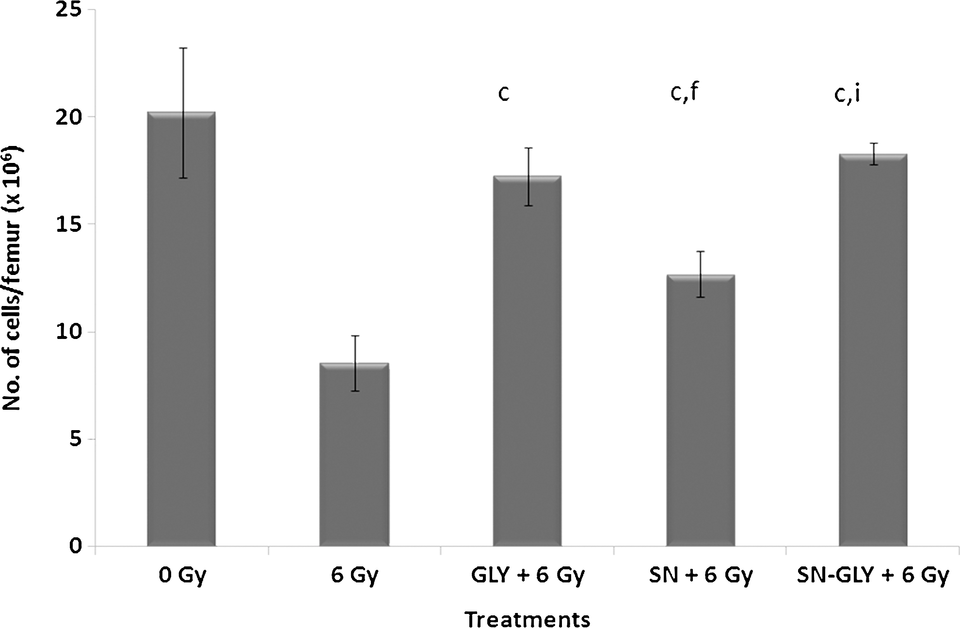
Effect of oral administration of GLY, SN, or SN-GLY (equivalent to 50 mg GLY/kg bw) on bone marrow cellularity in mice exposed to 6 Gy whole-body gamma radiation. Error bars indicate the standard deviation of the mean for n=5. c p<0.001 compared to respective radiation, f p<0.001 compared to respective GLY, i p<0.001 compared to respective SN. bw, body weight.
The activity of GPx and SOD, the major enzymes involved in the antioxidant defense system, were found to be decreased after irradiation in various tissues, such as intestine, liver, kidney, brain (Table 1). The administration of GLY, SN, or SN-GLY enhanced these enzyme activities in the tissues offering protection against radiation-induced antioxidant depletion. Similarly, the nonenzymatic cellular antioxidant GSH was also significantly maintained to near normal levels in the tissues of animals administered with GLY, SN, or SN-GLY 1 hour before whole body 6 Gy gamma irradiation, while in control irradiated animals, the levels of GSH were much depleted.
Errors indicate the standard deviation of the mean for n=4.
p<0.05 compared to respective radiation.
p<0.01 compared to respective radiation
p<0.001 compared to respective radiation.
p<0.05 compared to respective GLY.
p<0.01 compared to respective GLY.
p<0.001 compared to respective GLY.
p<0.05 compared to respective SN.
p<0.01 compared to respective SN.
p<0.001 compared to respective SN.
MDA, malonedialdehyde; GSH, reduced glutathione; Gpx, glutathione peroxidase; SOD, superoxide dismutase; bw, body weight; GLY, glycyrrhizic acid; SN, silver nanoparticles.
Exposure to 6 Gy whole-body gamma radiation significantly increased the membrane LPO levels monitored as malonedialdehyde. The administration of GLY, SN, or SN-GLY 1 hour before gamma radiation exposure significantly reduced the radiation-induced LPO (Table 1).
Administration of GLY, SN, or SN-GLY 1 hour before gamma radiation exposure also improved the number of visible endogenous spleen colonies per spleen on 12th day postirradiation, which indicates the improved hemopoetic system regeneration following radiation exposure. The number of spleen colonies was 3.20±1.76 in control irradiated animals, while it was 14.4±0.50, 11.5±2.48, and 17.55±0.94 in GLY-, SN-, or SN-GLY-administered animals (Fig. 3).

Effect of oral administration of GLY, SN, or SN-GLY (equivalent to 50 mg GLY/kg) on endogenous spleen colony formation in mice exposed to 6 Gy whole-body gamma radiation. Error bars indicate the standard deviation of the mean for n=5. c p<0.001 compared to respective radiation, e p<0.01 compared to respective GLY, i p<0.001 compared to respective SN.
Histopathological studies showed damage to the intestine of animals exposed to radiation (Fig. 4). Severe damage to intestinal villi and crypts could be seen in control animals compared to normal animals. Control animals showed maximum damage to intestinal mucosa 72 hours postirradiation. However, in GLY-, SN-, or SN-GLY-administered animals, the damage was much less than that observed in control mice. The crypt, which is the proliferative unit supplying cells for the maintenance of villus integrity, was maintained in these animals. SN-GLY-treated animals retained near normal villus architecture and crypt structure.

Effect of oral administration of GLY, SN, or SN-GLY (equivalent to 50 mg GLY/kg bw) on 6 Gy whole-body gamma radiation-induced gastrointestinal injury in mice. Sections (4 μm) were taken from a portion of the intestine fixed in 10% formalin and stained with hematoxylin–eosin and observed under the oil immersion microscope (100×).
Effect of GLY, SN, or SN-GLY on 8 Gy gamma radiation-induced body weight loss and mortality
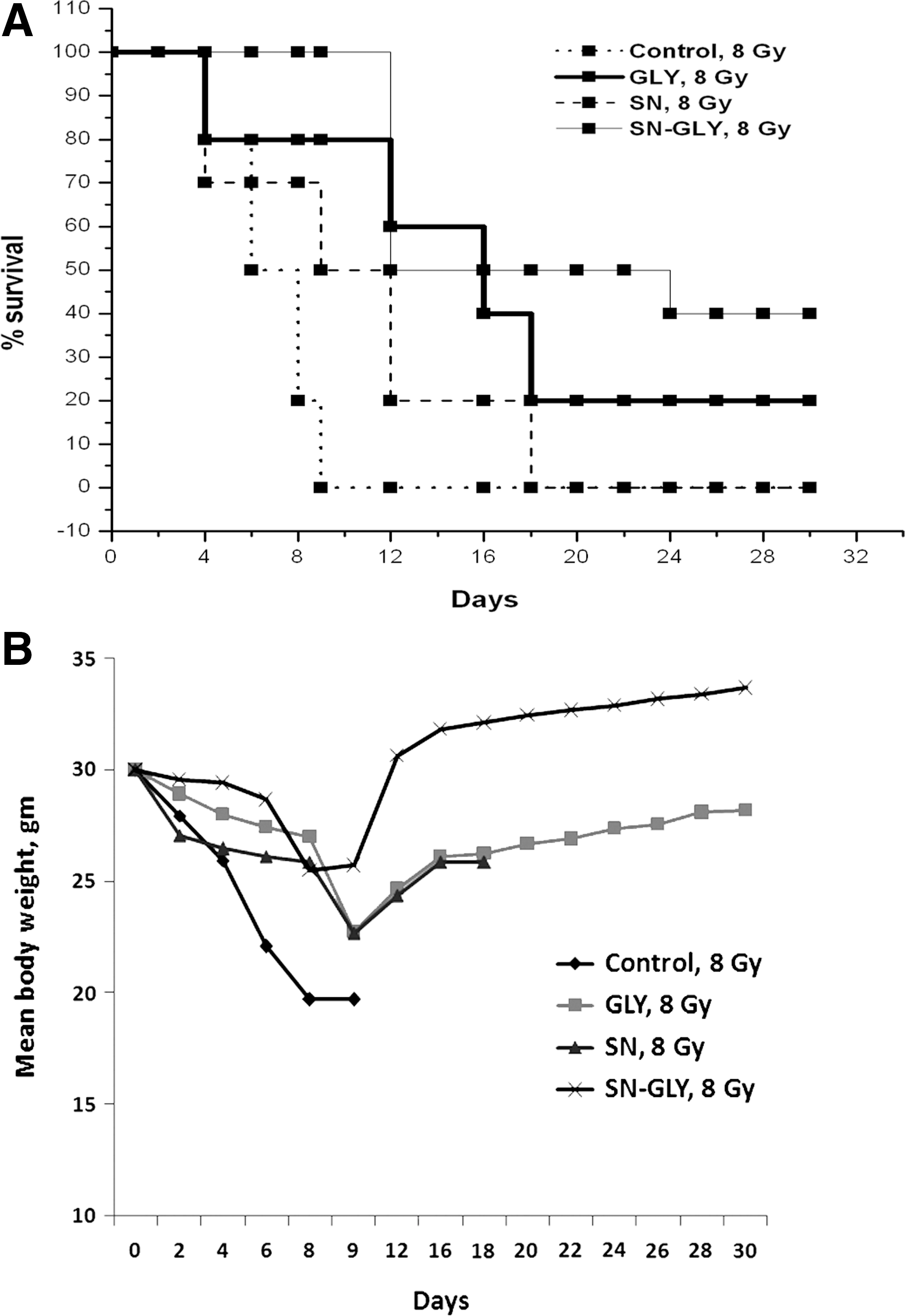
Figure 5A and B present the changes in body weight and survival of animals following exposure to acute lethal dose of 8 Gy whole-body gamma radiation. Animals in the irradiated group started dying from 4th day. On day 9, the mortality in the control untreated group was 100%, while it was only 20 in the GLY-treated group, 50% in the SN-treated group and all animals were surviving in the SN-GLY-treated group. The animals started dying only on the 12th day in the SN-GLY-treated group and 40% of animals survived in this group till the 30th day. Also, there was a marked decrease in body weight of the irradiated animals following 8 Gy gamma radiation exposure. The body weight of the animals in the control irradiated group continued to decrease till all the animals died on the 9th day. In GLY-, SN-, or SN-GLY-administered and irradiated groups, the body weight of the survivors started recovering after the 9th day.
Preferential protection by GLY, SN, or SN-GLY in tumor-bearing animals against gamma radiation
Whole-body exposure of tumor-bearing animals to gamma radiation (4 Gy) resulted in an increase in the comet parameters (such as % DNA in tail, TM, and tail migration) of cells of bone marrow and tumor as a result of damage to cellular DNA (Figs. 6 and 7).When GLY, SN, or SN-GLY was administered 1 hour before irradiation, there was a significant decrease in comet parameters in bone marrow cells, but not in the tumor cells of irradiated animals. The % DNA in tail, tail length, TM, and OTM were increased in the bone marrow cells of animals exposed to radiation (4 Gy), while the administration of GLY, SN, or SN-GLY 1 hour before irradiation brought down the comet parameters to near normal levels, respectively (Fig. 6).
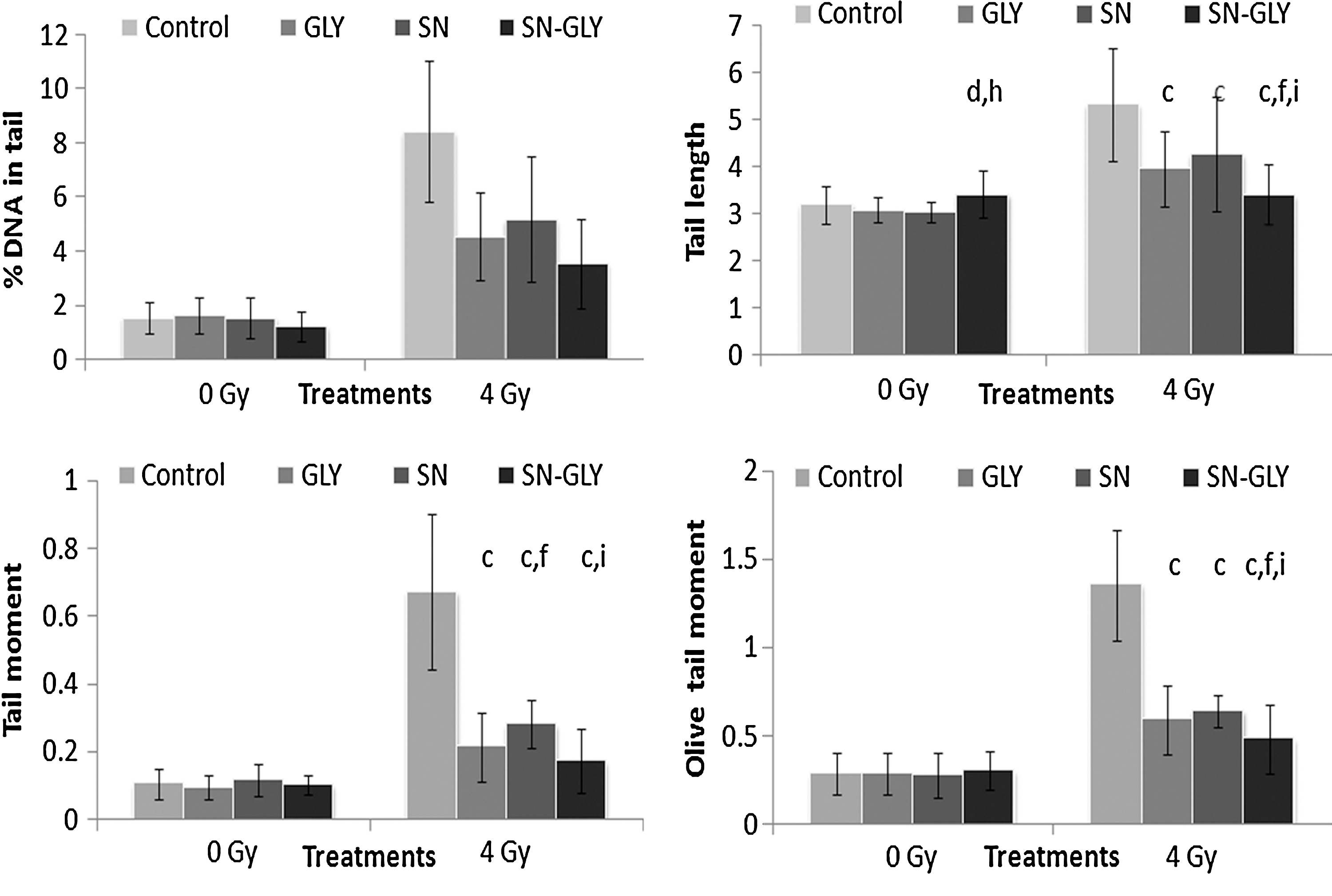
Effect of oral administration of GLY, SN, or SN-GLY (equivalent to 50 mg GLY/kg bw) on radiation-induced DNA damage in bone marrow cells of whole-body 4 Gy gamma irradiated tumor-bearing Swiss albino mice. Compared to respective control c p<0.001, compared to respective GLY d p<0.05, f p<0.001, compared to respective SN h p<0.01, i p<0.001.
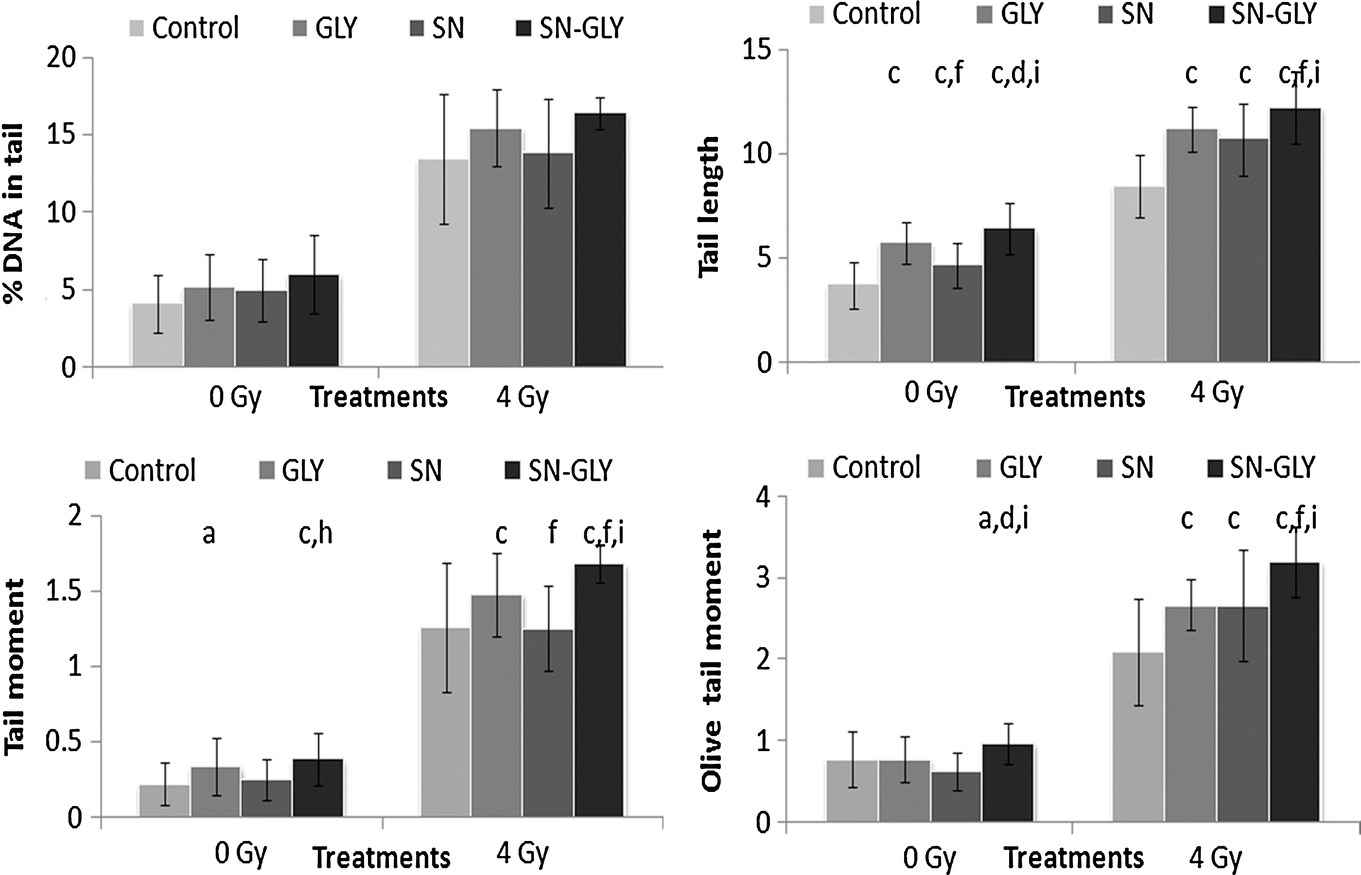
Effect of oral administration of GLY, SN, or SN-GLY (equivalent to 50 mg GLY/kg bw) on radiation-induced DNA damage in tumor cells of whole-body 4 Gy gamma irradiated tumor-bearing Swiss albino mice. Compared to respective control a p<0.05, c p<0.001, compared to respective GLY d p<0.05, f p<0.001, compared to respective SN h p<0.01, i p<0.001.
It can be seen in Figure 7 that after exposure to 4 Gy whole-body gamma radiation, the% DNA in tail, tail length, TM, and OTM were increased significantly in the tumor cells. In animals orally administered with GLY, SN, or SN-GLY 1 hour before irradiation, the % DNA in tail, tail length, TM, and OTM remained higher. Unlike the cells of bone marrow, there was no significant decrease in the comet parameters of tumor cells.
Figure 8 shows the results of the study on the combined effect of GLY, SN, or SN-GLY administration and 4 Gy gamma irradiation on solid tumor growth in Swiss albino mice. The growth of the tumor was found to be delayed in animals exposed to 4 Gy gamma radiation compared to unirradiated animals. The administration of GLY, SN, or SN-GLY reduced tumor growth to some extent. The tumor growth was found significantly reduced in the animals administered with SN-GLY and exposed to 4 Gy gamma radiation than the other treatment groups. This preferential protection could be attributed to poor vasculature of the tumor or peculiar characteristics of the tumor cells that either restricted the entry of the complexes inside the cells or resulted in inactivatation of the complexes. Further pharmacokinetic studies of the complex is needed to be done to determine whether the SN-GLY takes longer time to enter into the tumor compared to the normal tissues.
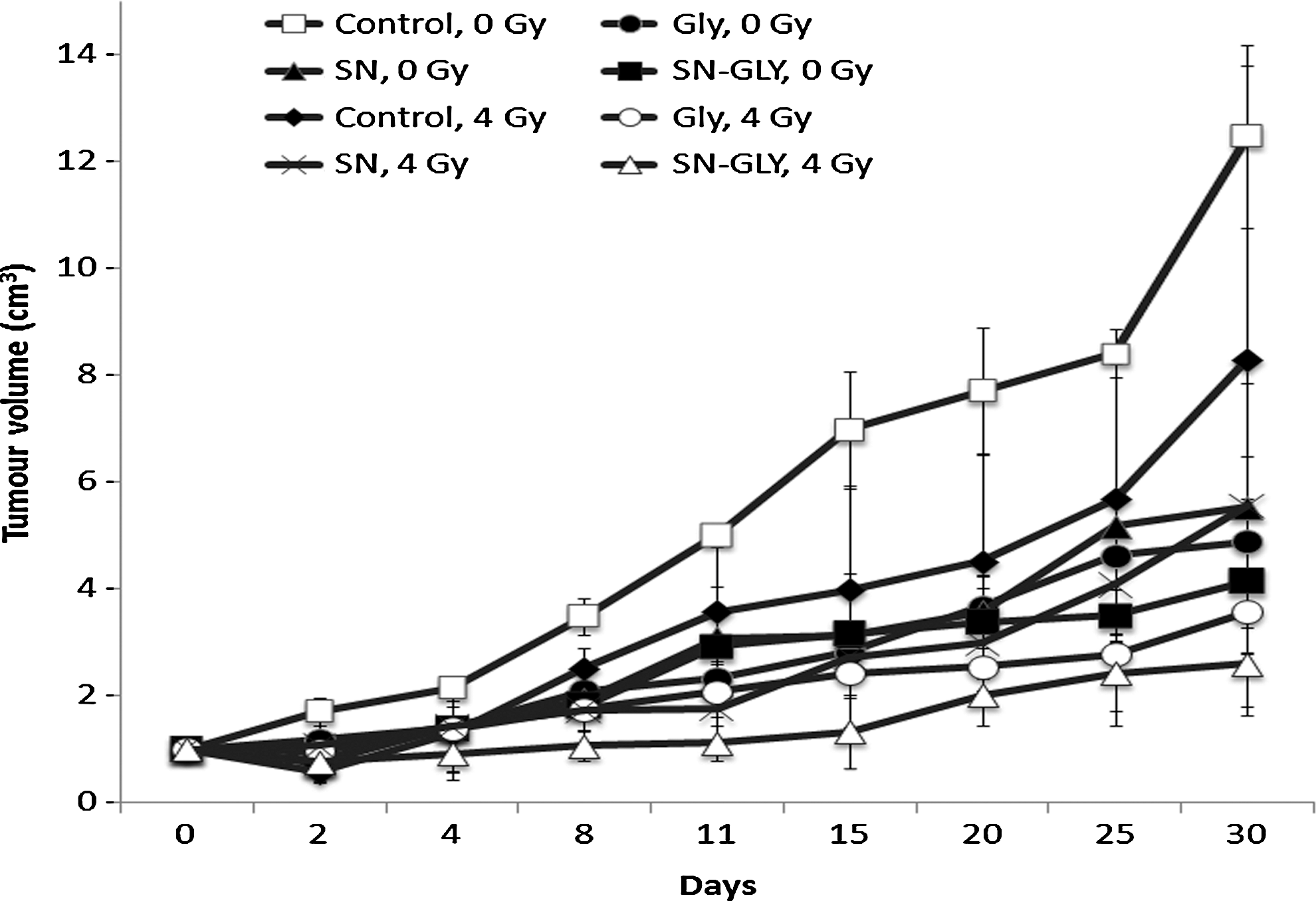
Effect of oral administration of GLY, SN, or SN-GLY (equivalent to 50 mg GLY/kg bw) on 4 Gy whole-body gamma irradiation-induced tumor growth delay.
Discussion
SN of less than 50-nm size used in the present study was obtained in a stable powder form from Dr. P.K Khanna, CMET. The UV–Visible absorption spectra, transmission electron microscopy and particle size distribution by dynamic light- scattering technique showed that the particles are in the range of less than 50 nm. 27 This nanoparticle could be easily resuspended into a colloidal suspension and be complexed with glycyrrhizic acid by means of ultrasonication.
The inherent cellular defense system that protects living cells against oxidative damage, consists of glutathione and antioxidant enzymes, such as GPx, SOD. 28 When the animals were administered with 0.15 mL of SN or SN-GLY complex for 7 continuous days to study the toxicity in Swiss albino mice by orally administering the animals with the complexes for 7 days, it was found that the levels of tissue antioxidants, LPO, and serum enzyme levels remained normal. This indicated that the complex at the specified dose is not toxic to animals even on 7-dose administrations; the animals were administered with a single dose to study the radioprotecting potential and ability to act as an adjunct in radiotherapy. A significant decrease in GSH content in liver, kidney, brain, intestine, blood, etc. was observed in animal tissues at 24 hours following 6 Gy gamma irradiation, while the oral administration of SN-GLY protected against the GSH depletion following irradiation. Administration of SN-GLY did not influence the endogenous GSH content significantly. The levels of antioxidant enzymes, such as GPx and SOD were also found to be restored in irradiated animals administered with SN-GLY.
The basic effect of radiation on cellular membranes is believed to be the peroxidation of membrane lipids, which is initiated by radiolytic products, including hydroxyl and hydroperoxyl radicals. The oxidative stress due to the radiation induced free radicals that cause a dramatic fall in the cellular GSH content overwhelms the cellular defense leading to membrane LPO. 29 Administration of SN-GLY helped to decrease the levels of membrane LPO in various tissues of irradiated animals.
Mortality occurring after irradiation is mainly attributed to gastrointestinal and hematopoietic syndromes. 30 The hematopoietic system is known to be one of the most radiosensitive systems, and its damage results in hematopoietic syndrome characterized by impairment of bone marrow hematopoietic function leading to leucopenia, erythropenia, and thrombocytopenia, which ultimately predispose to infection, hemorrhage, and finally, resulting in death. 31 The gastrointestinal mucosa also is an early response tissue, 6 where destruction of the crypt stem cells results in malabsorption, acute bowel reactions, and radiation proctitis. 32 Administration of SN-GLY to animals before radiation exposure helped to maintain the hemopoetic system as shown by the bone marrow cellularity, total WBC count, etc. and protected the animal from hematopoietic syndrome. The administration of SN-GLY to irradiated animals resulted in an increased number of spleen colonies indicative of an enhanced hemopoetic system regeneration, since each nodules or spleen colony arise from a single marrow stem cell. The gastrointestinal system was also protected from radiation-induced damage in animals administered with SN-GLY as shown by the antioxidant status and histopathological analysis. SN-GLY complex bestowed survival advantage and prevented weight loss of animals exposed to a lethal dose of gamma radiation. The increased survival of animals following the lethal dose of gamma irradiation might be due to the ability of the complex to protect various organ systems against gamma radiation-induced damages. There are literatures suggesting the anti-inflammatory, proapoptotic, and antitumor activity of SN 33 as well as literatures suggesting the use of Glycyrrhizic acid as an adjuvant for cancer chemotherapy. 34 So the presence of an anticancer agent along with an adjuvant may have played a positive role in increased tumor control as well as increased survival of animals by preventing damage to various radiation-sensitive systems.
To be used under clinical situations, such as during radiotherapy, a radioprotector compound should be one that preferentially protects normal cells from radiation damage, while sparing the tumor cells. Studies on the ability of the complex to act as an adjuvant in radiotherapy using tumor-bearing animals by comet analysis demonstrated its efficiency in offering protection to normal tissues against gamma radiation-induced DNA damage, while sparing tumor tissues. It was also found that there was an enhanced tumor regression in animals treated with the complex along with radiation. Thus, the present results indicated that there could be preferential protection, offered by the complex toward normal cells without affecting the therapeutic efficiency of gamma radiation.
Most radiation-induced damages arise from and interaction of the radiation-induced free radicals with the biomolecules. Molecules with the ability to scavenge free radicals, therefore, can prevent radiation damage. Evidences demonstrated that glycyrrhizic acid has strong antioxidant action and protects cellular DNA from ionizing radiation-induced damages. 35 Carbon nanoparticles, such as fullerenes, have been reported to display the potential to scavenge ROS, behaving as a free radical sponge. 36,37 Cerium oxide nanoparticles are reported to possess anti-inflammatory, radioprotective, and longevity-enhancing capabilities. 38 Thus, the radioprotecting ability of SN-GLY may be due to the free radical scavenging property of both GLY and SN in the complex. Our previous works have shown the radioprotecting property of the SN-GLY complex toward cellular DNA against radiation-induced strand breaks and genomic instability. 39,40
Conclusions
Our earlier works showed that SN and its complex with GLY offer protection against ionizing radiation-induced cellular DNA damage and genomic instability. The present works showed that this complex maintains cellular antioxidant levels, prevents membrane LPO, protects the hemopoetic system and the gastrointestinal system when exposed to a sublethal dose of ionizing radiation, and enhances survival of animals following exposure to a lethal dose of gamma radiation and offers preferential radioprotection to normal cells in tumor-bearing animals.
Footnotes
Acknowledgments
C.K.K.N. expresses his gratitude to BRNS, Department of Atomic Energy, Government of India, and D.K.C. thanks CSIR for the financial support.
Disclosure Statement
The authors report no declarations of interest.