
Abstract
Background and Purpose:
Radiotherapy is the mainstay of treatment modality for human nasopharyngeal carcinoma (NPC), but in some cases, the disease is radioresistant. This study aimed to identify proteins potentially responsible for radioresistance of NPC by a proteomic approach.
Material and Methods:
The radioresistant human NPC cell line CNE-2R and its parental cell line CNE-2 were analyzed by two-dimensional gel electrophoresis and matrix-assisted laser desorption/time-of-flight mass spectrometry analysis. The candidate proteins were validated by Western blot analysis.
Results:
In total, 16 differentially expressed proteins were identified; among them, Nm23 H1 was significantly increased, while Annexin A3 was significantly downregulated in CNE-2R cells, compared to CNE-2 cells.
Conclusions:
Our findings provide a platform for further characterization of the proteins implicated in radioresistance and suggest that these proteins could be exploited as a biomarker for predicting the NPC response to radiotherapy in the clinic.
Introduction
Nasopharyngeal carcinoma (NPC) is one of the most popular cancers in Southern China and Southeast Asia. In the Guangdong province of Southern China, the incidence rate for men is more than 20 per 100,000 each year and is as high as 25 to 40 per 100,000 each year in some areas bordering the Xijiang River and the Pearl River. 1 Epidemiologic studies show an interaction between chronic infection with Epstein-Barr virus and environmental carcinogens, especially the consumption of salt-preserved fish. 2 The current standard treatment for patients with NPC is radiation, or concurrent chemoradiotherapy with or without adjuvant chemotherapy for advanced nasopharyngeal cancer. 3 –5 Although the reported 5-year local control rates of NPC ranges from 80% to 90%, 6 the local recurrence and distant metastasis still represent the major causes of deaths and morbidity in advanced stages. Thus, the intrinsic radioresistance of NPC remains a serious obstacle to successful treatment in many cases.
Microarray has been used to compare the gene expression profiles between parental and radiation-treated subline cells of a variety of cancers. 7 –12 However, few overlapping genes have been identified to be involved in radioresistance, perhaps due to the distinct tissue specificity of the studied cancers. In this study, we employed a different approach to identify proteins potentially responsible for radioresistance of NPC. We established a radioresistant subline of the NPC cell line CNE-2R and performed matrix-assisted laser desorption/time-of-flight mass spectrometry (MALDI-TOF MS) analysis of CNE-2R and its parental cell line CNE-2.
Materials and Methods
Cell culture
The human NPC cell line CNE-2 was purchased from the Experimental Center of the Fudan University Cancer Institute (Shanghai, China). The CNE-2R cell line, a radioresistant subline of CNE-2, was established as described in our recent study. 13 Both cell lines were maintained in an RPMI 1640 medium (Invitrogen) supplemented with 10% heat-inactivated fetal calf serum, 100 U/mL penicillin, and 100 U/mL streptomycin. Cells were kept in an incubator at 37°C with a humidifying atmosphere of 5% CO2 and 95% air. Cells were passaged when they reached the logarithmic growth phase.
Two-dimensional electrophoresis
CNE-2 and CNE-2R cells at the exponential phase were collected and lysed in a lysis buffer containing 7 M urea, 2M thiourea, 4% (w/v) CHAPS, 40 mM Tris, 65 mM DTT, and 10 mM PMSF. After centrifugation at 15,000 rpm for 30 minutes at 4°C, the supernatant was collected, and the concentration of protein was determined by the 2D-Quant kit (Amersham Biosciences-GE Healthcare). About 500 μg of protein extractions were separated from CNE-2 and CNE-2R by two-dimensional electrophoresis, as described previously. 14 After electrophoresis, the protein spots were visualized by staining with silver nitrate staining. Triplicate gels were made for each cell line. The gel images were analyzed using Image Master 2D Platinum 5.0 software (Amersham Biosciences) according to the manufacturer's protocols. Spots differing by ≥2-fold variation were considered as proteins differentially expressed between CNE-2R and CNE-2.
MALDI-TOF MS analysis
All the differential protein spots showing a consistent difference between the two cell lines in triplicate experiments were excised from stained gels using punch were in-gel trypsin digested. Peptide mass fingerprinting (PMF) was performed on a Bruker reflex III MALDI-TOF MS. All MALDI spectra were externally and internally calibrated using two standard peptide mixtures. For PMF identification, the Mascot search engine (
Western blot analysis
The protein samples were separated by 15% SDS-PAGE and transferred to polyvinylidene difluoride membranes. Membranes were blocked with 5% nonfat dry milk for 2 hours at room temperature, and then incubated with a monoclonal rabbit Nm23 H1 antibody (1:1000 dilution; Epitomics), polyclonal rabbit anti-Annexin A3 antibody (1:1000 dilution; Abcam), or monoclonal mouse GAPDH antibody (1:1000 dilution; Abcam) for 2 hours at room temperature, followed by incubation with a 1:3000 dilution of horseradish peroxidase-conjugated secondary antibody (Abcam) for 1 hour at room temperature. The signal was visualized with an enhanced chemiluminescence detection reagent.
Statistical analysis
Significant differences in protein expression levels were determined by the Student's t-test with a set value of p<0.05.
Results
Comparison of two-dimensional gel electrophoresis patterns between the CNE-2 and CNE-2R cell lines
Under the same conditions, the protein samples from CNE-2 and CNE-2R cells were subjected to two-dimensional polyacrylamide gel electrophoresis (2-DE) and repeated three times. The resulting six gel sets were stained and subjected to the Image Master 2D Platinum 5.0 software imaging analysis. Around 95% of all spots were matched on triplicate gels, and the intensity of the identical spot from different triplicate gels showed no significant change. All the images showed a great similarity of 90% between the radioresistant subline CNE-2R and the parental cell line CNE-2. The PI of the differentially expressed spots mostly ranged between 4 and 7, and the molecular weight was about 20–70 kDa. Totally, 32 differential protein spots (≥2-fold difference in spot intensity) in the two cell lines were clearly observed (Fig. 1).
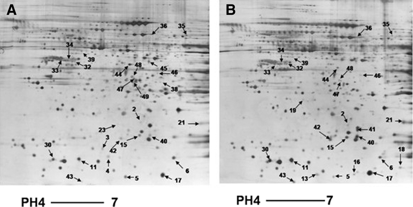
The representative two-dimensional polyacrylamide gel electrophoresis (2-DE) gel images of proteins from CNE-2 and CNE-2R cells. The total proteins were extracted from CNE-2 cells
Protein identification of the spots
Spots of interest were excised from the gels, trypsin digested, and analyzed by PMF and tandem mass spectrometry (MS/MS). The representative peptide-mass map and MS/MS map for one spot are shown in Figure 2. Among the 32 differentially expressed spots, 16 were successfully identified by the MALDI-TOF MS analysis. Protein identification was repeated at least once with spots from different gels to ensure the reliability.

Mass spectrometry analysis of spot 48 in a 2-DE gel. The spot 48 was excised from the gel, trypsin digested, and mixed with the matrix, and then subjected to MALDI-TOF mass spectrometry and tandem TOF/TOF. The results were shown as the peptide mass fingerprinting
Three spots corresponding to galectin-1, stathmin, and S100-A11 were found upregulated in the radioresistant cell line CNE-2R. Eight spots were found downregulated in the radioresistant cell line CNE-2R cells, including C protein, human elongation factor-1 delta, nucleolar phosphoprotein B23, laminin M-chain, succinyl-CoA:3-ketoacid-coenzyme A transferase 1, protein disulfide isomerase, 60S acidic ribosomal protein P0, and Annexin A3. Four spots were found exclusively in CNE-2 cells, including dUTPase Chain A, Three Crystal Structures of Human Coactosin-Like Protein, profilin-2 isoform b, and isocitrate dehydrogenase [NAD] subunit-α. Nm23 H1 protein was only identified in CNE-2R cells (Table 1). Interestingly, stathmin was identified twice in two separate spots. Although both spots had a similar molecular weight, their pIs were different, suggesting that the post-translational modifications of stathmin may have been selected in the radioresistant CNE-2R cells.
Expression ratio indicated the ratio of CNE-2R/CNE-2. aExpressed only in CNE-2.↑Expressed only in CNE-2R.
MALDI-TOF MS, matrix-assisted laser desorption/time-of-flight mass spectrometry; MW, molecular weight; pI, isoelectric point; ↓, downregulation.
Validation of the identified proteins
To validate the differential expression of the proteins identified, we detected the protein levels of two proteins Nm23 H1 and Annexin A3 by the Western blot analysis. The results showed that the protein level of Nm23 H1 was low in CNE-2 cells, but increased significantly in CNE-2R cells. In contrast, the Annexin A3 protein level was decreased in CNE-2R cells compared to CNE-2 cells (Fig. 3). These results are consistent with the results of 2-DE and prove the reliability of our analysis.
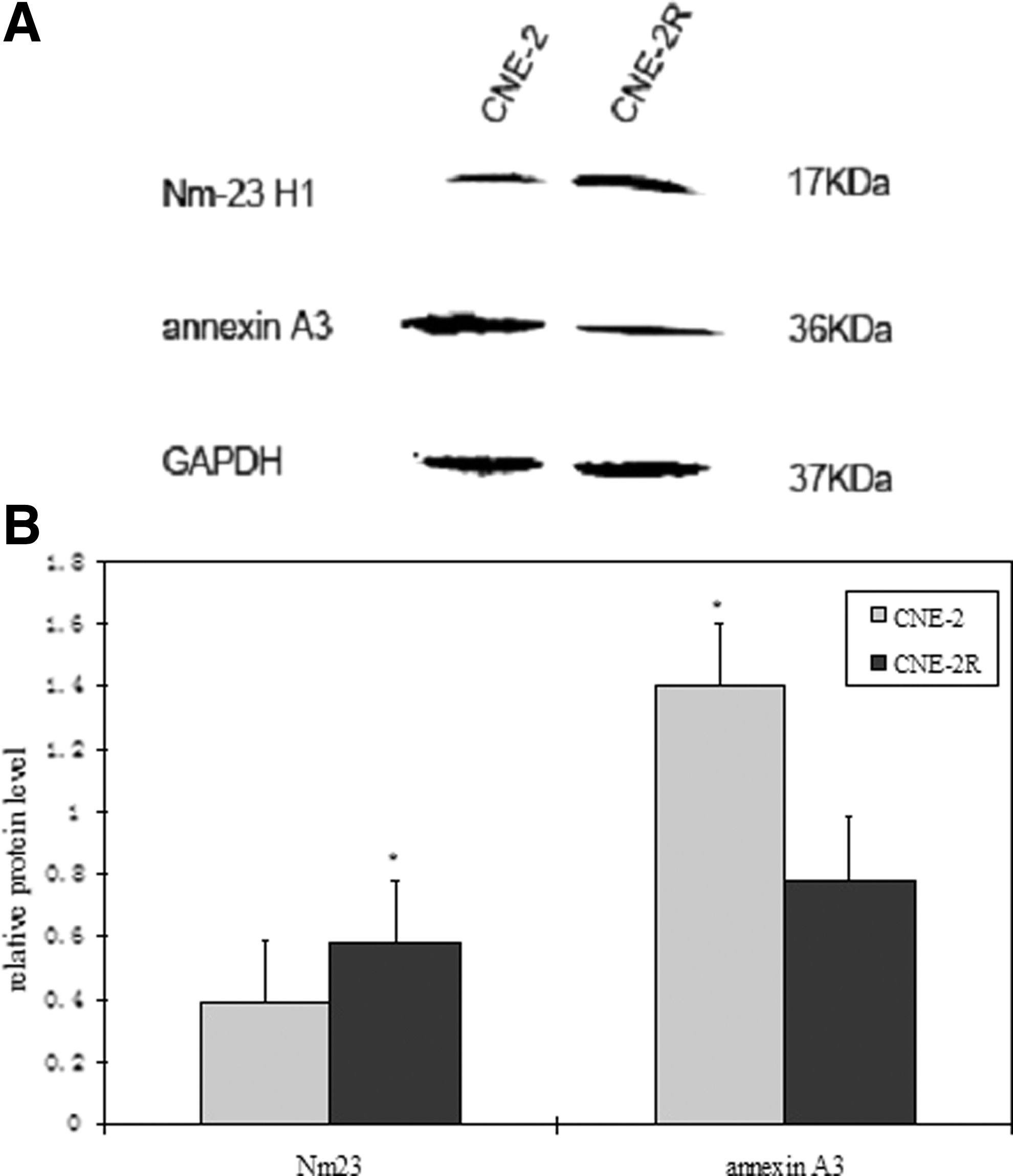
Expression of Annexin A3 and Nm23 in CNE-2 and CNE-2R.
Discussion
Proteomic techniques have been widely applied in many preclinical and clinical investigations due to their advantage of revealing the dynamics of protein expression and protein–protein interactions from a global perspective, which contributes greatly to our understanding of the gene function.
In this study, we used a comparative proteomic approach to screen the differential proteins in radioresistant NPC CNE-2R cells and their parental CNE-2 cells. We found 32 protein spots with significant alterations in the expression levels between the two cell lines. The MALDI-TOF MS analysis successfully identified 16 proteins, most of which were classified into cytoskeletal proteins, metabolic enzyme, signal transducer, and chaperones, based on their molecular functions.
Among the 16 proteins, we selected 2 representative proteins Nm23 H1 and Annexin A3 for further characterization due to their biological function. Nm23 H1 is a metastasis suppressor gene, which is implicated in tumor invasion and metastasis. 15 Many studies have shown that Nm23 H1 plays a role in DNA damage repair or the induction of DNA synthesis to maintain the genomic stability after irradiation or UV irradiation. 16,17 Overexpression of Nm23 H1 in HeLa cells provides cells with a higher resistance to oxidative stress possibly due to increased intracellular levels of p53 and GPX1. 18 Moreover, overexpression of Nm23, specifically its nuclear translocation, may be a powerful predictor of radiation resistance in HNSCC. 19 Consistent with these previous studies, in the present study, we showed that the Nm23 H1 protein was significantly increased in CNE-2R cells compared to CNE-2 cells as verified by the Western blot analysis, suggesting that the upregulation of Nm23 H1 in NPC increases the radiation resistance.
Annexin A3 is a member of the lipocortin/Anx family, which binds to phospholipids and membranes in a Ca2+-dependent manner. 20 Annexin A3 exhibits anticoagulant and anti-phospholipase A2 properties in vitro. 21 Annexin A3 has been shown to mediate the membrane–membrane contact during biological processes such as phagosome–lysosome fusion, vesicular trafficking, plasma membrane binding, and degranulation. 22 However, so far, no studies have reported the role of Annexin A3 in radioresistance. By the Western blot analysis, we confirmed that the expression of Annexin A3 was significantly downregulated in radioresistant CNE-2R cells compared to CNE-2 cells, indicating that Annexin A3 could function to increase the radiosensitivity of cancer cells. Further mechanistic studies are necessary to explore the role of Annexin A3 in the regulation of NPC radioresistance.
Recently, Wu reported that there are identified 12 proteins that were aberrantly expressed in radioresistant NPC tissues compared to radiosensitive NPC tissues. 23 Surprisingly, the list of these proteins did not overlap with the list of the proteins identified in this study. These may be due to the heterogeneity between cancer tissue samples and cancer cells.
In summary, in this study, we performed a proteomic analysis of a radioresistant NPC cell line CNE-2R and validated Nm23 H1 and Annexin A3 as two proteins with significant changes in the expression level in the cells compared to parental CNE-2 cells. Our findings provide a platform for further characterization of the proteins implicated in radioresistance and suggest that these proteins could be exploited as a biomarker for predicting the NPC response to radiotherapy in the clinic.
Footnotes
Acknowledgments
The authors thank Dr. Jinzi Wang at the Guangxi University for the technical assistance. This research was supported by the National Nature Science Foundation of China (No. 30860329) and the Nature Science Foundation of Guangxi Zhuang Autonomous Region (No. 0832229). It was also supported by the Major Scientific Research Foundation of Guangxi Colleges (No. 201101ZD004).
Disclosure Statement
No competing financial interests exist.