
Abstract
Aims:
To investigate the antitumor activity of cytokine-induced killer (CIK) cells combined with epidermal growth factor receptor (EGFR) monoclonal antibody (mAb) against gastric cancer cell line SGC7901.
Materials and Methods:
Immunocytochemistry assay was performed to detect the expression of EGFR in SGC7901 cell lines. The cytotoxicity activity of CIK cells combined with EGFR mAb was analyzed by the 51Cr release assay. Then, the comparison of the cytotoxicity activity between CIK cells combined with EGFR mAb and CIK cells combined with CD3 mAb and CIK cells was conducted. Antitumor activity of CIK cells combined with EGFR mAb in vivo was analyzed by tumor growth assay and tumor reduction assay.
Results:
The cell lysis rate of CIK cells combined with EGFR mAb was higher than those of CIK cells combined with CD3 mAb and CIK cells only (p<0.05). The lysis rates of the latter two groups were not different. The antitumor activity of CIK cells combined with EGFR mAb was higher than those of other groups in vivo (p<0.05).
Conclusion:
It was suggested in the current study that EGFR mAb could enhance the antitumor ability of CIK cells to bind and kill the gastric cancer cells in vitro and in vivo.
Introduction
Globally, gastric cancer is a very common malignancy. Although the incidence of the disease has been declining for the past few decades, advanced gastric cancer continues to have a very poor prognosis and with limited treatment options. 1 The adjuvant treatments such as chemotherapy and radiotherapy are frequently used; their overall impact on the prognoses of these patients is still limited and their adverse reaction can influence the patients' life quality frequently. 2 Some new therapeutic strategy could be an emergency request for gastric cancer therapy.
The epidermal growth factor receptor (EGFR) is the most important member of the epidermal growth factor receptor family. The overexpression of EGFR is found on many human malignancies, including lung, breast, colon, and gastric carcinomas. 3 –5 It is well known that EGFR is related to malignant transformation and tumorigenesis; so, this molecule was regarded as an important target in many preclinical and clinical researches. The overexpression of EGFR in gastric cancer has been confirmed in many researches in these years. 6,7 Some EGFR monoclonal antibodies (mAbs) have been generated and applied against a variety of malignancies.
The anti-EGFR mAb (cetuximab) have been used against gastric cancer in some preclinical studies or phase I and II studies with considerible results. Hotz et al. 8 have assessed the effect of cetuximab on three gastric cancer cell lines (AGS, MKN-45, and NCI-N87) with different phenotypes in vitro and in a therapeutic orthotopic murine gastric cancer model. Their results suggested that cetuximab is effective in K-ras wild-type, EGFR-expressing gastric cancer cell lines, and xenografts. Liu et al. 9 and Luo et al. 10 have assessed the treatment effects of cetuximab/irinotecan combination or cetuximab/oxaliplatin combination on gastric cancer cells respectively and confirmed that cetuximab combined with irinotecan or oxaliplatin could enhance the antitumor activity in vitro. Chan et al. 11 have performed a multi-institutional phase II study of cetuximab in patients with unresectable or metastatic esophageal or gastric adenocarcinoma. They thought that cetuximab administered as a single agent had minimal clinical activity in patients with metastatic esophageal and gastric adenocarcinoma. Moehler et al., 12 Kim et al., 13 and Han et al. 14 have conducted phase II clinical trials to assess the antitumor activity of cetuximab combined with chemotherapy (IF, XELOX, and modified FOLFOX6, respectively) in advanced gastric cancer. Their results showed that efficacy data of cetuximab plus chemotherapy were encouraging. According to these reports, the antitumor activity of cetuximab is considerable in vitro, but the treatment effects as a single-agent is limited. So, it is important that cetuximab should be combined with other agents in clinical therapy.
At present, the cellular immunotherapy against solid tumors has become a promising treatment following many preclinical and clinical trials. Cytokine-induced killer (CIK) cells has received considerable attention. CIK cells are expanded in vitro from peripheral blood mononuclear cells (PBMCs) by addition of interferon gamma (IFN-γ), interleukin-2 (IL-2), IL-1, and a mAb against CD3. 15,16 They are highly efficient cytotoxic effector cells with co-expression of CD3 and CD56 and NK activity. 17 They have recently been used as effector cells in adoptive cell therapy against some cancers and have shown a promising effect. 18,19
EGFR mAb could recognize the EGFR epitope on tumor cells as a target epitope, on the other hand, Fc of EGFR mAb could redirect immune effector cells to kill tumor cells by antibody dependent cellular cytotoxicity (ADCC). So, EGFR mAb could directly link immune effector cells to tumor cells and activate immune-mediated destruction of cancer cells theoretically. In comparison with conventional chemotherapy or radiotherapy, the treatment of EGFR mAb combined with CIK cells (immune effector cells) could yield more mild adverse effect. At present, the therapeutic strategy of a combination between EGFR mAb and CIK cells against gastric cancer is still not be reported. So, we investigated the antitumor activity of CIK cells combined with EGFR mAb against gastric cancer cell line SGC7901 in vitro and in vivo for preclinical assessment of the effect of this therapeutic strategy against gastric cancer.
Materials and Methods
Materials
EGFR mAb was purchased from Beijing Zhongshan Biotechnology Co., Ltd (Zhongshan, P.R. China), the product number is ZM-0083. Rabbit antimouse HPR (1:1000; Dako, Copenhagen, Denmark) was used to recognize corresponding proteins. Common cell culture plates were purchased from Orange Company (Braine-l'Alleud, Belgium). CD3 mAbs were purchased from Biosynthesis Co., Ltd (Beijing, China). Mouse IgG-isotype control was purchased from Abcam Company Ltd (Hong Kong, China). Forty 8–10-week-old female nude mice weighing 18–22 g (nu/nu; Iffa Credo, l'Arbresle, France) were purchased from Biotechnology Co., Ltd (Beijing, China) and housed in self-contained filter-top cages (5 mice/cage).
Cell lines and culture
The human gastric adenocarcinoma cell line (SGC7901) was obtained from Shanghai Cell Research Institute of Chinese Scientific Academy, stored, and transfer cultured in our laboratory. DMEM containing 10% calf serum, 100 IU/mL penicillin, and 100 IU/mL streptomycin were used as conventional culture medium. The culture procedures were taken under 37°C, 5% CO2, and saturation humidity.
Investigation of EGFR expression in SGC7901 cell line
The expressions of EGFR in SGC7901 cells were detected to determine whether the cell lines could be used in the research. Immunocytochemical assay and reverse transcription polymerase chain reaction (RT-PCR) were performed and the results showed that there was expression of EGFR in the cell line.
Preparion of CIK cells
CIK cells were prepared according to the method in Freddy's study. 20 Peripheral blood mononuclear cells (PBMCs) were isolated from healthy donors via the blood bank of Beijing. PBMCs were isolated by Ficoll density gradient centrifugation, washed by RPMI-1640, and then resuspended in RPMI-1640 containing 10% calf serum, 100 IU/mL penicillin, 100 IU/mL streptomycin, and IFN-γ (500 U/mL). On day 1, IL-2 (400 U/mL), IL-1α (100 U/mL), and anti-CD3 mAb (20 ng/mL) were added. The cultures were changed every 2 days and the concentration of cells was regulated to 2×10⁶. The cells were cultured for 21 days and detected by flow cytometry assay.
Cytotoxicity assays
51Cr release assays were performed to detect the cytotoxicity of CIK cells against gastric cancer cells combined with EGFR mAb as described by Hoyle et al. 21 and John et al. 22 previously. SGC7901 cells (1×10⁶) were labeled with 300 μCi sodium chromate (Dupont-NEM, Boston, MA). Then, the labeled cells were washed twice with PBS and suspended by RPMI-1640, then plated in 96-well plates at 1×10⁴/well. CIK cells and antibodies were added as follows: A: EGFR mAb (20 μg/mL)+CIK cells (5×10⁶/mL)+SGC7901 cells (1×10⁵/mL); B: Mouse IgG-isotype control (20 μg/mL)+CIK cells (5×10⁶/mL)+SGC7901 cells (1×10⁵/mL); and C: CIK cells (5×10⁶/mL)+SGC7901 cells (1×10⁵/mL). CIK cells were added as effector cells at E:T cell ratios (50:1) and incubated at 37°C, 5% CO2 for 4 hours. The radioactivity of supernatant was measured in a gamma counter (Cobra/AII; Packard BioScience, Meriden, CT). The lysis rate was calculated according to the formula: lysis rate (%)=[(sample release)−(spontaneous release)/(maximum release)−(spontaneous release)]×100%. Five reduplicate wells were used for each group and the mean value of each group was calculated.
Cytotoxicity assays at different E:T cell ratios
The cytotoxicity of CIK cells combined with EGFR mAb against gastric cancer cells was analyzed by 51Cr release assays at different E:T cell ratios. The assays were performed as described in “Cytotoxicity assays” part. CIK cells were added as effector cells at E:T cell ratios (1:1, 10:1, 20:1, 30:1, 40:1, 50:1, 60:1, 70:1, 80:1, 90:1, and 100:1) and incubated at 37°C, 5% CO2 for 4 hours, then the lysis rates of all groups were measured. The results reported were mean values of three independent experiments done in triplicate and a curve was established based on the mean lysis rates.
Cytotoxicity analysis in vivo
The cytotoxicity of CIK cells combined with EGFR mAb against gastric cancer was explored by therapeutic experiment on the xenograft mouse model. Forty mice were used in the in vivo assay and each mouse was injected s.c. in the right flank with 5×10⁶ SGC7901 cells in 0.2 mL PBS. After 10 days, the 30 mice with tumors (diameter ≥0.5 cm) were selected for therapeutic experiment. Then, the selected mice were assigned to 6 different treatment groups (5 mice per group), as follows: group A: EGFR mAb (1 mg)+CIK cells (1×10⁹/mL); group B: CD3 mAb (1 mg)+CIK cells (1×10⁹/mL); group C: CD3 mAb (1 mg); group D: EGFR mAb (1 mg); group E: CIK cells (1×10⁹/mL); and group F: 0.9% NaCl i.v. injection alone (0.2 mL/injection). All antibodies and effector cells were i.v. injected in the heat-dilated tail vein, and the day of beginning treatments was day 0. The treatments were performed and tumor dimensions were measured twice weekly, and tumor volumes (mm3) were estimated by the formula V=a×b 2/2, where a is the length, b is the width. The tumor growth curve of each group was established based on the mean tumor volume at different time. At day 35, tumors were dissected and weighed, then the tumor inhibition rates were estimated by the following formula: tumor inhibition rate (%)=[(mean tumor weight of group F)−(mean tumor weight of each experimental group)/(mean tumor weight of group F)]×100%.
Investigation of EGFR and CD3 expression in tumor tissues after treatment
The expressions of EGFR and CD3 in the tumor tissues of the xenograft mice after treatment were detected to determine whether the CIK cells could migrate into the tumor tissues. Tumors were dissected and embedded in paraffin. Tissue sections of 5 μm thickness were cut from paraffin-embedded tissue blocks, then H&E staining and immunohistochemical assay were performed. For the semiquantification of CD3 immunostaining, the results were analyzed based on the criteria that were presented by Kase et al. 23 and Tadahiro et al. 24 The CD3-labeling index (CD3-positive proportion) was expressed as the percentage of the number of CD3-labeled cells divided by the total number of cells examined under a microscope (20 objective) and the average value of 10 fields was calculated.
Results
Expression of EGFR in SGC7901 cell line
The expression of EGFR in gastric adenocarcinoma cell strain SGC7901 was detected by RT-PCR and immunocytochemical analysis. The RT-PCR results in SGC7901 cell were positive. The results of immunohistochemical assay showed that the expression of EGFR in SGC7901 cells was mainly distributed in cell membrane and cytoplasm and there was no obvious positive signal in cell nucleus (Figs. 1 and 2).
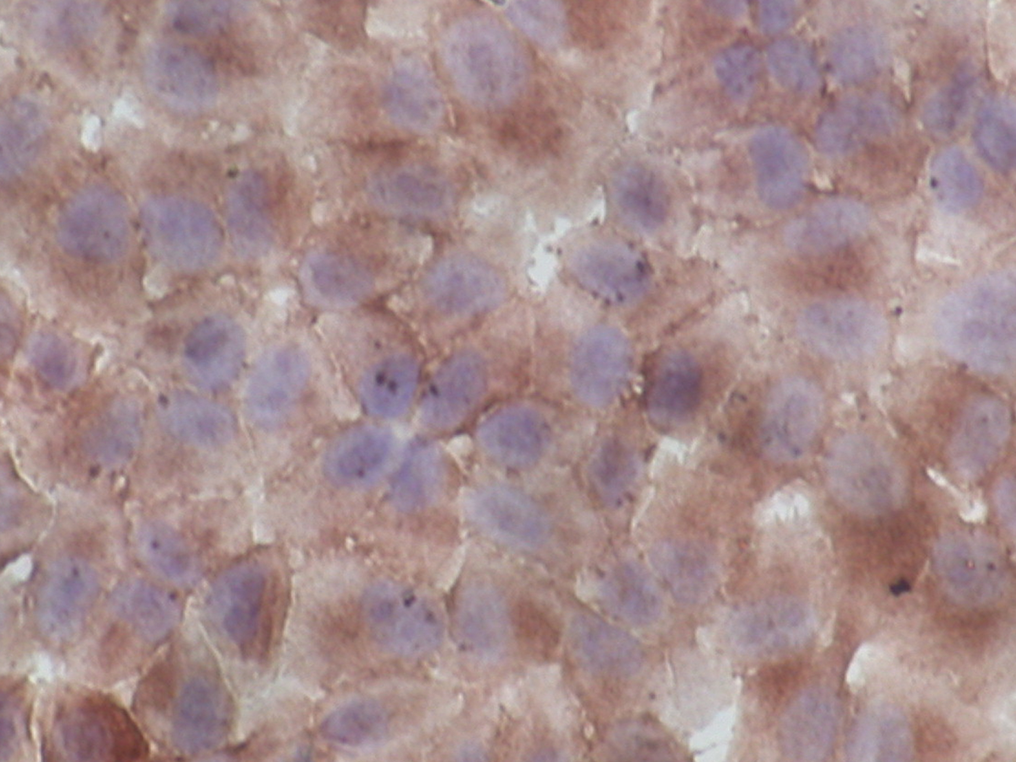
The immunostaining of epidermal growth factor receptor (EGFR) in SGC7901 cell line. The expressions of EGFR in SGC7901 cells were detected by immunocytochemical assay. Cell sections were prepared and treated with 2% bovine serum albumin (BSA) to block non-specific staining. Then, they were treated with 3% H2O2 for 10 minutes to block endogenous peroxidase activity. After washing in PBS for 10 minutes, EGFR monoclonal antibody (mAb) was applied for 1 hour, the secondary antibody—biotin-labeled rabbit antigoat HPR was added for 20 minutes after removing primary antibody by washing in PBS thrice. The sections were washed in PBS for 10 minutes, then 3,3′-diaminobenzidine (DAB) was used as the chromogen. Finally, the sections were counterstained with Harris's hematoxylin for 3 minutes and coverslips applied with a xylene-based mounting medium. The result showed that positive signals were found in most of the cells and brown positive signals, which can be seen in color online, were mainly distributed in the cell membrane and cytoplasm, and there was no obvious positive signal in the cell nucleus (×400). Color image available online at
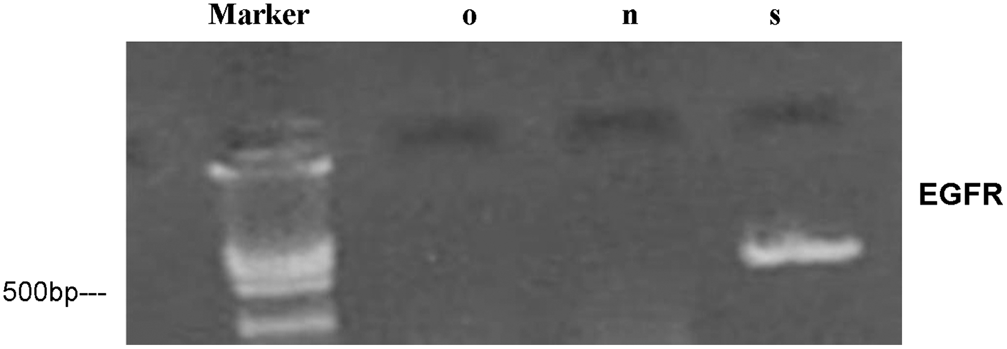
The result of reverse transcription polymerase chain reaction (RT-PCR) assay for EGFR expression in SGC7901 cell line. The total RNAs of SGC7901 cells were extracted by homogenization in TRIzol (Invitrogen, Carlsbad, CA), and cDNA synthesis was performed in 20 μL reaction system of reverse transcription including 5 μg RNA. Amplification of EGFR was then carried out in DNA thermal cycler (PerkinElmer, Waltham, MA) using equal cDNA as a template. PCR products were separated by 1.5% agarose gel electrophoresis, then scanned and analyzed by VDS imagemaster system (Pharmacia, New York, NY). The result showed that there was EGFR expression in SGC7901 cell line in mRNA level.
Investigation of characterization of CIK cells
The different populations of cells in expanded CIK cells were analyzed by flow cytometry analysis at day 21. The results suggested that populations of CD4+CD8+ T cells accounted for about 7.2%±1.3%, concurrently, populations of CD3+CD56+ T cells accounted for about 25.0%±6.4% (Fig. 3).

The result of flow cytometry assay of cytokine-induced killer (CIK) cell. The different populations of cells in expanded CIK cells were analyzed by flow cytometry analysis at day 21. CD3, CD4, CD8, and CD56 were detected by flow cytometry. The result showed that the populations of CD4+CD8+ cells accounted for about 7.2%±1.3%, concurrently, populations of CD3+CD56+ cells accounted for about 25.0%±6.4%. Color images available online at
The different cytotoxicity at different E:T cell ratios
The cytotoxicity of CIK cells combined with EGFR mAb at different E:T cell ratios was tested by 51Cr release assays. The results of the assay were shown in Figure 4. At an E:T cell ratio of about 50:1, CIK cells combined with EGFR mAb could yield a considerable cytotoxicity. The cytotoxicity of CIK cells did not significantly increase when the E:T cell ratio increased beyond 50:1. On the other hand, CIK cells could yield a stable cytotoxicity at an E:T cell ratio of about 70:1. The result suggested that EGFR mAb could enhance the cytotoxicity of CIK cells at a lower E:T cell ratio.
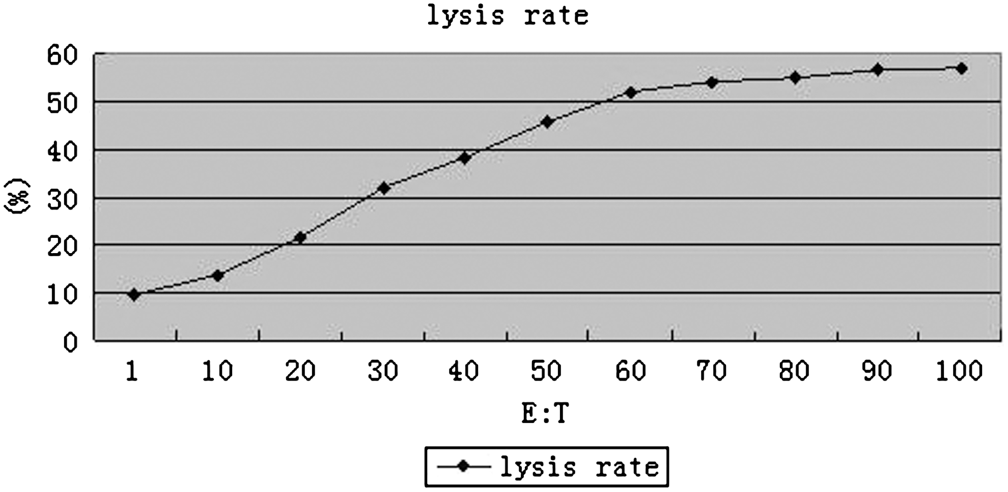
The different cytotoxicity at different E:T cell ratios. SGC7901 cells (1×10⁶) were labeled with 300 μCi sodium chromate (Dupont-NEM, Boston, MA). Then the labeled cells were washed twice with PBS and suspended by RPMI-1640, then plated in 96-well plates at 1×10⁴/well. CIK cells and antibodies were added as follows: EGFR mAb (20 μg/mL)+CIK cells+SGC7901 cells. CIK cells were added as effector cells at E:T cell ratios (1:1, 10:1, 20:1, 30:1, 40:1, 50:1, 60:1, 70:1, 80:1, 90:1, and 100:1) and incubated at 37°C, 5% CO2 for 4 hours, then the lysis rate was measured. The results reported were mean values of three independent experiments done in triplicate and a curve was established. The result showed that CIK cells targeted by EGFR mAb could yield a considerable cytotoxicity at the E:T cell ratio of about 50:1.
Enhanced cytotoxicity of CIK cells by EGFR mAb
The cytotoxicity of CIK cells combined with EGFR mAb was tested by cell lysis assay and compared with that of CIK cells combined with CD3 mAb or CIK cells only. The results of the assay were shown in Figure 5. At the E:T cell ratio of 50:1, CIK cells combined with EGFR mAb yielded a cell lysis rate of 51.3%±6.1%, which was higher than those of CIK cells combined with CD3 mAb and other control groups (p<0.05).

The result of 51Cr release assays. SGC7901 cells (1×10⁶) were labeled with 300 μCi sodium chromate (Dupont-NEM). Then the labeled cells were washed twice with PBS and suspended by RPMI-1640, then plated in 96-well plates at 1×10⁴/well. CIK cells and antibodies were added as groups: A: EGFR mAb (20 μg/mL)+CIK cells (5×10⁶/mL)+SGC7901 cells (1×10⁵/mL); B: Mouse IgG-isotype control (20 μg/mL)+CIK cells (5×10⁶/mL)+SGC7901 cells (1×10⁵/mL); and C: CIK cells (5×10⁶/mL)+SGC7901 cells (1×10⁵/mL). CIK cells were added as effector cells at E:T cell ratios (50:1) and incubated at 37°C, 5% CO2 for 4 hours. The radioactivity of supernatant was measured in a gamma counter (Cobra/AII; Packard BioScience). At an E:T cell ratio of 50:1, CIK cells directed by EGFR mAb yielded a cell lysis rate of 51.3%±6.1%, which was higher than those of other groups (17.5%±2.5% and 25.2%±4.1%).
The influence of CIK cells combined with EGFR mAb on xenograft tumor growth
Antitumor activity of CIK cells combined with EGFR mAb was analyzed by tumor growth curve and tumor reduction assay. The results are shown in Table 1 and Figure 6. According to the results, the mice treated with CIK cells combined with EGFR mAb showed a significant enhancement of antitumor activity of CIK cells. The results of tumor growth curve assay showed that the tumors in groups treated with CIK cells combined with EGFR mAb grew significantly slower than other groups (p<0.05) (Fig. 6). The administration of CIK cells combined with EGFR mAb had a mean tumor reduction of 52.3%, which was higher than those of other groups (p<0.05) (Table 1). The administration of CD3 mAb alone without CIK cells had no significant antitumor effect compared with the control group treated with 0.9% NaCl.
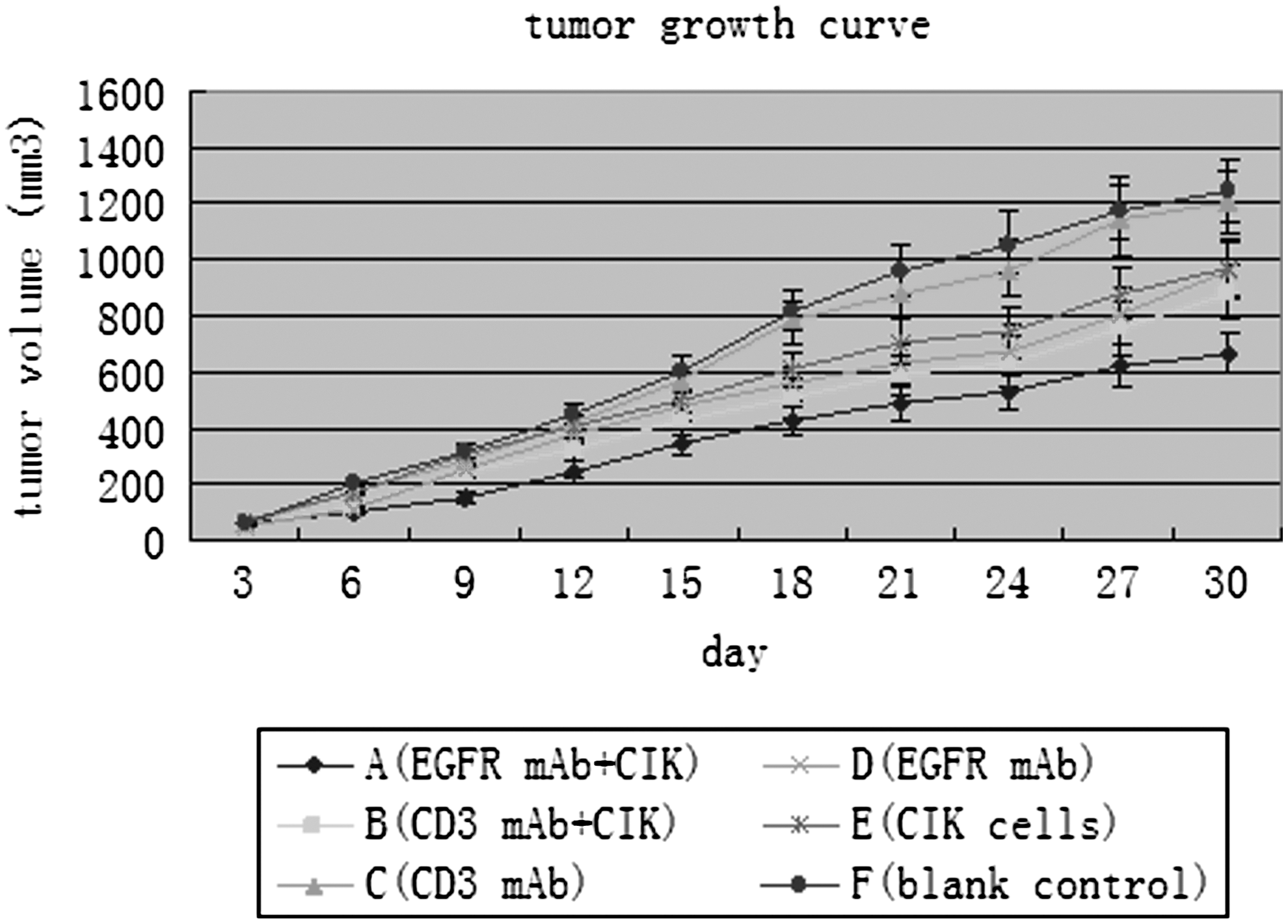
The result of tumor growth curve assay. The tumor dimensions were measured twice weekly, and tumor volumes (mm3) were estimated by the formula V=a×b 2/2, where a is the length, b is the width. The tumor growth curve of each group was established based on the mean tumor volume at different time. The results of tumor growth curve assay showed that the tumors in group treated with CIK cells directed by EGFR mAb grew significantly slower than those in other groups (p<0.05).
Compared with group F, adenoting p<0.05.
Compared with group B, bdenoting p<0.05.
Expression of EGFR and CD3 in tumor tissues after treatment
The results of immunohistochemical analysis showed a strong positive expression of EGFR in the tumor tissues of all groups and the positive signals were distributed in cytoplasm and membrane of tumor cells (Fig. 7). On the other hand, the results of immunohistochemical assay showed that the expression of CD3 (CD3-positive proportion) in the tumor tissues of the mice treated with EGFR mAb combined with CIK cells was 14.7%±3.2%, which was significantly higher than that of other groups (p<0.05) and the positive signals were mainly distributed in membrane and cytoplasm of cells (Fig. 8). There was no positive signal in the tumor tissues of the mice treated with CD3 antibodies or EGFR antibodies alone. These results indicated that EGFR mAb could direct CIK cells to tumor tissues to some extent in vivo.
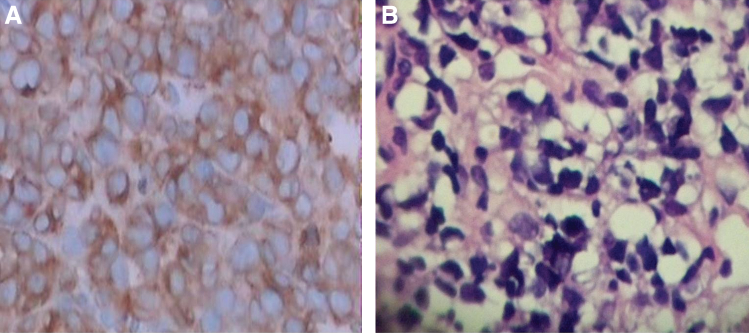
The immunostaining of EGFR in tumor tissues after treatment.

The immunostaining of CD3 in tumor tissues after treatment. Tumors were dissected and embedded in paraffin. Tissue sections of 5 μm thickness were cut from paraffin-embedded tissue blocks; then, the immunostaining of CD3 in tumor tissues were performed. For the semiquantification of CD3 immunostaining, the CD3-labeling index (CD3 positive proportion) was expressed as the percentage of the number of CD3-labeled cell divided by the total number of cells examined under a microscope (20×objective) and the average value of 10 fields was calculated.
Statistical analysis
In this research, the data were expressed as mean±SD. One-way ANOVA test and Student's t-test were performed. All data were analyzed with the SPSS 11.0 (SPSS, Chicago, IL) statistical software package. Statistical significance was defined as p<0.05.
Discussion
In China, gastric cancer still remains a common disease and its morbidity and mortality increase consecutively. Because of the deficiency of mass survey and early diagnosis, the prognosis of gastric cancer is very poor. The therapeutic effect of traditional chemotherapy or radiotherapy for the patients with unresectable gastric cancer is not satisfactory; on the other hand, the adverse reaction of these treatments could be distressful. The pathogenesis and molecular controls of gastric cancer are poorly understood and the involvement of a number of different influencing factors has been proposed. Cellular immunity is very important for restraining the growth of tumor cells. Cellular immune defect could be connected with a poor prognosis in patients with gastric adenocarcinoma and other malignancies. Cellular immunity could include immunologic recognition, immunologic regulation, cytotoxicity, and others, which could play very important roles in regulating critical antitumor functions. Therefore, cellular immunotherapy has been thought as an important adjunctive therapy for gastric cancer and yielded some promising effect. As most common effector cells, CIK cells can express the T cell marker CD3 and NK cell marker CD56, and they develop cytotoxic activity against various cancer cells including gastric cancer cells. It was reported 25 –29 that CIK immunotherapy is a safe and effective therapy for many malignancies. Some agents have been used to enhance the cytotoxicity and antitumor specificity of CIK cells in previous researches. 20,22
In theory, EGFR mAb can direct CIK cells to tumor cells by linking Fcγ receptor on effector cells with EGFR on tumor cells by ADCC. Pievani et al. 30 have investigated combining immunotherapy with CIK cells and anti-CD20 mAb (GA101 or rituximab) to optimize B-cell non-Hodgkin lymphoma (B-NHL) therapy. They confirmed that anti-CD20 mAb could enhance the antitumor activity of CIK cells and thought this was due to the combine action of complement-mediated cytotoxicity, ADCC, and CIK-mediated NC. On the other hand, EGFR mAb could close EGFR and restrain tumor cells growth in many preclinical and clinical researches. 31,32 EGFR mAb can bind to EGFR and Fcγ receptor at the same time, so it should not only direct T cells/CIK cells but also restrain tumor cells growth. In the present study, the cytotoxicity of CIK cells against gastric cancer cells targeted by this EGFR mAb was investigated. The results of cytotoxicity assays in vitro suggested that redirected lysis could significantly increase when CIK cells combined with EGFR mAb; otherwise, Mouse IgG-isotype could not enhance tumor cell lysis. For the expression of Fc receptors (CD16, CD32, or CD64) on CIK cells, EGFR mAb could also mediate effector cells to EGFR-positive target cells by ADCC. Sabbatino et al. 33 confirmed that the significant improvement of the ADCC could enhance the antitumor activity of EGFR-specific mAb. There are some preclinical and clinical evidence that the interaction of the ADCC is very important for the activity of therapeutic CD20 mAbs against tumor cells. 34 It was demonstrated that CD20 mAb therapy is no longer effective in FcRγ-chain knockout (KO) mice (the lack of surface expression of all activating FcγRs in FcRγ-chain KO mice) 35 –39 In contrast to FcRγ-chain KO mice, CD20 mAb therapy was enhanced in mice lacking the inhibitory FcγRIIb in both a xenograft and a syngenic tumor model. 35,37,38 Lefterova et al. 40 reported that the CIK-lysis of various lymphoma and leukemia cell lines could be partially blocked by preincubation of target cells with anti-CD16 and anti-CD32 antibodies. These evidences could suggest that ADCC is very important for the antitumor activity of mAbs. In the present research, mouse IgG-isotype could weaken the cytotoxicity of CIK cells against gastric cancer cells to some extent. We thought that mouse IgG-isotype could block some Fc receptors on CIK cells, which could also weaken the interaction of the ADCC. So, we thought that the cytotoxic activity could be caused by Fc receptor-mediated binding. The ability of EGFR mAb to enhance cytotoxicity could be limited by the expression level of Fc receptors. Leemhuis et al. 41 reported their efforts for the large-scale expansion of CIK cells and the results of their phase I clinical trial. They confirmed that the ratio of CD16+CD56+ CIK cells ranged from 0.2% to 7.7%. Though CD32 and CD64 could account for a part of cytotoxicity activity of CIK cells, this low expression level of CD16 could restrain antibody-dependent cellular cytotoxicity. No research on this therapeutic strategy for gastric cancer was reported previously, so, it is first reported that EGFR mAb could enhance the cytotoxicity of CIK cells against gastric cancer cells in vitro and in vivo.
The results of experimental treatment in vivo showed that antitumor activity of CIK cells could be enhanced to some extent by EGFR mAb when compared with CD3 mAb in nude mouse models. Our results are in agreement with other investigators who have used the anti-CD20 mAb to direct CIK cells to B-NHL cells. 30 These findings confirmed the promising therapeutic effect and clinical potential of this strategy on some malignant tumors. Bioluminescent imaging should be used to serially observe the response to therapy without the need for sacrifice. There was no equipment in our laboratory to perform bioluminescent imaging, but we thought that credible data could be provided with current test methods.
By immunohistochemical assay, we found that the positive immunohistochemical staining of CD3 was distributed in the cell membrane and cytoplasm of some cells in tumor tissues of A, B, and E groups and there was no obvious positive signal in tumor tissues of other groups. Our results showed that CD3-positive cells in mice treated with EGFR mAb-redirected CIK cells were significantly more than those in the mice treated with CD3 mAb-redirected CIK cells or with CIK cells only. The results suggested that EGFR mAb could direct CD3-positive CIK cells to tumor cells; so, the antitumor ability of CIK cells could be enhanced by EGFR mAb.
Conclusions
Given the poor prognosis of patients with advanced gastric cancer, to investigate some new therapeutic strategy to improve the outcome of these patients is very important. The results of the current research demonstrated that EGFR mAb significantly enhanced the antitumor activity of CIK cells against gastric cancer cells in vitro and in vivo; so, the combination between EGFR mAb and CIK cells could be an effective therapeutic strategy in cellular therapy.
Footnotes
Acknowledgments
The authors wish to thank Drs. Junshan Zhai and Yanmei Wang, and Nurse Weidi You, Weihua Wang et al., for handling patient contacts. We wish to thank the Fourth Military Medical University of PLA for providing means for the current investigation.
Authors' Contributions
L.Z. and G.Z. conceived the study, carried out experiments on the cellular therapy, and drafted the article. Y.H. and G.Z. carried out experiments on the cell culture and cytotoxicity analysis. J.Z. and J.H. participated in the study design and revised the article. K.Z. used flow cytometry to complete some analysis.
Disclosure Statement
The authors declare no conflict of interest in this research.