
Abstract
MicroRNAs (miRNAs) are a class of endogenous small noncoding regulatory RNAs, which are new regulators of gene expression. They interfere with multiple biological processes involved in tumorigenesis such as cell proliferation, cell cycle, apoptosis, angiogenesis, invasion, and metastasis. A large body of evidence shows that the aberrant expression of miRNAs in cancer patients provides numerous underlying merits as diagnostic, clinical pathological, prognostic markers, and as promising therapeutic targets of lung cancer, providing an insight into the clinical application for lung cancer. Here, we focus on specific miRNAs as biomarkers in lung cancer and briefly introduce the biological function and modification of miRNAs.
Introduction
Lung cancer is the leading cause of cancer-related deaths worldwide. Lung cancer accounts for 13% of all cancers and 18% of the deaths in 2008 around the world. 1 The 5-year overall survival rate strongly correlates with the time of diagnosis and ranges around 70% in clinical stage I, but only 30% in stage III of nonsmall-cell lung cancer (NSCLC), 2 which accounts for 80% of lung cancer. Unfortunately, lung cancer is mostly diagnosed in late stages. The traditional diagnostic methods (e.g., iconography, sputum cell screening, biopsy, and tumor biomarkers) have the limitations on accurate diagnosis of lung cancer. Currently, no appropriate biomarker exists to detect and predict lung cancer.
MicroRNAs (miRNAs) are a class of endogenous small noncoding regulatory RNAs of ∼22 nucleotides in length, which are known to regulate gene expression by repressing translation or decreasing the stability of mRNAs. 3 Altered expression of miRNAs results in aberrant expression of their corresponding target genes, which can influence cell behaviors associated with malignancy, such as cell proliferation, cell cycle, apoptosis, angiogenesis, invasion, and metastasis. 4 –9 This has rendered the potential of specific miRNAs as biomarkers to characterize cancer types, augment the accuracy of diagnosis, and predict prognosis, ultimately exerting a great impact on the treatment of cancer, especially lung cancer (Fig. 1).
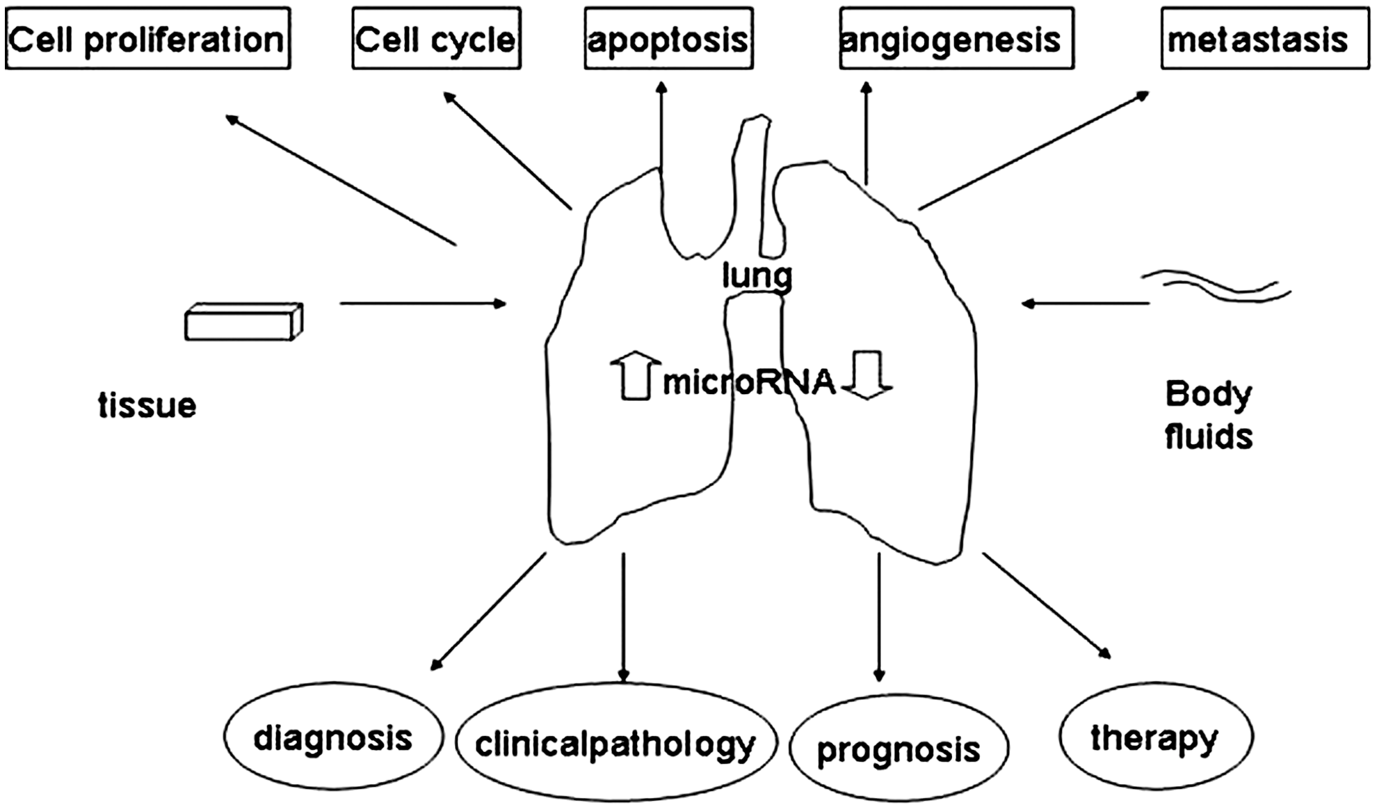
The clinical implication of miRNA in lung cancer. The altered expression levels of miRNAs from tissues and body fluids have been described in many cancers (including lung cancer), resulting in aberrant expression of proteins that influence the malignant behavior such as cell proliferation, cell cycle, apoptosis, angiogenesis, invasion, and metastasis, and rendering the value of specific miRNAs as biomarkers for diagnosis, clinical pathology, and prognosis, ultimately bring great significance to the treatment of cancer, especially in lung cancer. miRNA, microRNA.
The Biogenesis and Function of miRNA
Most miRNA genes are derived from primary miRNA transcripts containing a cap and a poly(A) tail and produced by RNA polymerase II from the miRNA genes. 10,11 The primary miRNAs are further cleaved into ∼22-nt mature miRNAs by the consecutive function of RNAse III Drosha-DGCR8 and Dicer, present in the nucleus and cytoplasm, respectively 12 –16 (Fig. 2). These 22-nt RNA molecules are able to bind to the specific sites typically present in the 3′-untranslated region (3′-UTR) of their target genes and mediate either mRNA decay with perfect base pairing or translational blockade with imperfect base pairing. 17
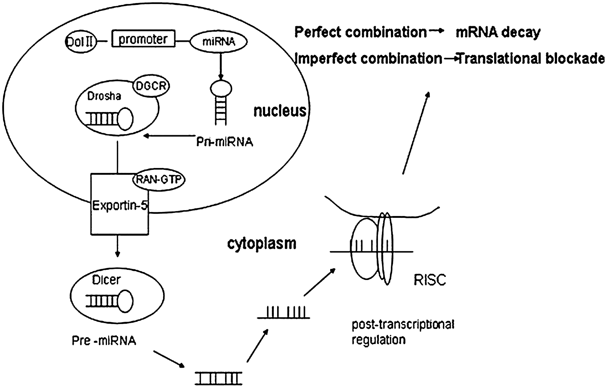
The biogenesis of miRNA. Most miRNAs are derived from primary miRNA transcripts (pri-miRNAs) containing a cap and a poly(A) tail produced by Pol II from the miRNA genes. The pri-miRNA is cleaved within the nucleus by a multiprotein complex, which is composed of the RNAase III enzyme Drosha- DGCR8 into a 70-nt pre-miRNA with hairpin and is exported by Expor-tin-5 with a Ran-GTP into the cytoplasm, where pre-miRNA further be cleaved into ∼22-nt miRNA duplex by an RNAse III enzyme, Dicer. The mature miRNA is selectively incorporated with RISC, which guides the complex specifically to its mRNA targets through the base–pairing interaction. The modulation mode of miRNA relies on the degree of homology to its target sequence. miRNAs with a perfect combination to the 3′-UTR of mRNA induce the mRNA decay, but an imperfect interaction leads to degradation of the translational protein.
Increasing evidence indicates that miRNAs play critical roles in tumorigenesis by functioning as tumor suppressors or oncogenes (oncomiRs) depending on the target genes. The ability of a single miRNA to target multiple mRNAs, as well as the possibility of one mRNA to be targeted by multiple miRNAs, adds a rich layer of regulation to gene expression 18 (Fig. 3). miRNAs act as the tumor suppressor by downregulating the expression of oncogenes and other specific genes involving in tumorigenesis, contributing to the inhibition of tumor growth. Tumor-suppressive miRNAs are downregulated in lung cancer, such as the let-7 family of miRNAs, which target p53, MYC, RAS, and other oncogenes, thereby suppressing the occurrence and growth of tumor cells. 4 Kumarswamy et al. 19 found that miR-30a could target Snail, leading to upregulated E-cadherin expression and inhibition of the tumor invasion and metastasis. Moreover, in lung cancer cell lines, miR-378 can downregulate vascular endothelial growth factor (VEGF), an important factor for angiogenesis leading to inhibition of tumor growth. 8 In contrast, oncomiRs promote tumor growth mainly through inhibiting the tumor suppressor genes or regulating factors that control differentiation and apoptosis. miR-17-5p may inhibit the expression of the target gene RB2 to promote lung cancer proliferation and tumor growth. 6 Ivanovska et al. 7 found that miR-106b downregulated its target gene p21 to promote tumor cell proliferation. Phosphatase and tensin homolog (PTEN), an inhibitor of the survival signaling pathway, is downregulated by miR-21, possibly contributing to cell transformation and increased tumor cell survival in lung cancer. 20,21

The biological function of miRNA. The miRNAs acting as tumor suppressors or oncogenes influence the malignant behavior such as cell proliferation, cell cycle, apoptosis, angiogenesis, invasion, and metastasis by targeting the related factors.
The Modification of miRNA
At present, most studies focus on the function of miRNA at the post-transcriptional level. However, the genetic and epigenetic changes of miRNAs also affect their expression levels, leading to a significant variation in the physiological aspect of miRNAs, and may serve as specific biomarkers for diseases. About 50% of miRNA genes are located in the fragile areas in chromosome, and presumably regulated through gene amplification, deletion, or translocation to exert its altered expression in the tumor development process. 22 miRNA-128b is located in 3p, whose allelic loss in lung cancer is the most common and earliest genetic events. The miR-17–92 family is located in 13q31, the chromosome fragments abnormally amplified in lung cancer.
miRNA-related single-nucleotide polymorphisms (miR-SNPs) can affect miRNA biogenesis and targeting. The SNP can occur in the 3′-UTR of the mRNA target genes and in the seed sequence of miRNAs. Yang et al. 23 selected the case–control cohorts from the southern and eastern of China and found that NBS1 gene's 3′-UTR rs2735383CC variant genotypes can significantly increase the risk of lung cancer in 1559 patients with lung cancer compared to 1679 healthy controls. Campayo et al. 24 examined 11 miR-SNPs (including three kinds of sites), and significant differences in the time to recurrence were found with KRT81 rs3660 and XPO5 rs11077. In addition, studies showed that the SNP in hsa-miR-196a2 rs11614913 can increase the risk of lung cancer significantly and influence the expression of the mature hsa-miR-196a2, thus affecting the function of miR-196a2 to bind to the mRNA. 25 –27 These results suggest that miR-SNPs were a novel class of SNPs that can add useful prognostic information to the clinical outcome of lung cancer patients and may be a potential key tool for selecting high-risk patients. Further characterization of miRNA SNPs may open new avenues for the study of cancer and therapeutic interventions.
In addition to genetic changes, epigenetic modulations are also responsible for aberrant miRNA expression. DNA methylation and histone modification, which are involved in carcinogenesis, could affect the expression and function of miRNA. Watanabe et al. 28 selected qualified miRNAs with CpG islands in six kinds of lung cancer cell lines, and found that miR-34b and miR-126 were silenced by DNA methylation. In addition, they found H3K9me2 and H3K9me3 in the miR-34b and EGFL7 genes, and H3K27me3 in EGFR7 by immunoprecipitation, indicating that histone methylation plays a role in silencing miRNA independent of the DNA methylation. Additionally, the term epi-miRNAs refers to a subgroup of miRNAs that target, directly or indirectly, the effectors of the epigenetic machinery. The miR-29 family was found to directly target DNMT3a and DNMT3b, the two key enzymes involved in DNA methylation, in a lung cancer cell line. 29 miR-449b can function as a tumor suppressor in lung cancer by directly targeting HDAC1, which plays a crucial role in lung cancer. 30 Zhang and coworkers 31,32 reported that overexpression of miR-101 or knockout of EHZH2 protein (a kind of methyltransferase) can modify H3K27 methylation to silence miRNA expression, resulting in reduced tumor cell growth and invasion. Taken together, the modulation is reciprocal between miRNA and epigenetic elements.
miRNA as Diagnostic Biomarkers
Recently, a wide variety of reports found that the aberrant miRNA profiles can be a more reliable, noninvasive diagnostic tool in screening and diagnosing lung cancer with a relatively higher specificity and sensitivity than traditional tumor biomarkers of lung cancer.
Boeri et al. 33 identified miR-7, miR-21, miR-200b, miR-210, miR-219-1, miR-324 (upregulated), miR-126, miR-451, miR-30a, and miR-486 (downregulated) in tumor compared to normal lung tissues by microarray hybridization with qRT-PCR validation. The same study suggested that the miRNA signature in plasma samples can be used to identify the circulating biomarkers for the risk, diagnosis, and prognosis collected before or with a present CT-detected disease, with the diagnostic sensitivity of 75% and a specificity of 100% in the validation cohort. Chen et al. 34 reported the use of 10 serum miRNAs to diagnose NSCLC, and that the sensitivity and specificity can reach 93% and 90%, respectively. Xie et al. 35 collected the sputum of patients with NSCLC and verified that the sensitivity of miR-21 was significantly higher than the sputum cytology.
Early diagnosis of lung cancer has drawn increasing attention, as the survival of stage I lung cancer patients is much longer than those at advanced stages. Current studies show that miRNA can be as a screening marker in a high-risk population of early-stage lung cancer. Chen et al. 34 reported that aberrant expression of 10 miRNAs could be used to screen the high-risk population before diagnosed as lung cancer in a retrospective analysis, indicating that the miRNA can be used as the biomarker for early diagnosis. Keller et al. 36 revealed that the serum miRNA expression spectrum of lung cancer patients can change before the diagnosis, and the closer to the point diagnosed as lung cancer, the more obvious the changes of miRNA expression profile are observed. Bianchi et al. 37 reported the detection of miRNA from serum in the asymptomatic high-risk groups of early-stage lung cancer, and the accuracy can reach 80%.
miRNA as a Clinicopathological Biomarker
A myriad of studies found that miRNA was associated with some clinicopathological features, and specific miRNAs were closely related to tumor stages, pathological types, and metastasis.
Clinical stage
Evidence for the association of miRNA expression with clinical stages is supported by a variety of studies. The serum miR-21 expression level and TNM stage in NSCLC show statistical significance. 38,39 The same results come from the comparison between the stage III–IV and stage I–II, with the AUC of 0.775, the sensitivity of 76.2%, and the specificity of 70%. 39 Recently, it was shown that the levels of miR-126 and miR-183 in the serum exhibit statistical differences between the stage I–II patients and stage IV patients of lung cancer, suggesting that the status of the two miRNAs may predict the procedure of lung cancer metastases. 40
Subtype of lung cancer
Conventional methods to subclassify lung cancer such as immunohistochemistry and histomorphological analyses with complex progress and low specificity and sensitivity have been a clinical obstacle for a long time. Evidence showed that miRNAs may be incorporated into clinical decision making by classifying the subtypes of patients with NSCLC. Lu et al. 41 showed that miRNA expression profiles strongly differentiated lung adenocarcinomas (ADC) from lung squamous cell carcinomas (SqCC) in stage I patients of lung cancer, including 145 miRNAs differentially expressed between the two subtypes and 53 overexpressed in SqCC and 92 overexpressed in ADC. The expression of miR-205 in NSCLC could highly distinguish SqCC and ADC (miR-205 mainly expressing in SqCC) with a higher specificity and sensitivity than the traditional methods, 42 and can identify SqCC from NSCLC with the specificity and sensitivity of 90% and 96%, respectively, in an independent blinded validation set. 43 Additionally, Shen et al. 44 demonstrated that miR-21, miR-126, miR-210, and miR-486-5p in the plasma can reach a significantly higher sensitivity to diagnose pulmonary ADC (91.67%) than lung SqCC (82.35%).
Metastasis
Metastasis is a main cause of death in lung cancer patients, and is largely undermining the beneficial effects of treatment and patient prognosis. Thus, looking for the biomarkers that can accurately detect early tumor changes and characterize metastasis is a principle warrant in the NSCLC management. Jiang et al. 45 indicated that in lung cancer tissues, both miR-125a-5p and miR-125a-3p were poorly expressed and associated with lymph node metastasis. However, in lung cancer cell lines, the two had inverse effects on invasion and migration. Wang et al. 9 reported that miR-206 can significantly inhibit the metastasis of lung cancer cells, as a decreased level of miR-206 expression was detected in highly metastatic tumors, and induced expression of miR-206 in tumor cells can significantly attenuate cell growth and promote apoptosis.
Much direct evidence for the links between miRNAs and metastasis has been demonstrated by several studies. The epithelial–mesenchymal transition (EMT), a key step of metastasis, is an evolutionarily conserved process in which cells undergo conversion of epithelial cells to mesenchymal cells. The most well-known miRNA with an EMT-relevant role is the miRNA-200 family, which reciprocally regulates expression of ZEB transcriptional factor in a negative feedback loop. 46 In addition, miR-365 forms a signaling loop with transforming growth factor-β and can induce EMT by targeting HMGA2, an EMT promoter in lung cancer. 47 Kim et al. 48 found that in the absence of wild-type p53 function, a Snail1-dependent EMT was activated in lung carcinoma cells through downregulation of miR-34, which suppresses Snail1 activity by binding to 3′-UTR in Snail1 itself as well as those of key Snail1 regulatory molecules.
Moreover, recent reports on the role of miRNAs in lung cancer with brain metastasis (BM) have caused increasing attention. BMs can affect the survival of about 25% of patients with lung cancer and cause significant cognitive, neurologic, and emotional difficulties and ultimately poor survival. 8 Arora et al. 49 reported that miR-328 can significantly distinguish BM-positive and BM-negative patients and is involved in conferring the migratory potential to NSCLC cells working partly through regulating PRKCA. Interestingly, miR-378 is overexpressed in BM+ lung cancer patients, and may effect through promoting cell migration, invasion, and tumor angiogenesis, as a potential tool to characterize BM in lung cancer and a new target for intervention that may mitigate BM development. 8 In another report, 10 miRNAs were identified to be associates with BM. Among them, miR-145 had previously been shown to suppress cell invasion and metastasis by directly targeting the metastasis gene mucin 1 (MUC1). 41
miRNA as Prognostic Biomarkers
Nowadays, the poor prognosis of lung cancer has raised a major clinical challenge, and robust prognostic markers are in urgent need for improved disease management and therapeutic options. More and more researchers show that the expression level of miRNA can significantly predict the prognosis of patients.
Gao et al. 50 indicated that high expression of miR-21 in lung cancer patients is associated with a poor prognosis and shorter survival. Univariate and multivariate analyses showed that the miR-21 was an independent prognostic factor of NSCLC. 38 Luo et al. 32 found that downregulated miR-101 in NSCLC or upregulated Mcl-1 was associated with poor prognosis. The prominent role of miR-126 relating to tumor angiogenesis by targeting VEGF-a is an independent negative prognostic factor in lung cancer patients with coexpression of VEGF-a. 51 Patnaik et al. 52 revealed that miR-146b is an indicator of recurrence in lung cancer. Comparing postoperative patients with and without recurrence, miR-146b-5p was overexpressed, but miR-146b-3p was diminished, and thus the combination of miR-146b-5p and miR-146b-3p can be used to predict the postoperative recurrence. The analytic results of Lu's report showed that different miRNAs were associated with recurrence-/relapse-free survival in SqCC and ADC patients, respectively. Lu et al. 41 further determined the efficacy of miRNA profiles to predict survival, which suggests that the Cox model with risk scores estimated by miRNA expression gives a better predictive performance than stages.
miRNA as Tumor Therapeutic Targets
Surgery is still considered as the best treatment option for patients with NSCLC. However, lung cancer is usually not diagnosed until advanced stages, and only a small fraction of patients with NSCLC can be appropriate candidates for surgical therapy. Chemotherapy is another important therapeutic strategy for cancer treatment, but the drug and radio resistance show disturbing effects.
miRNAs exert function on lung cancer treatment mainly by affecting the sensitivity to chemotherapy through promoting or inhibiting apoptosis of tumor cells. Bian et al. 53 found that the expression of miR-451 was significantly increased in NSCLC comparing to adjacent normal tissue; ectopic expression of miR-451 can induce apoptosis, and may increase sensitivity to cisplatin by inhibiting the Akt signaling pathway. Zhang et al. 54 found that miR-98 and miR-34a-c can target the 3′-UTR of TP53, and cisplatin might inhibit A549 cell growth by regulating the p53 pathway through miR-98, indicating that p53-relevant miRNAs might be the new targets for therapy or new drug design.
However, miRNAs do not always add a positive effect to chemotherapy. Wei et al. 39 found that the expression of miR-21 in plasma is related to platinum-based chemotherapy sensitivity. After two to three cycles of platinum-based chemotherapy in patients, the miR-21 levels in the plasma of progressive disease and stable disease patients were significantly elevated than partial-respond patients. Galluzzi et al. 55 found that pre-miR-181a can induce, and pre-miR-630 reduces, cisplatin-trigged cell death in A549 cells, and more recently found that miR-34c may protect lung cancer cells from paclitaxel-induced apoptosis. 56
Besides miRNAs' function in regulating chemosensitivity, miRNAs themselves may be the perfect therapeutic targets. In cancer, the loss of tumor-suppressive miRNAs reinforces the expression of target oncogenes, whereas increased expression of oncogenic miRNAs can suppress target tumor suppressor genes, which enlighten us a new strategy for cancer therapy by restoring the tumor-suppressive miRNAs and targeting oncogenic miRNAs. 57 Wiggins et al. 58 have developed the miRNA replacement therapy, which uses the chemically synthesized miR-34a and a lipid-based delivery vehicle that blocks tumor growth in the mouse models of NSCLC. The antioncogenic effects are accompanied by an accumulation of miR-34a in the tumor tissue and downregulation of the direct miR-34a targets without an immune response. On the other hand, the carcinogenetic miRNA inhibitors can significantly repress tumor cell growth and metastasis. Wang et al. 59 indicated the role of antisense oligonucleotide (ASO) of specific miRNAs to induce proliferation or apoptosis of A549 cells by suppressing the expression of miRNAs. They concluded that ASO (specific to oncogenic miRNAs) could induce A549 apoptosis by inhibiting oncogenic miRNAs, and could increase the chemotherapy sensitivity of A549 cells to an anticancer drug, which holds great promise to lung cancer therapy. Chen et al. 8 found that tumors formed by A549-anti-miR-378 cells contained smaller and less blood vessels than those formed by the A549-miR-378 cells in mice. Their findings reveal a new method to inhibit the blood vessel by anti-miR-378 in lung cancer. Collectively, these findings open up a new field of therapeutic targets for lung cancer.
Conclusion
Over the last decade, miRNAs have emerged as new and fundamental players in the gene regulation scenario and tumorigenesis accompanying with thousands of articles published. These former junk molecules become a focus of life sciences and point to a new direction for the development of the epigenetic era.
Lung cancer is a serious threat to human health; a large body of studies has shown that miRNAs are closely related to diagnosis, clinical pathology, prognosis, and treatment of lung cancer (see Table 1). In recent years, miRNAs from body fluids have become a booming area, which is expected to serve as new tumor markers with a high sensitivity and specificity. It is worth mentioning that the therapeutic value of miRNA in lung cancer, the miRNA replacement, or the anti-miRNAs will possibly become a new strategy for cancer treatment. However, integration of miRNAs into clinical applications still requires multicenter clinical trials of large-sample cohorts and detailed mechanistic studies. The genetic (e.g., miR-SNP) and epigenetic modulations of miRNA can significantly affect the expression and function of miRNA, and also can be useful biomarkers for lung cancer diagnosis. Further characterization of these fields may open new avenues for the study of lung cancer and therapeutic interventions.
The specific miRNAs act as biomarkers of diagnosis, clinical pathology, and prognosis of lung cancer, and miRNAs that have therapeutic value are listed in this table with the sample type used in each article and reference number of the corresponding report.
↑Stands for the upregulation in lung cancer.
↓Stands for the downregulation in lung cancer.
miRNAs, microRNAs.
Footnotes
Acknowledgment
This work was supported by the National Natural Key Scientific Research Projects (973) (No. 2012CB9333004).
Disclosure Statement
No competing financial interests exist.