
Abstract
We report the development of a 18F-labeled 4-ipomeanol (4-IM), which is metabolized by the CYP4B1 enzyme, to image tumors and monitor enzyme-activating anticancer prodrugs. The fluorine-substituted derivative, 1-(3-furyl)-4-hydroxy-5-fluoro-1-pentanone (F-4-IM,
Introduction
Gene prodrug activation therapy (GPAT), also known as suicide gene therapy for cancer, is an approach in which the activation of a prodrug by an exogenously expressed enzyme in tumor cells leads to tumor cell death
1
Among the many enzyme-prodrug systems, cytochrome P450 (CYP) enzymes play a role in the oxidative metabolism of numerous drugs, chemical carcinogens, and environmental contaminants.
2
The CYP4B1 isozyme efficiently converts the prodrugs, 4-ipomeanol (4-IM),
Among the radionuclides, positron-emitting fluorine-18 has adequate physical properties for applications that require imaging gene expression. Here we designed fluorine-18 labeled 4-IM (Fig. 1) to monitor the mechanism of rabbit CYP4B1-mediated prodrug conversion in rat C6-glioma cells (C6). To label the proximal end of carbon (1-(3-furyl)-4-hydroxy-5-[18F]fluoro-1-pentanone, [18F]4-IM), we prepared the mesylated precursor that could be readily labeled with the fluoride-18 ion. Subsequently, we reduced it at the carbonyl group and hydrolyzed at the ketal group. In this report, we describe the details of the radiofluorination of ligand-targeting CYP4B1 expression, as well as its metabolic stability, in vivo tissue biodistribution in normal rats, and the uptake in CYP4B1 gene transfected C6-glioma cells (C6-CYP4B1).
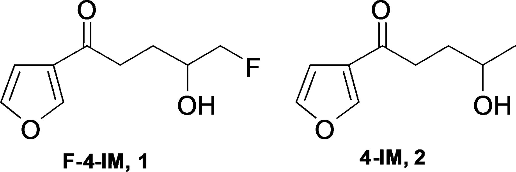
Structure of F-4-IM and 4-IM.
Materials and Methods
Reagents and equipment
Chemicals and reagents were purchased from Sigma-Aldrich and High-performance liquid chromatography (HPLC) solvents from Fisher Scientific. 1H and 13C NMR spectra were recorded on a Varian Gemini–200 or Varian Gemini–400 and chemical shifts were reported in parts per million (ppm, δ units). Electron impact (EI) and chemical ionization (CI) mass spectra were obtained on a GC/MS QP5050A spectrometer (Shimadzu). Fast atom bombardment (FAB) mass spectra were obtained on a JMS 700 (Jeol Ltd.). HPLC was carried out on a Waters Co. system with a semipreparative column (Alltech, Econosil silica gel, 10 μ, 10×250 mm), and simultaneously monitored by a Waters 2487 instrument (254 nm) and Raytest GABI γ-detector. Radio-TLC was performed on Merck F254 silica plates and analyzed on a Bioscan AC-3000 scanner. An automated gamma counter (1480 Wizard 3′′; Perkin Elmer Life Sciences) was used to measure radioactivity in the cell binding uptake and biodistribution studies. Total RNA Extraction Kit (Intron Biotechnology) was used for the preparation of RNA in cells. MTT cell growth and colorimetric cell growth assays were performed using an MTT assay kit (Sigma-Aldrich). The 2-aminoanthracene (2-AA) used in prodrug treatments was purchased from Sigma-Aldrich.
Synthesis
The preparation of target compound, 1-(3-furyl)-4-hydroxy-5-fluoro-1-pentanone (

(a) 1,3-propanedithiol, BF3
.Et2O, THF, N2, 0 to 70°C, 1.5 hours; (b) n-BuLi, THF, 4-bromo-1-butane, −78°C, 2.5 hours; (c) Cu(II)O, CuCl2, DMF, acetone, 60°C, 2 hours; (d) ethylene glycol, toluene, 120°C, 12 hours; (e) K3Fe(CN)6, K2CO3, K2OsO4·2H2O, (DHQ)2PHAL, t-BuOH:H2O (v/v=1:1), sodium sulfite, 0 to r.t., 2 hours; (f) TBDPSCl, imidazole, CH2Cl2, 0°C to r.t., 1 hour; (g) Dess-Martin oxidation, 0°C, 2 hours; (h) nBu4NF, THF, 0°C, 30 minutes; (i) MsCl, CH2Cl2, 0°C, 0.5 hours; (j) 1 M nBu4NF in THF, 100°C, 0.5 hours for
Structural parameters
2-(3-Furyl)-1,3-dithiane (4 )
To a solution of 1,3-propanedithiol (2.08 mL, 20.73 mmol) in dried tetrahydrofuran (30 mL), was carefully treated with borontrifluoride-diethyletherate (2.63 mL, 20.73 mmol) under a nitrogen atmosphere at 0°C. After 3-furaldehyde (1.79 mL, 18.84 mmol) was added, the reaction mixture was refluxed for 1 hour. The reaction was quenched by crashed-ice, and then saturated sodium bicarbonate was poured. The crude product was extracted by methylene chloride (20 mL×3), dried over anhydrous sodium sulfate. The product
2-(3-Furyl)-2-(3-butene)-1,3-dithiane (5 )
A solution of 2-(3-furyl)-1,3-dithiane (
1-(3-Furyl)-4-penten-1-one (6 )
To a stirred solution of 2-(3-Furyl)-2-(3-butene)-1,3-dithiane (
1-(3-Furyl)-4-penten-1-one, ethylene ketal (7 )
To a stirred solution of
1-(3-Furyl)-4,5-dihydroxy-1-pentanone, ethylene ketal (8 )
The solution of potassium ferricyanide (1.5 g, 4.57 mmol), potassium carbonate (632 μL, 4.57 mmol), and (DHQ)2PHAL in 10 mL of t-BuOH:H2O (v/v 1:1) was stirred for 10 minutes, and then added potassium osmate dihydrate (5.6 mg, 1 mol%), methanesulfonamide (244 mg, 1.52 mmol), and
1-(3-Furyl)-4-hydroxy-5-(tert-Butyl-diphenyl-silanyloxy)-1-pentanone, ethylene ketal (9 )
To a stirred solution of
1-(3-Furyl)-5-(tert-Butyl-diphenyl-silanyloxy)-pentan-1,4-dione, ethylene ketal (10 )
To a solution of
1-(3-Furyl)-5-hydroxy-pentan-1,4-dione, ethylene ketal (11 )
To a stirred solution of
1-(3-Furyl)-5-(methanesulfonyloxy)-pentan-1,4-dione,ethylene ketal (12 )
To a stirred solution of
1-(3-Furyl)-5-fluoro-pentan-1,4-dione, ethylene ketal (13 )
To a stirred solution of
1-(3-Furyl)-4-hydroxy-5-fluoro-1-pentanone, ethylene ketal (14 )
To a stirred solution of
1-(3-Furyl)-4-hydroxy-5-fluoro-1-pentanone (1 )
To a stirred solution of
Fluorine-18 labeling procedure for [18F]1
[18F]Fluoride was produced in a MC-50 cyclotron by the
18
O(p,n)18F reaction. A volume of 100–200 μL [18F]fluoride (8.0–17.0 GBq) in water was added to a vacutainer containing n-Bu4NHCO3 (40% aq., 2.70 μL, 3.65 μmol). The azeotropic distillations were carried out each time with 300 μL aliquots of CH3CN at 85°C under a stream of nitrogen. A fluorine-18 ion displacement reaction of
In vitro stability assessment
The stability of [18F]
Cells
Rat C6-glioma cells (C6) were purchased from the American Type Culture Collection (ATCC). The cells were cultured in Dulbecco's Modified Eagle's Medium (DMEM) containing high glucose (WelGENE), supplemented with 10% heat-inactivated fetal bovine serum (FBS; GIBCO) and antibiotics (100 units/mL penicillin G and 10 μg/mL streptomycin; GIBCO) at 37°C in a humidified 5% CO2 atmosphere.
Construction of CYP4B1 retroviral expression vector
cDNA for CYP4B1 was obtained from rabbit lung tissue and cloned into the pcDNA3.1/Hygro vector (Invitrogen) generating pcDNA-CYP4B1. 20 The nucleotide sequence for rabbit CYP4B1 was referred to GenBank No. NM_001082103.1. To construct the retroviral expression vector, the expression of CYP4B1 gene was introduced downstream of the long terminal repeats in the MFG vector backbone (kindly provided by Dr. Kwon, KIRAMS, Republic of Korea) by using NotI/BamHI and named as pMFG-CYP4B1.
Establishment of C6-CYP4B1 using the retroviral vector
The pMFG-CYP4B1 was introduced into H29D retrovirus packaging cell culture. The retroviral titers were measured between 1×10⁵ and 5×10⁵ infection units/mL. C6 were seeded in a six-well plate at a concentration of 3×10⁵ cells/well 1 day before retrovirus infection and infected by exposing the cell monolayer to 700 μL of retroviral vector in 300 μL of serum free media for 4 hours in the presence of 8 μg/mL of polybrene (Sigma-Aldrich). The transfected C6 cells were plated at a density of 1 cell/well and kept for 10 days. These stably transfected cells were selected with CYP4B1 expression by reverse transcription polymerase chain reaction (RT-PCR).
Evaluation of CYP4B1 expression in C6-CYP4B1
Reverse transcription polymerase chain reaction
Total RNA extraction was performed from 5×10⁵ cells of C6-CYP4B1 by using Easy-spin™ (DNA free) Total RNA Extraction Kit (iNtRON Biotechnology), according to the manufacturer's protocol. The total RNA was used as a template to produce cDNA using the SuperScript III First-Strand Synthesis kit (Invitrogen), according to the manufacturer's protocol. The resulting cDNAs were amplified using 2.5 units of Taq polymerase and 10 pmol of each primer (Table 1) in a GeneAmp PCR System (Applied Biosystems). The PCR conditions were as follows: for 35 cycles of denaturation (95°C for 30 seconds), annealing at 50°C for 30 seconds, and extension at 72°C for 30 seconds. The final extension was performed at 72°C for 7 minutes. Amplification products were confirmed by ethidium bromide staining after products were subjected to electrophoresis on a 2% agarose gel.
Prodrug activation cytotoxicity
MTT assay
C6 and C6-CYP4B1 were seeded in 96-well plates in triplicate at a density of 2×10³ cells/well. After 24 hours, cell culture media containing various concentrations of 2-aminoanthracene (2-AA; Sigma-Aldrich) ranging from 0 to 0.1 mM were added and incubated for 96 hours. The number of surviving cells was determined using the MTT assay (In vitro Toxicology Assay Kit MTT based; Sigma-Aldrich). Next, 10 μL of MTT reagent was added to each well and incubated for 2 hours at 37°C. When a purple precipitate was clearly visible under the microscope, 100 μL of Detergent Reagent was added to all wells, and the plate was placed in the dark at room temperature for 30 minutes. MTT color development was measured and analyzed using an ELISA reader (Bio-Rad) with a single filter (595 nm).
In vivo biodistribution in normal rats
Fisher 344 rats (180–200 g body weight, n=5) were obtained from Charles River laboratory and handled in accordance with the guidelines of the Korea Institute of Radiological and Medical Sciences. The animals were anesthetized with ethyl ether before injection of radiotracer and remained anesthetized through the study. Rats were injected with 7.4 MBq of [18F]
Comparative cellular uptake of [18F]F-4-IM and [3H]4-IM
C6 and C6-CYP4B1 were seeded in six-well plates in triplicate at a density of 1×10⁶ or 5×10⁵ cells/well 1 day before cellular uptake of [18F]F-4-IM or [3H]4-IM. After 24 hours, cells were incubated with [18F]F-4-IM (0.037 MBq) or [3H]4-IM (0.037 MBq; Moravek Biochemicals and Radiochmicals) with 10 μM of 4-IM, at a concentration of 1–4 μCi/well for an additional 0, 10, 30, 60, 180, and 360 minutes. After incubation, the supernatants were removed; cells were immediately rinsed with cold phosphate-buffered saline (PBS), and stripped with 1% trypsin-EDTA or cells were lysis with 0.2% SDS. [18F]F-4-IM-trapped cells were harvested in plastic test tubes by centrifugation at 1,000 rpm for 3 minutes. The supernatant was removed, and cell pellets were washed twice with cold PBS. [3H]4-IM-trapped lysates were harvested in plastic counter tubes and 10 mL of liquid scintillation cocktail (ULTIMA GOLD; PerkinElmer, Inc.) was added, followed by vortexing at 4°C. The CYP4B1 expression level was determined by gamma counting or beta counting of [18F]F-4-IM or [3H]4-IM uptake in cells for 1 minute, respectively. The level of binding of [18F]F-4-IM and [3H]4-IM was expressed as the percentage of binding to C6-CYP4B1 relative to that of C6.
Statistical analysis
Comparisons between groups were performed using IBM SPSS Statistics 19 software. Data were analyzed using the two-way factorial analysis of variance (ANOVA) followed by Bonferroni post-hoc tests with a p-value less than 0.05 were considered as significant.
Results
Chemistry
Using 1,3-propandithiol as a protecting group of aldehyde in 3-furnaldehyde (
Radiolabeling
The process for preparation of 1-(3-furyl)-4-hydroxy-5-[18F]fluoro-1-pentanone ([18F]F-4-IM, [18F]
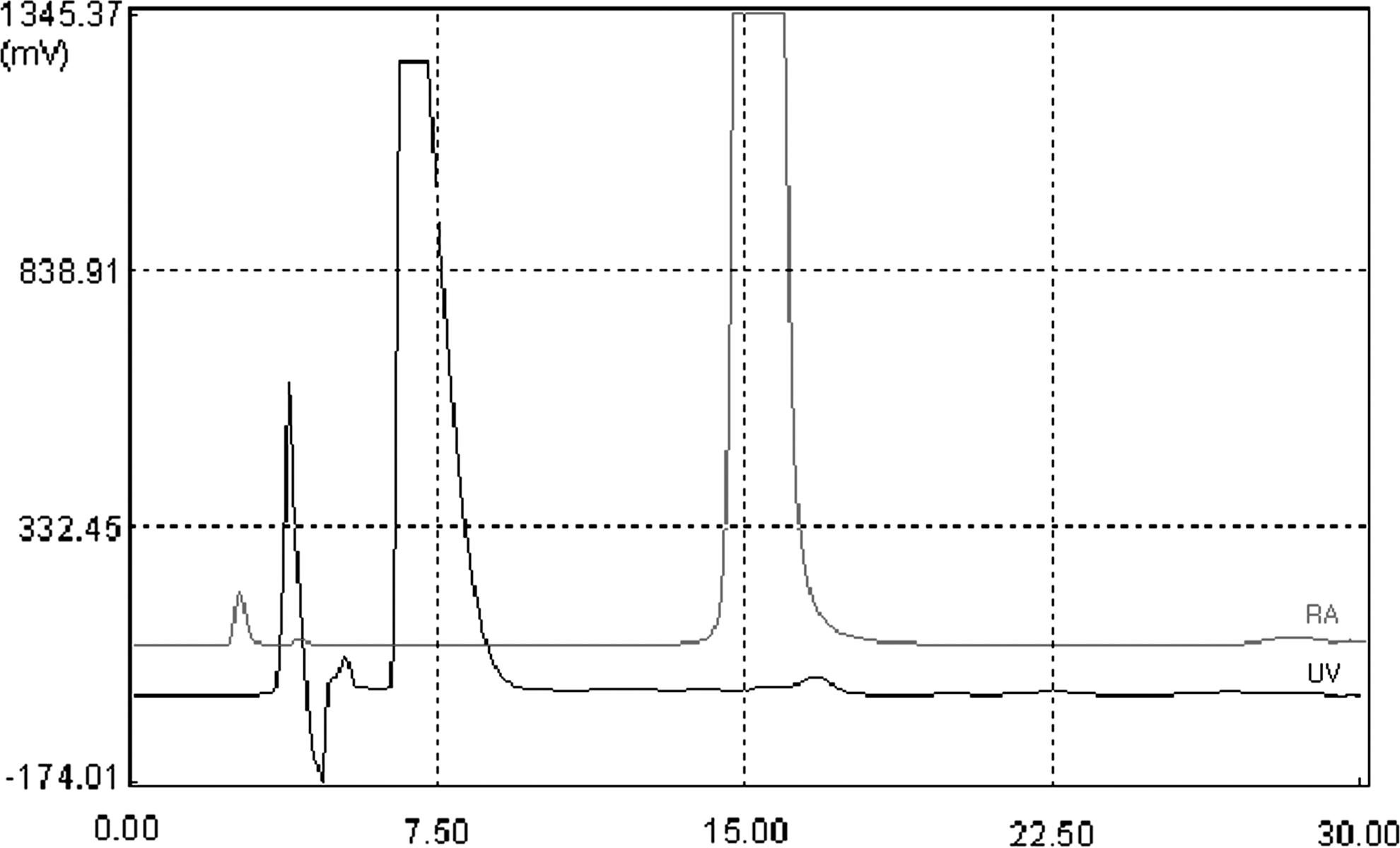
High-performance liquid chromatography (HPLC) profile of the reaction mixture.
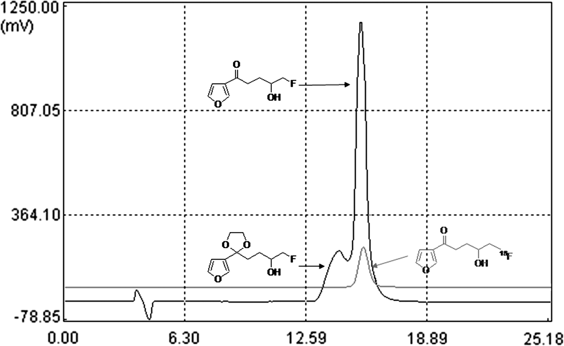
HPLC profile of the collected [18F]
Stability of [18F]F-4-IM ([18F]1 ) in human serum
The results of [18F]F-4-IM analysis in human serum showed that the intact has over 96% stability for up to 120 minutes (Fig. 4). This indicated that [18F]
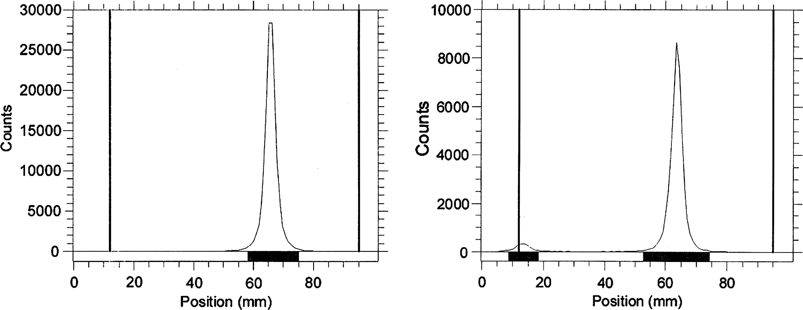
Radio-TLC chromatogram of stability in human serum (left: 0 min, right: 120 min).
In vivo studies
The biodistribution data of radioactivity of [18F]F-4-IM bound to tissues in normal rats are summarized in Table 2. The data are presented as percentage of injected dose per gram of tissues (%ID/g). After 1 hour postinjection, tissue distribution studies showed the uptake of [18F]F-4-IM to be highest in the lung (%ID/g=25.83%±2.90%), followed by the liver (%ID/g=6.43%±0.87%). The initial level of tissues in gastrointestinal and urinary system was high and decreased in a time-dependent manner. An exception was in the lung, in which the tracer was retained for the duration of the experimental. In addition, bone uptake (%ID/g=1.73%±0.17% at 1 hour) was low and similar to those of other tissues, such as muscle, heart, stomach, and blood. PET images in normal rat also showed that [18F]F-4-IM uptake was high in liver, lung, and kidney but radioactivity of other tissues was relatively low (Fig. 5).
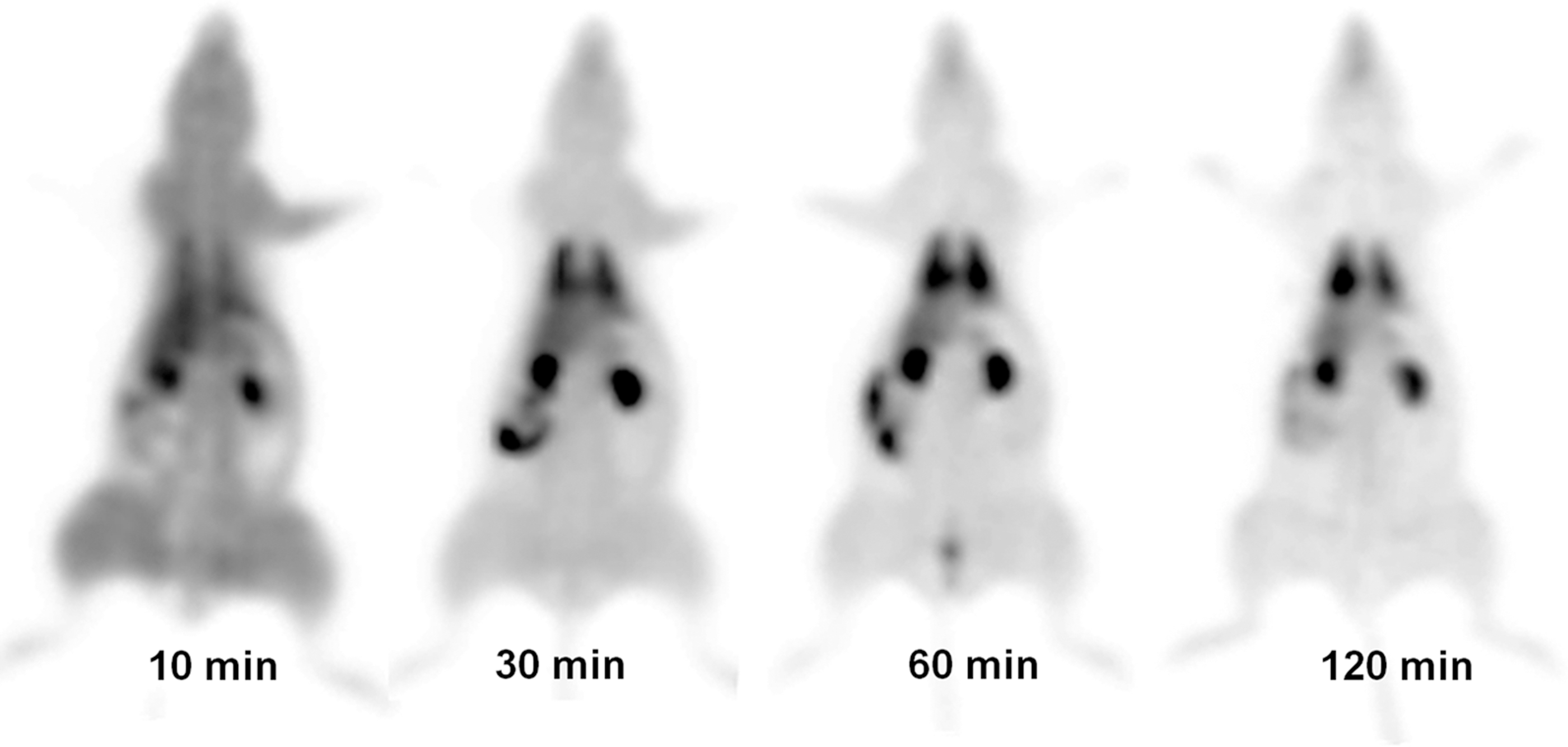
PET images of [18F]F-4-IM in normal rat.
Confirmation of CYP4B1 gene expression
To investigate the gene expression of the CYP4B1, we performed RT-PCR. CYP4B1-specific primers were designed to produces PCR products of 650-bp (540-bp of human and murine beta-actin). A specific band was detected in C6-CYP4B1 but not in parental cells (Fig. 6). This indicated that the CYP4B1 gene is expressed following retroviral transduction.
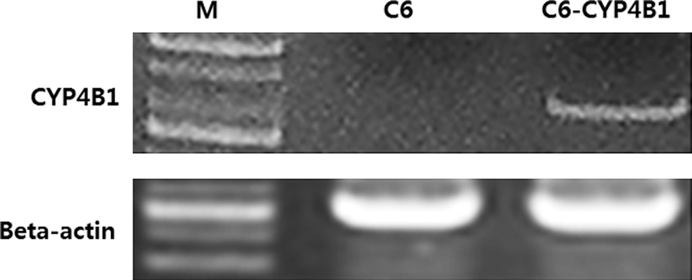
Confirmation of CYP4B1 gene expression in C6-CYP4B1.
Comparison of prodrug activation therapy between C6 and C6-CYP4B1
To evaluate the therapeutic efficacy of prodrug targeting CYP4B1, the survival rates of C6 and C6-CYP4B1 treated with 2-AA were determined using an MTT assay. As shown in Figure 7, the survival rate of C6-CYP4B1 was lower compared with C6 in a dose-dependent manner. Specifically, the 50% lethal dose (LD50) of C6-CYP4B1 was 0.03 mM following 96 hours treatment with 2-AA. In two-way factorial ANOVA test, a significant difference showed in all group-specific and concentration differences (with F-value 2028.162, 477.024, and p<0.005). Interaction effect of group and concentration was significant difference (with F-value 92.403 and p<0.005).
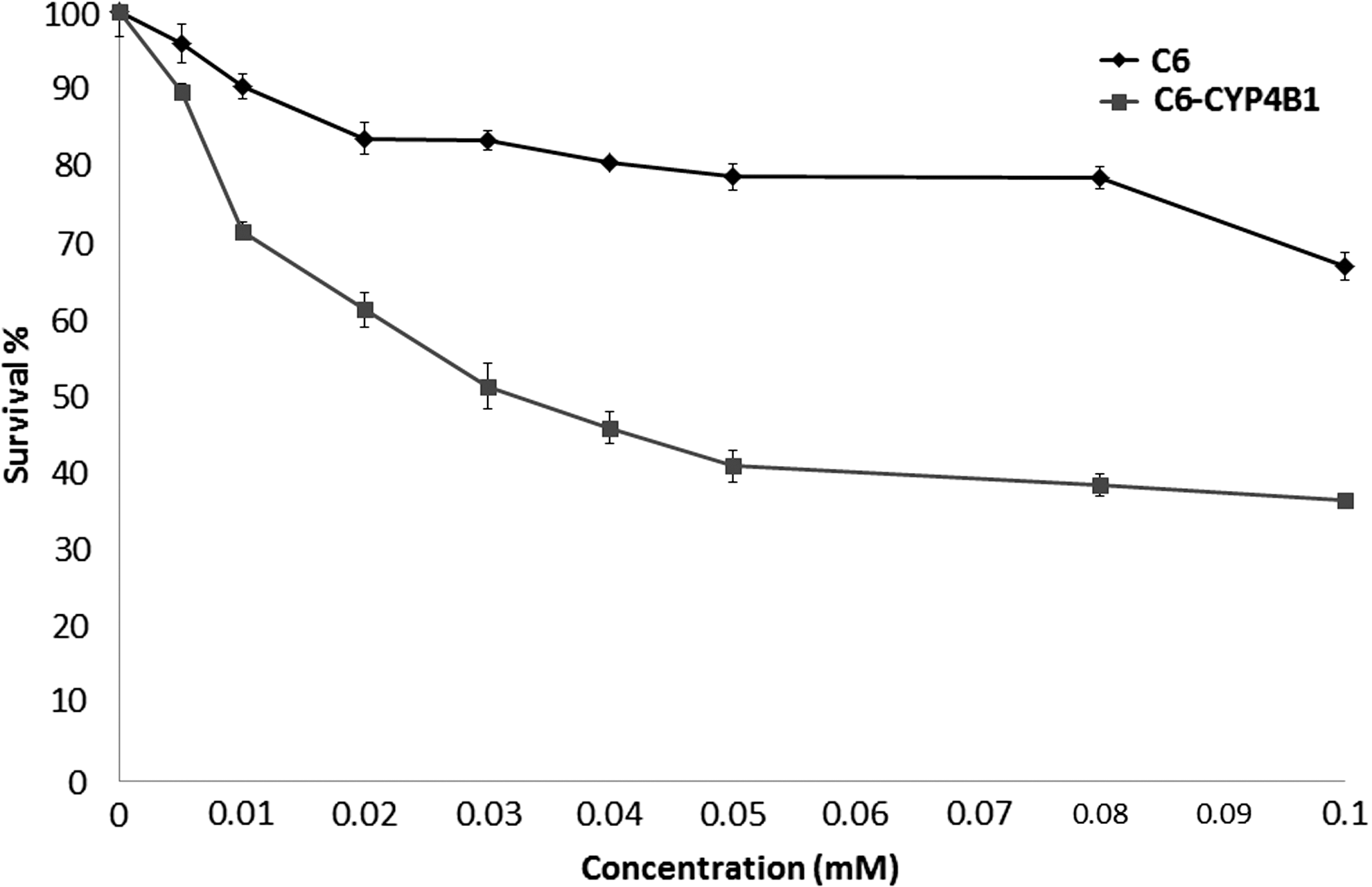
Prodrug activation therapy as measured by the MTT assay. p<0.001 (>0.01 mM).
Measurement of CYP4B1 activity by [18F]F-4-IM or [3H]4-IM
To evaluate the sensitivity of radiolabeled 4-IM analogs for GPAT, a cellular uptake assay was carried out using C6 and C6-CYP4B1 in a paired format to compare [18F]F-4-IM and [3H]-4-IM. In the in vitro cell uptake study, [18F]F-4-IM and [3H]4-IM showed comparable results as shown in Figure 8. In C6-CYP4B1, the increase in uptake of both compounds was proportional to the incubation time. However, the specific uptake of [18F]F-4-IM in C6-CYP4B1 was about 2.8-times higher than that in C6 at 1 hour (p<0.01), whereas it required up to 3 hours for [3H]4-IM to achieve a similar effect (p<0.001). In two-way factorial ANOVA test, a significant difference showed in all group-specific and time differences (with F-value 128.394, 49.769, and p<0.005 for [18F]-4-IM; F-value 441.110, 149.292, and p<0.005 for [3H]-4-IM). Interaction effect of group and time was significant difference (with F-value 50.502 and p<0.005 for [18F]-4-IM; F-value 151.166 and p<0.005 for [3H]-4-IM).
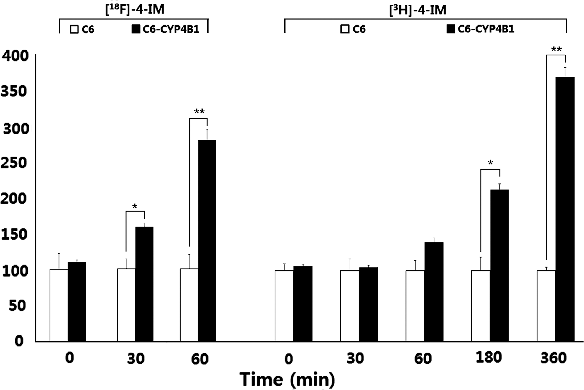
In vitro cellular uptake of [18F]F-4-IM (left) or [3H]4-IM (right) in C6 and C6-CYP4B1 cells. *p<0.01, **p<0.001.
Discussion
In this study, a 4-IM derivative was designed for imaging of CYP4B1-transfected tumor cells and investigated as a potential ligand for fluorine-18 labeling. The successful GPAT study depends on effective transfer and expression of the desired genes into the target cell and tissues. Among viral and nonviral techniques, the simplest way of therapeutic transgene delivery is injecting naked DNA but this method is relatively less efficient than those of viral methods. 21 Viral techniques use various classes of viruses as a tool for gene delivery by genetic modification of retroviruses, adenoviruses, herpesviruses, and others. Because the best established vector system for achieving stable transduction due to integration into the target cells is retrovirus vector, we selected retroviral system as CYP4B1 gene delivery. As examples of the concept for GPAT, stably expressing clones of 9L and U87 glioma cells with the CYP4B1 plasmid DNA were sensitized about 20-fold to 4-IM with an efficient bystander effect. 19
The radiolabeling method we have developed here lay the groundwork for the synthesis of the fluorine substituted 4-IM and fluorine radiolabeled form. As shown in Scheme 1, we prepared the mesylated precursor (
[18F]4-IM was synthesized via the same route as the preparation of F-4-IM. Various conditions were explored for the preparation of [18F]4-IM from the mesylated precursor. The results from reaction conditions optimized to give [18F]4-IM in 20%–35% radiochemical yield (decay corrected for 120 minutes): first step–nBu4N[18F]F in acetonitrile at 100°C for 10 minutes, second step–NaBH4 in ethanol at room temperature for 10 minutes, third step–PPTS in a mixture of acetone-water at 100°C for 10 minutes. The reaction mixture was then purified by HPLC (Econosil silica gel; 10 μ; 10×250 mm), and the retention time of [18F]4-IM was 15 minutes using a 36:65 mixture of ethyl acetate:hexane as the eluants (Fig. 2). As shown Figure 3, the collected HPLC fraction was analyzed by HPLC. This was identified as the fluorine-18 labeled 4-IM by comparison with authentic samples that we had prepared (cf., Scheme 1).
Percentage of the remaining [18F]F-4-IM was about 98% after 1 hour and over 96% even after 2 hours when the radiotracer was incubated with human serum at 37°C and analyzed by radio-TLC, indicating high in vitro stability.
In biodistribution studies, the radioactivity of [18F]F-4-IM was greater in the lung, in which CYP4B1 is preferentially expressed, than in all other organs at all time points. The uptake of [18F]F-4-IM shows a similar biodistribution pattern as that of [3H]4-IM. 22 In addition, Low levels of fluoride incorporation into bone implied that the injected compounds remained largely intact throughout the course of the experiment and is consistent with the results of the in vitro serum stability study. The gene expression of CYP4B1 in C6-CYP4B1 was confirmed with RT-PCR (Fig. 6). Using this C6-CYP4B1, we measured the cell survival in the presence of 2-AA with MTT assay. As shown in Figure 7, the survival rates of C6-CYP4B1 was more rapidly decreased with increasing 2-AA dose compared with C6. This result indicated that the therapeutic efficacy of 2-AA is dependent on CYP4B1 expression, and that the metabolic conversion of this prodrug under these conditions resulted in enhanced tumor cell death.
To evaluate the sensitivity of radiolabeled 4-IM analogs to the CYP4B1 expression, a cellular uptake assay was performed using [18F]F-4-IM and [3H]4-IM. Levels of [18F]F-4-IM in C6-CYP4B1 was rapidly increased about 2.8-fold, compared to that in C6 at 1 hour. [3H]4-IM, on the other hand, had low levels of accumulation in C6-CYP4B1 at 1 hour, and then reached the similar level at 3 hours. Despite the same electron configuration of the fluorine and hydrogen atom, cell uptake of two radiolabeled 4-IM analogs may not be the same in our comparison studies because of their different electronegativity. Therefore, 18F]F-4-IM selectively bound to the CYP4B1 protein and could be used to identify transfected cells within a short period of time (∼1 hour) postinjection.
Conclusion
In conclusion, F-4-IM (
Footnotes
Acknowledgments
This study was supported by grants from the National Research Foundation of Korea (20120006392, 2012014020, 2012013480, 20120004883 (BAERI), 2012K001119 (Brain Research Center of the 21st Century Frontier Research Program)) funded by the Ministry of Education, Science and Technology of Republic of Korea.
Disclosure Statement
No competing financial interests exist.