
Abstract
Objective:
To study the expression and regulatory effects of CD146 protein in colorectal cancer and the correlation between CD146 protein expression and the prognosis of colorectal cancer.
Materials and Methods:
The CD146 protein level was detected by immunohistochemistry staining. The relationship between CD146 expression and clinicopathological parameters of colorectal cancer was determined.
Results:
It was observed that 216 (20.00%) of the 1080 cases positively expressed CD146 protein. Univariate analyses indicated that CD146 expression was related to histological grade, Duke's stage, and liver metastasis (p=0.001, 0.001, and 0.001, respectively). Spearman correlation analysis showed that CD146 expression has line correlation to histological grade, Duke's stage, and liver metastasis (p=0.02, 0.01 and 0.001, respectively). After multivariate analysis, Duke's stage and CD146 were related to liver metastasis (p=0.01 and 0.001, respectively). In the Cox regression test, histological grade, Duke's stage, and CD146 were detected as the independent prognostic factors (p=0.045, 0.01, and 0.001, respectively).
Conclusions:
CD146 protein may be a potential biomarker for the postoperative liver metastasis of colorectal cancer.
Introduction
Although cancer is a multifactorial disease, most of the tumors have similar molecular and cellular mechanisms. 1 Although screening, early diagnosis, and curative operation have significantly increased the survival rates, recurrence and metastasis remain a serious challenge for patients with colorectal cancer. 2,3 In recent years, many researchers have studied the relationship between CD146 and tumor development. 4,5
CD146, also known as melanoma cell adhesion molecule (MCAM) or cell surface glycoprotein MUC18, is a 113-kDa cell adhesion molecule currently used as a marker for the endothelial cell lineage. In humans, the CD146 protein is encoded by the MCAM gene. 6 CD146 plays a critical promigratory role in the vascular system, normal development, and tumor progression patterning. 7 It was reported that CD146 has been discovered in many cancers, including melanoma, prostate cancer, epithelial ovarian cancer, and breast cancer, and is associated with tumor progression. 8,9 However, the underlying mechanisms of CD146 involved in cancer progression are still unclear. 10
In 2012, Liu et al. showed that CD146 expression correlated positively with lymph node involvement and a poor prognosis, and remained an independent prognostic factor for patients with gastric cancer. 5 These findings suggest that CD146 might promote epithelial-mesenchymal transition (EMT) and progression in gastric cancer, and thus may be a potential therapeutic target for patients with gastric cancers. While no report has investigated, the expression status and clinical implication of CD 146 in colorectal cancer, the relationship between the CD146 expression and biological behavior, and prognosis of colorectal cancer are still unclear. In the present study, we investigate the clinical implications of CD146 protein in colorectal cancer to lay a foundation for the management of colorectal cancer.
Materials and Methods
Patients and tissue specimens
The sample consists of 1080 patients who were histologically confirmed with colorectal cancer, and underwent radical operations in the First Affiliated Hospital of the Liaoning Medical University and Liaoning Tumor Hospital between January 2001 and January 2007. The mean age of the enrolled patients was 50.82±7.65 (mean±SD). The inclusion criteria were as follows: (1) curative operations were performed; (2) resected specimens were pathologically examined; (3) more than 10 lymph nodes were pathologically examined after operation; and (4) Duke's D-stage caners were excluded. The Ethics Committee of Liaoning Medical University and Liaoning Tumor Hospital approved this study's protocol.
Thin slices of tumor tissue of all cases received in our histopathology unit were fixed in 4% formaldehyde solution (pH 7.0) for periods not exceeding 24 hours. The tissues were processed routinely for paraffin embedding, and 4-μm-thick sections were cut and placed on glass slides coated with 3-aminopropyl triethoxysilane for immunohistochemistry. Tissue samples were stained with hematoxylin and eosin to determine the histological type and grade of tumors.
Immunohistochemical analysis
Briefly, immunohistochemical staining was performed using the standard streptavidin–peroxidase method with the UltraSensitive TM S-P Kit (Maixin-Bio) according to the manufacturer's instructions, and the signals were visualized using the 3,3′-diaminobenzidine (DAB) substrate, which stains the target protein yellow. Briefly, one paraffin-embedded block of the tissue was cut at 4 μm and placed on poly-
CD146 expression was classified semiquantitatively according to the following criteria: 0 if <1% of neoplastic cells discretely expressed CD146 in their cell cytoplasmic and membranous; 1+ if ≥1 and <10% of morphologically unequivocal neoplastic cells discretely expressed CD146 in their cell cytoplasmic and membranous; and 2+ if ≥10% of morphologically unequivocal neoplastic cells discretely expressed CD146 in their cell cytoplasmic and membranous. Samples scored as 1+ or 2+ were considered positive.
Statistical analysis
All data were analyzed with SPSS statistics software (Version 13.0). Relationships between tumor markers and other parameters were studied using chi-square test, Fisher's extract test, or independent t-tests. Disease-specific survival was analyzed using the Kaplan–Meier method. The log-rank test was used to analyze survival differences. Multivariate analysis was performed using the Cox proportional hazard model selected in forward stepwise. A p-value of <0.05 was considered statistically significant.
Results
The expression of CD146 in colorectal cancer and the relationship between CD146 expression and clinicopathological characteristics
Immunohistochemical examination showed that CD146 was located in the cytoplasm and the membrane of colorectal cancers, and CD146 was higher expressed in the colorectal cancers compared to the adjacent nontumor tissues (Fig. 1). In total, 216 (20.00%) of the 1080 cases were positively expressed CD146.
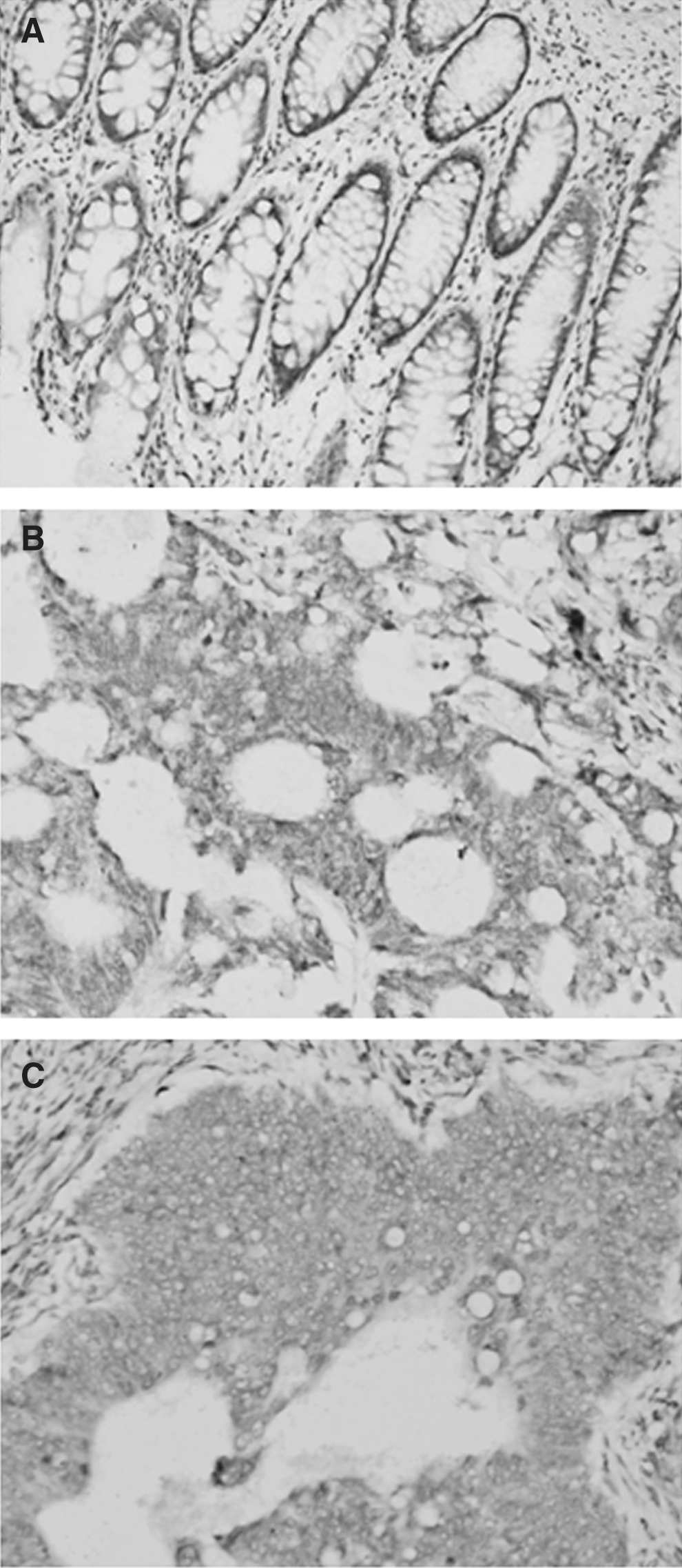
CD146 was highly expressed in human adjacent-tumor tissue
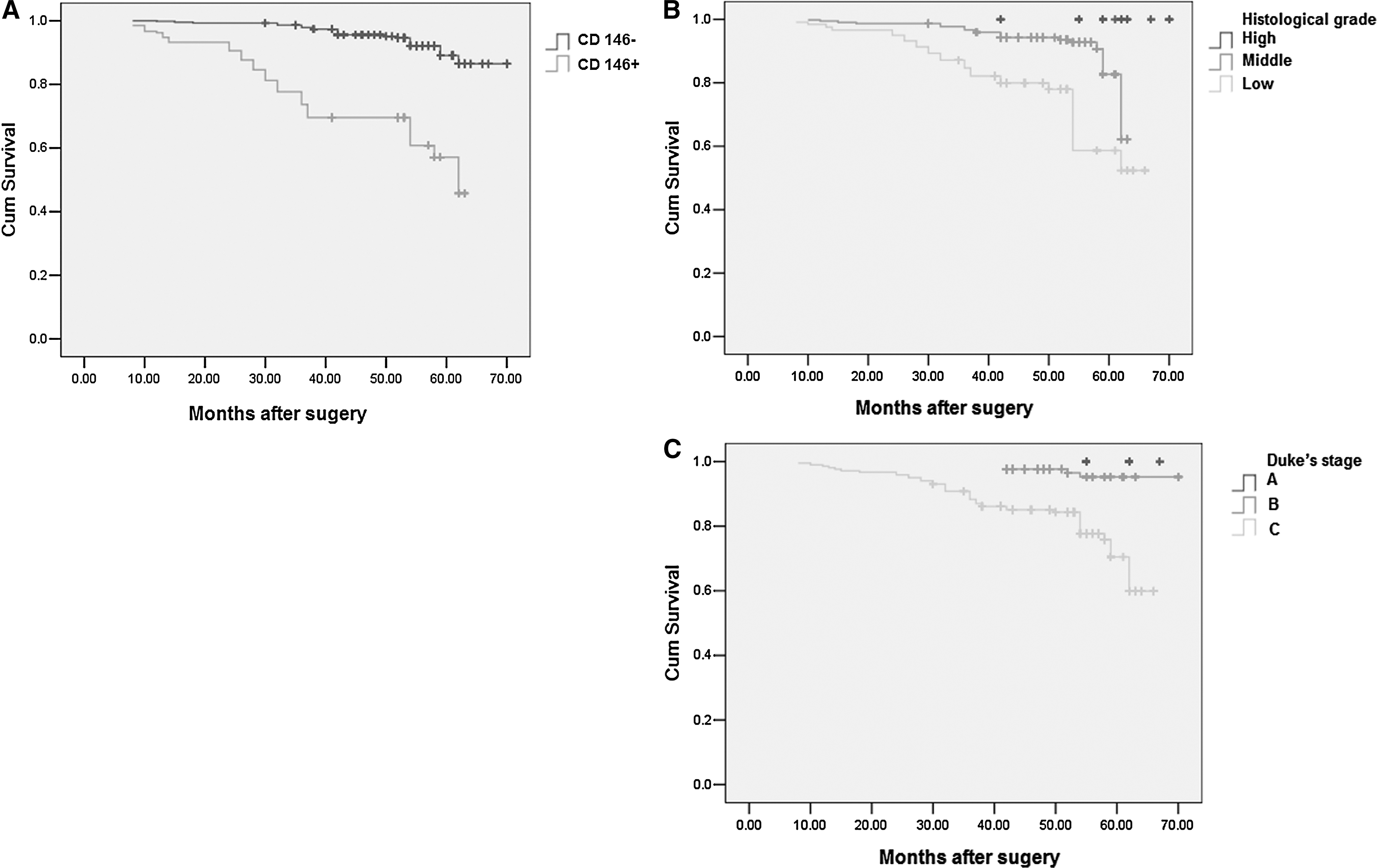
Univariate analyses indicated that CD146 expression was related to histological grade, Duke's stage, and liver metastasis. In the Cox regression test, histological grade, Duke's stage, and CD146 were detected as the independent prognostic factors (p=0.045, 0.01, and 0.001, respectively) (Table 1).
Spearman correlation analysis showed that CD146 expression has line correlation to histological grade, Duke's stage, and liver metastasis (p=0.02, 0.01, and 0.001, respectively) (Table 2). After multivariate analysis, Duke's stage and CD146 were related to liver metastasis (p=0.01 and 0.001, respectively) (Table 3).
OR, odd ratio; CI, confidence interval.
Prognostic analysis
Survival analysis revealed that the cases with positive CD146 expression exhibited a significantly higher postoperative liver metastasis rate compared to those without CD146 expression (39.22% vs. 18.00%, p=0.001). Survival analysis revealed that the cases with CD146 expression exhibited a worse postoperative survival than did those without CD146 expression (p=0.001) (Fig. 2). Moreover, histological grade and Duke's stage were associated with colorectal cancer-specific survival in all 1080 cases (p=0.01 and 0.001; log-rank test, Fig. 2). In the Cox regression test, histological grade, Duke's stage, and CD146 were detected as the independent prognostic factors (p=0.045, 0.01, and 0.001, respectively) (Table 4).
Survival analysis revealed that the cases with CD146
Discussion
CD146, a cell–cell or cell–matrix adhesion molecule, was first described in colorectal cancer. 11 CD 146 is distributed throughout normal and benign proliferative mammary ductal epithelium, but it is frequently lost in carcinomas; it functions as a heterophilic cell–cell adhesion molecule in epithelium, and loss of Mel-CAM expression in breast carcinoma may be an important step for tumor progression. 11 –13 However, accumulating evidence supports that CD146 acts as a metastatic factor. 12 CD146 was reported to be expressed on a subset of epithelial cells in the malignant breast. Changes in the molecular signatures after downmodulation of CD146 expression suggest that CD146 downmodulation is associated with the reversal of several biological characteristics associated with the epithelial-to-mesenchymal transition, and the phenomenon associated with the metastatic process. 13 In another study, Wu et al. CD146 is an important determinant in increasing metastasis of human prostate cancer LNCaP cells to distant organs in a nude mouse model. 14 More recent studies have associated the high expression of CD146 with metastatic progression in various cancers. 8 –10 However, there are still few studies investigating the expression status of CD 146 in colorectal cancer. In the present study, we investigate the clinical implications of CD146 protein in 1080 colorectal cancer to lay a foundation for the management of colorectal cancer.
We observed that 20.00% of the enrolled cases positively expressed CD146 protein. CD146 expression was related and has line correlation to histological grade, Duke's stage, and liver metastasis. After multivariate analysis, Duke's stage and CD146 were related to liver metastasis. Hence, CD146 might be a new potential predictor of liver metastasis of colorectal cancer and a new potential target in the management of colorectal cancer, though there is still no evidence in colorectal cancer. In the Cox regression test, histological grade, Duke's stage, and CD146 were identified as independent prognostic factors.
Colorectal cancer affects more than 150,000 people a year and is responsible for the death of about 50,000 people a year. 15 The most common site of metastatic disease seen in patients with colorectal cancer is the liver. The effective therapy for advanced colorectal cancers with liver metastases is still controversial. 16,17 Therefore, early screening of the distant metastasis in colorectal cancer is very important. The outcome of the study demonstrated that CD 146 protein was related to the liver metastasis of colorectal cancer, which may be used as a potential early liver metastasis-screening factor for colorectal cancer.
Authors' Contributions
Study conception and design: Buxian Tian and Yuhong Zhang
Acquisition of data: Buxian Tian, Yuhong Zhang, and Nan Li
Analysis and interpretation of data: Buxian Tian
Writing manuscript: Buxian Tian, Yuhong Zhang, and Nan Li
Footnotes
Disclosure Statement
No competing financial interests exist.