
Abstract
In this study, a microemulsion system was evaluated for delivery of mitomycin C (MMC). To track the distribution of the formulated drug after intravenous administration, radiochemical labeling and gamma scintigraphy imaging were used. The aim was to evaluate a microemulsion system for intravenous delivery of MMC and to compare its in vivo behavior with that of the MMC solution. For microemulsion formulation, soybean oil was used as the oil phase. Lecithin and Tween 80 were surfactants and ethanol was the cosurfactant. To understand the whole body localization of MMC-loaded microemulsion, MMC was labeled with radioactive technetium and gamma scintigraphy was applied for visualization of drug distribution. Radioactivity in the bladder 30 minutes after injection of the MMC solution was observed, according to static gamma camera images. This shows that urinary excretion of the latter starts very soon. On the other hand, no radioactivity appeared in the urinary bladder during the 90 minutes following the administration of MMC-loaded microemulsion. The unabated radioactivity in the liver during the experiment shows that the localization of microemulsion formulation in the liver is stable. In the light of the foregoing, it is suggested that this microemulsion formulation may be an appropriate carrier system for anticancer agents by intravenous delivery in hepatic cancer chemotherapy.
Introduction
Mitomycin C (MMC) is a potent antineoplastic antibiotic naturally synthesized by the fungus Streptomyces caespitosus. It is used as a single drug or in combination with other anticancer agents to treat malignancies such as colorectal and anal cancer, bladder cancer, cervix cancer, gastric cancer, lung cancer, and pancreas cancer. 1 MMC has been shown to be metabolized by xanthine oxidase and microsomal enzymes such as NADPH-cytochrome P450 and cytochrome b5, which are abundant in the liver, 2 –4 while approximately 10% of the drug is excreted in the urine. 5 It has the ability to bind and alkylate nucleic acids, but to show anticancer effect metabolic activation by reductive enzymes under hypoxic conditions is needed. 6,7 When activated, MMC forms crosslinks between neighboring guanine bases on the complementary DNA strands. 8 It also forms free radicals that contribute to the elimination of cancer cells 9 and induces apoptotic pathways in the cells. 10
One of the main drawbacks of MMC in clinical application is its very short biological half-life, only approximately 50 minutes. 5 Furthermore, systemic administration of this drug is associated with severe side effects, such as bone marrow depression, renal and gastrointestinal damage. 11 To overcome these side effects of MMC, and to obtain higher efficacy of the treatment, many attempts have been made to incorporate and deliver the drug in different delivery systems. These attempts include intratumoral injection of MMC-dextran conjugates, 12 arterial chemoembolization with polyvinylalcohol or degradable starch microspheres, 13,14 and incorporation of MMC in polylactic acid nanoparticles. 15
Microemulsions (M) are self-aggregated thermodynamically stabile systems in which oil and water are homogenously dispersed in each other with the help of a suitable surfactant or surfactant/cosurfactant blend. They differ from conventional emulsions by their ease of preparation without a requirement of energy input, and their much smaller structural size, also their thermodynamic stability makes these systems particularly interesting as they allow for a long-lived stabilization of mixed oil/water systems. 16 Their benefits as drug delivery systems have led to the development of long-term stable systems for parenteral administration. 17,18
The aim of this study was to evaluate an o/w microemulsion formulation as a carrier system for intravenous delivery of MMC and to compare its in vivo behavior with that of the MMC solution in rabbits.
Materials
Soybean oil was donated by Orkide Oils, Inc. MMC was purchased from Onko-Kocsel Co. Soybean lecithin (L) (90% phosphatidylcholine) was obtained from AppliChem GmbH, Tween® 80 (T80) originated from Merck-Co., ethanol (E) (assay>99.9%), methanol, and potassium dihydrogen phosphate from Merck KGaA, di-sodium hydrogen phosphate from Carlo Erba Reagenti, sodium chloride was from Atabay, and dialysis bag (Spectra/por® dialysis membrane, M.W. cut off 12,000–14,000, 32×104-mm flat width by flat length, volume length: 3.3 mL/cm) from Spectrum Laboratories, Inc.
99mTechnetium-pertechnetate (Na99mTcO4) was obtained from the Department of Nuclear Medicine of Ege University. Labeling was achieved in the presence of stannous chloride dihydrate (Merck-Schuchardt) as the reducing agent. Silica gel instant thin-layer chromatography sheets (ITLC-SG) were purchased from Pall Life Science. Cellulose acetate membrane filters with a 0.2-μm pore size were obtained from Sartorius to remove impurities of colloids and sterile filtration. Scintigraphic images were obtained by a gamma camera (Toshiba Digital GCA 602 A).
Methods
Preparation of oil-in-water microemulsion
For microemulsion formulation, soybean oil was used as the oil phase. Lecithin and Tween 80 at a ratio of 1:2 (S) were surfactants and ethanol was the cosurfactant (coS). Lecithin was dissolved in predetermined amount of ethanol by stirring with a vortex at 1000 rpm. Then, oil and Tween 80 were added and mixed. Finally, distilled water was added drop by drop until the turbidity was observed, while stirring with a magnetic stirrer (IKA Labortechnik). Ideal S/coS weight ratios and micromulsion existence areas were determined with the aid of phase diagrams drawn using a computer program developed in the computer center at the Ege University Faculty of Pharmacy. 19 The HLB value of the S/coS mixture was 11.33, which is suitable for O/W microemulsion formulation. In the final, MMC was added to the microemulsion formulation (1 mg/1g). 20 MMC-loaded microemulsion is abbreviated as MMC-M.
Preparation 99mTc labeled MMC solution and 99mTc labeled MMC-M
MMC was radiolabeled with radioactive technetium (99m Tc) at the Radiopharmacy Department of the Pharmacy Faculty of Ege University as follows: to a 5-mL vial, 1 mL of the MMC solution (1 mg MMC in 1 mL normal saline), 25 μL of freshly prepared solution of stannous chloride dihydrate (1 mg/mL) in 0.1N HCl, and 74 MBq (2 mCi) Na99mTcO4 were added. The mixture was vortexed for 30 seconds and filtered through a membrane filter (0.22 μm) directly into a sterile vial. The solution was incubated in a closed container for 10 minutes at room temperature. The radiochemical purity was evaluated by instant thin layer chromatography (ITLC).
99mTc labeled MMC-M was prepared as follows: 1 mL of radiolabeled MMC mixture was incorporated into 0.92 mg oily phase (surfactant and oil). Then, 0.095 mL distilled water was added and stirred with a magnetic stirrer.
Radiochemical analysis
Radiochemical analysis was performed on ITLC-SG strips with the acid citrate dextrose buffer as a mobile phase. The 99mTc labeled MMC remained at the point of spotting, whereas hydrolyzed 99mTc and free 99mTc impurities moved with the solvent front (Rf: 0.8–1.0).
Gamma camera imaging of in vivo MMC distribution
To understand the whole body localization of therapeutic dose of MMC-M, gamma scintigraphy was used. The in vivo study conducted was approved by the Ege University Local Ethique Committee (Protocol no: 2009/2). The 99mTc labeled MMC-M and 99mTc labeled MMC solution as control were evaluated in New Zealand white male rabbits (2 kg) by recording static images at different time points. Three rabbits were used to observe in vivo behavior of MMC-M and one as the control for MMC solution. The radiolabeled formulations were injected intravenously through the right marginal ear vein of each rabbit using volumes of 0.120 mL and activities of approximately 0.120 mCi. After delivery of formulations, static images were obtained by a gamma camera (Toshiba digital GCA 602 A) equipped with a low-energy general-purpose parallel-hole collimator, which was used for static image acquisition at 15th, 30th, 60th, 90th minutes. Energy windows were set at 140 keV (20% width).
Results and Discussion
Microemulsion preparation and its properties
The droplet size of microemulsions with and without MMC was determined as 57.67±3.16 nm and 34.65±5.86 nm, respectively. The pH values of MMC unloaded microemulsions were between 6.80–6.95. These pH and the viscosity value (32.4 cPs) were appropriate for i.v. application. Incorporation of MMC into the M system did not significantly affect the pH value of the microemulsion formulation. The addition of MMC slightly increases the electrical conductivity because of its amphiphylic character. 20 As shown in the previous data, the addition of an amphiphylic drug increases the conductivity value of a microemulsion system. 17,20,21
According to previous reported data on the hemolitic activity, microemulsion formulations are safe and appropriate for i.v. application. 22 –24 Previously, we showed that the microemulsion has the ability to reduce the hemolytic potential of T80, which is used as the hydrophilic surfactant for microemulsion production. Besides, cytotoxicity tests were carried out in Calu1 and A-549 cell lines with the microemulsion formulation. It was observed that, microemulsion showed no significant cytotoxic effect at all applied doses on Calu1 cells. For A549 cells, the viability was over 81% up to 210 μM dose. 20
Gamma camera imaging of in vivo behavior of MMC
The radiolabeling efficiency of 99mTc labeled MMC was found to be greater than 90%. Gamma camera images of the rabbits after the i.v. administration of 99mTc labeled MMC-M and 99mTc labeled MMC solution are shown in Fig. 1. Radioactivity in the bladder observed in 30 minutes after injection of the MMC solution shows that urinary excretion of the latter starts very rapidly (Fig. 1A). On the other hand, following administration of MMC-loaded microemulsion, no radioactivity appeared in the bladder during 90 minutes (Fig. 1B). The unabated radioactivity in the liver during the experiment shows that this formulation is stably localized in the liver.
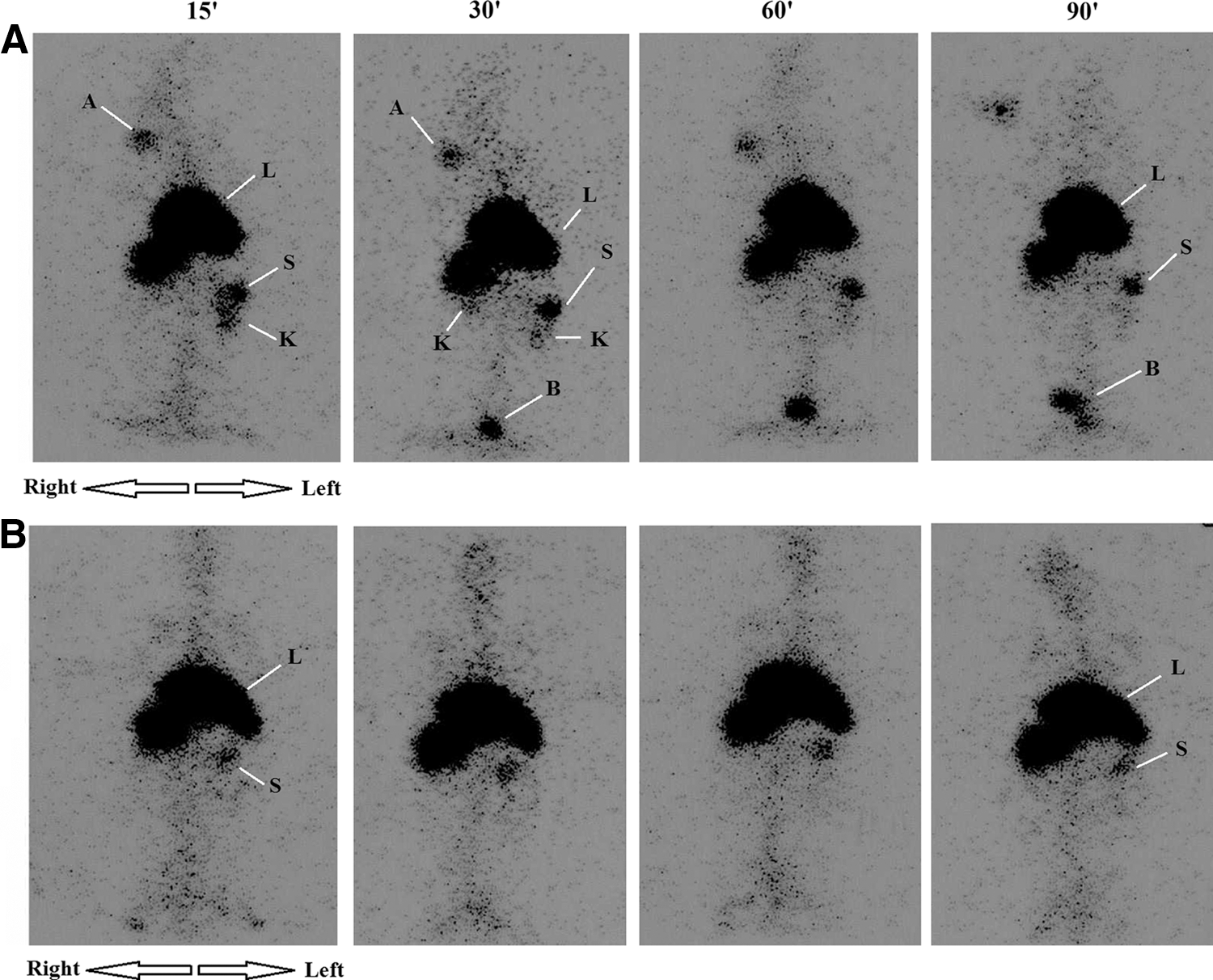
Gamma camera images of rabbits administered with radiolabeled drug.
Radiopharmaceuticals, which are used for imaging either the biliary tract or phagocytic cells also provide liver imaging. Due to the bile circulation, these radiopharmaceuticals usually appear in the intestine approximately in 1 hour after administration. In the present study, no activity was observed in the intestine during imaging experiments, only liver hepatocytes, which are of phagocytic character were observed.
Also, radioactivity is clearly seen in the liver in the images obtained after both the MMC solution and MMC-M, while later only the MMC solution is eliminated via urine. The liver is an organ composed of hepatocytes, which have phagocytic property for certain size of particles. Targeting of the liver by 99mTc labeled MMC-M is to be expected, considering that MMC-M is a colloidal carrier system containing about 50 nm lipid droplets. Following the administration of 99mTc labeled MMC solution, radioactivity was seen in the bladder alongside the liver. Although no visual change was observed in the system after radiolabeling, and also MMC was below its solubility maximum, it may have formed colloidal molecules with 99mTc in the solution. It could be interesting to see if such a formation took place, to explain the high radioactivity in the liver after MMC solution administration.
Another possible explanation for the high radioactivity in the liver after administration of the drug solution is that MMC has previously been shown to be metabolized by xanthine oxidase and microsomal enzymes such as NADPH-cytochrome P450 and cytochrome b5, which are abundant in the liver, 2 –4 while approximately 10% of the drug is excreted in the urine. 5 As to the octanol/water partition coefficient definition, MMC was determined as 76% in the water phase and 24% in the octanol phase. Also, it has been reported that MMC tends to be located at the lipid/water interface, thus causing the drug-loaded microemulsion droplets to be larger in size than the ones of unloaded microemulsion. 20 Even though the external phase in the MMC-M system is water, no excretion was seen via urine for 90 minutes after administration. This finding confirms that a majority of MMC in the system is located at the interface of the droplets. In conclusion, the microemulsion system ensures a longer drug presence in the liver.
It is definitely difficult to foresee the biological behavior of the drug in case of liver failure. Given that the solution of MMC may be excreted more actively via urine it may be preferable to treat with the MMC solution in cases of liver failure, considering that MMC-M may not be eliminated from the body. So, another set of experiments, including both healthy animals and animals with liver failure model could give valuable information about the elimination and the tolerability of the developed system.
Nevertheless, the results of gamma camera imaging and our previous in vitro release studies 20 indicate that the MMC-M system is more appropriate than the MMC solution to treat liver cancers because of its better targeting and its longer stable location in the target organ.
Conclusion
The radiolabeled MMC-loaded microemulsion system was observed to reach the liver without being eliminated via urine following administration, and to remain longer in the liver than the MMC solution. It is therefore proposed that the microemulsion formulation is a potentially appropriate carrier system for MMC to target the liver intravenously for therapeutic purposes.
Footnotes
Acknowledgment
This study was supported by the Scientific Research Fund of Ege University by project number 08.ECZ.018.
Disclosure Statement
No competing financial interests exist.