
Abstract
Doxorubicin (Dox) is widely used for the treatment of solid tumors but its clinical utility on glioma is limited. In this study, we developed a novel nano-scale drug delivery system employing biodegradable nanoparticle (NP) as carriers to load Dox. Transferrin (Tf) was conjugated to the surface of NP to specifically target the NP to glioma. Tf-NP-Dox was prepared via emulsification-solvent evaporation method, and characterized for the size, Drug loading capacity (DLC), entrapment efficiency, and Tf number on the surface. The antitumor efficiency in vitro was evaluated via CCK-8 assay. The transmembrane transportation was evaluated via HPLC assay. The antitumor efficiency in vivo was assessed in C6 glioma intracranial implant rat model. The average diameter of Tf-NP-Dox was 100 nm with ∼32 Tf molecules on the surface. DLC was 4.4%. CCK-8 assay demonstrated much stronger cytotoxicity of Tf-NP-Dox to C6 glioma cells compared to NP-Dox or Dox. HPLC assay showed that Tf-NP-Dox transported Dox into C6 cells with high efficiency. In vivo, Tf-NP-Dox could transport Dox into tumors compare to contralateral part, with tumor inhibitory ratio and survival higher than NP-Dox or Dox. Taken together, our results suggest that Tf-NP-Dox exhibits better therapeutic effects against glioma both in vitro and in vivo, and is a potential nano-scale drug delivery system for glioma chemotherapy.
Introduction
Glioma is a form of aggressive malignant brain tumors. Despite recent developments in invasive surgery, radiotherapy, and chemotherapy, median survival of patients with glioma has increased little during the past decades. The blood brain barrier (BBB) restricts the penetration of agents into glioma. 1 Furthermore, chemotherapy agents may damage surrounding tissues for the lack of targeting specificity. Such factors contributed to the poor prognosis of glioma. Nanoparticles (NPs) have been regarded as a potential alternative for glioma therapy, which enhanced therapeutic efficacy and reduced toxicity of many drugs. 2 –4 NPs based on diblock PEG-PLA copolymer have been extensively investigated for their potential as controlled and slow-releasing drug carriers with many advantages over liposomes, such as the low number of excipients used in the formulations, the simple procedures for preparation and high physical stability. 5 Furthermore, both PEG and PLA are materials approved by FDA; thus, endowing good safety.
An effective approach for targeting the NPs is to modify their surface with ligands, such as transferrin (Tf), which can be recognized by Tf receptor (TfR) that is highly expressed on the cell surface of glioma and brain capillary endothelium. 6,7 Doxorubicin (Dox), a well-known chemotherapy agent, is widely used for the treatment of breast, prostate, cervix, ovarian and lung cancers. Dox can hardly pass through the BBB, which limited its clinical utility on glioma. Therefore, in this study we developed a novel nano-scaled drug delivery system employing biodegradable NP as carriers to load Dox. Tf was conjugated to the surface of NP to specifically target the NP to glioma.
Materials and Methods
Preparation of NP and Tf-NP
MPEG-PLA (Sigma-Aldrich) and maleimide-polyethylenegly-col-poly (lactic acid) (maleimide-PEG-PLA; Sigma-Aldrich) diblock copolymers were synthesized by ring opening polymerization. Next, PEG–PLA NP were prepared using the double emulsion and solvent evaporation method. 8 Tf (Sigma-Aldrich) was thiolated by mixing for 60 minutes with a 40:1 molar excess of 2-iminothiolane in 0.15 M sodium borate buffer, pH8.0 supplemented with 0.1 mM EDTA. Tf-NP was prepared by incubating the purified thiolated Tf with the NP at room temperature for 9 hours. The products were then subjected to a 1.5×20 cm sepharose CL-4B column and eluted with 0.01 M phosphate buffered saline (PBS) buffer pH 7.4 to remove unconjugated proteins.
Preparation of Dox-encapsulated NP and Tf-NP
Blank NP and Tf-NP were prepared in the same way as described above. Dox was encapsulated via equilibrium dialysis method as described previously. 9 The hydration solution was citrate buffer (0.2 M, pH 4.0). After hydration and filtration, the pH of the vesicle dispersion was neutralized to 7.4 by Na2CO3 (1 M), and 1 mL of 1 mg/mL Dox solution (Beijing Huafeng United Technology) was added and mixed at room temperature for 24 hours. After ultrafiltration, unencapsulated Dox was eluted and the vesicle dispersion was concentrated to yield Dox-encapsulated NP (NP-Dox) or Dox-loaded Tf-NP (Tf-NP-Dox). All procedures were performed in the dark.
Characterization of NP-Dox and Tf-NP-Dox
The concentration of NP was determined by turbidimetry using UV2401 spectrophotometer at 350 nm (Shimadzu). For morphology observation, blank NP or Tf-NP was concentrated and the buffer was exchanged with saline via ultrafiltration to avoid the reaction of uranyl ions with citrate ions during negative staining. After staining with 1% urany lacetate solution, blank NP was observed by transmission electron microscopy (TEM) (H-600; Hitachi). NP size was tested by dynamic light scattering analysis via particle size analyzer (PSS.NICOMP PARTICLE SIZE SYSTEM). Drug loading capacity (DLC) and entrapment efficiency (EE) of Dox were determined by HPLC (Ex: 480 nm and Em: 580 nm) (waters1525). The average number of Tf on Tf-NP was calculated as follows: N=M/n, M was the number of Tf determined by using Tf ELISA kit (Bethyl), and n was the number of NP measured as described previously. 10 Finally, NP-Dox or Tf-NP-Dox was suspended in a dialysis system and Dox released into 0.01 M PBS (pH 4 and 7.4) was periodically taken out, and the supernatant was analyzed for the released Dox by HPLC assay.
In vitro cytotoxicity assay
C6 glioma cells were seeded into 96-well plates at a density of 1×10⁴ cells/well and cultured for 24 hours before exposure to different Dox formulations (Dox, NP-Dox or Tf-NP-Dox) for another 24 hours at 37°C. Five wells for untreated cells were prepared as controls. After exposure, the cytotoxicity of these formulations was evaluated by using CCK-8 kit (Yes Service Biotech, Inc.). Furthermore, cytotoxicity of blank NP and Tf-NP to C6 cells was also evaluated by CCK-8 assay. In competition assay, C6 glioma cells were seeded into 96-well plates and pretreated with excessive amount of Tf (50 μg) dissolved in 100 μL culture medium for 30 minutes, the medium was exchanged, and then the cells were treated with Dox, NP-Dox or Tf-NP-Dox, the cytotoxicity was evaluated by using CCK-8 kit. The experiments were performed in triplicate, dose-effect curves were made and IC50 values were calculated.
Uptake of Dox formulations by C6 glioma cells in vitro
C6 cells were seeded into 24-well plates at a density of 5×10⁵ cells/well and cultured for 24 hours before exposure to different Dox formulations (Dox, NP-Dox or Tf-NP-Dox, with the final Dox concentration at 4 μg/mL per well) for 1, 2, 4, 8, or 12 hours at 37°C. At each time point, cells in each well were washed twice with ice-cold PBS to remove surface-bound agents and cells were collected for HPLC detection of intracellular Dox, total cell protein was calculated by using BCA protein assay kit (Shenergy Biocolor Bioscience and Technology). The uptake index was expressed as Dox (μg)/cellular protein (mg) as described previously. 11,12
Biodistribution of Dox in the brain of glioma-bearing rats
The rats were anesthetized with 10% chloral hydrate (0.4 g/kg), and 1×10⁶ of C6 glioma cells were injected into right caudate nucleus on a stereotactic apparatus. Ten days after tumor intracranial implantation, nine rats were randomly divided into three groups in which free Dox, NP-Dox, and Tf-NP-Dox (dose of Dox 3 mg/kg) were injected intravenously, respectively. After 3 hours, the animals were sacrificed and the brain tissues were collected and washed by cold PBS to remove the surface blood. Left caudate nucleus (normal brain tissue) and right caudate nucleus (tumor tissue) were separated from the brain tissue. After homogenization, the concentration of Dox was analyzed by HPLC as reported previously. 12
Xenograft tumor model
Fifty-two glioma bearing rats were randomly divided into four groups (n=13), on the day 7th postintracranial implantation of C6 glioma cells. Animals in control group were administered with physiological saline. Animals in other three groups were treated with free Dox, NP-Dox and Tf-NP-Dox via tail vein at a dose of 1.5 mg/kg Dox, respectively. Administration was made every 2 days with total four doses per rat. On the 15th days postimplantation, three rats in each group were sacrificed and the brain tissue was collected and fixed in 4% formaldehyde solution overnight. The tumors were excised and the volume was calculated as follows: volume (mm3)=l (mm)×w (mm)×h (mm), where l was the major anteroposterior diameter, w was the major diameter from left to right, and h was the major diameter from up to down. The l, w, and h values were measured by a vernier caliper. The other 10 rats in each group were maintained to monitor the survival curves. Kaplan-Meier survival curves were plotted for each group. The tumor volume inhibitory ratio was calculated with the formula: Rv=(Vdrug/Vsaline)×100%, where Vdrug was glioma volume after treatment with free Dox, NP-Dox or Tf-NP-Dox, and Vsaline was glioma volume after treatment with saline. 13
Statistical analysis
Statistical analysis was performed using Student's t-test. Survival data were presented using Kaplan-Meier plots and analyzed using a log-rank test. The differences were considered significant for p<0.05.
Results
Characterization of NPs
TEM micrographs showed that the NP vesicles were generally round with a diameter around 100 nm (Fig. 1A). The intensity-based vesicle size of NP-Dox and Tf-NP-Dox was 125.9±33.0 and 128.4±36.5 nm, respectively. DLC of Dox in the two formulations was about 4.4% with EE above 96%. Zeta potential of the NPs ranged from −10 to −9 mV. ELISA analysis showed that there was about 32 Tf molecules on the surface of each Tf-NP-Dox on average. The in vitro release analysis demonstrated that no more than 8% of Dox was released from NP-Dox or Tf-NP-Dox after 24 hours incubation in pH 4.0 and 7.4 at 37°C (Fig. 1B).

Characterization of NPs.
In vitro cytotoxicity of NPs
The cytotoxicity of NP and Tf-NP was evaluated by CCK-8 assay. Even at the highest concentration (1 mg/mL) of NPs, the viability of C6 cells was still over 85%. Moreover, there were no significant differences in cellular viability between the NP and Tf-NP (p>0.05, Fig. 2A). The cytotoxicity of Dox, NP-Dox, and Tf-NP-Dox was concentration dependent. IC50 (Dox concentration of 50% inhibition) of Dox, NP-Dox, and Tf-NP-Dox calculated from viability-concentration curves was 21.8, 45.2, 9.3 μg/mL, respectively (Fig. 2B). In Tf-competitive inhibition assay, pretreatment with Tf led to markedly decreased cytotoxicity of Tf-NP-Dox, but had little effect on the cytotoxicity of NP-Dox or Dox (Fig. 2C).
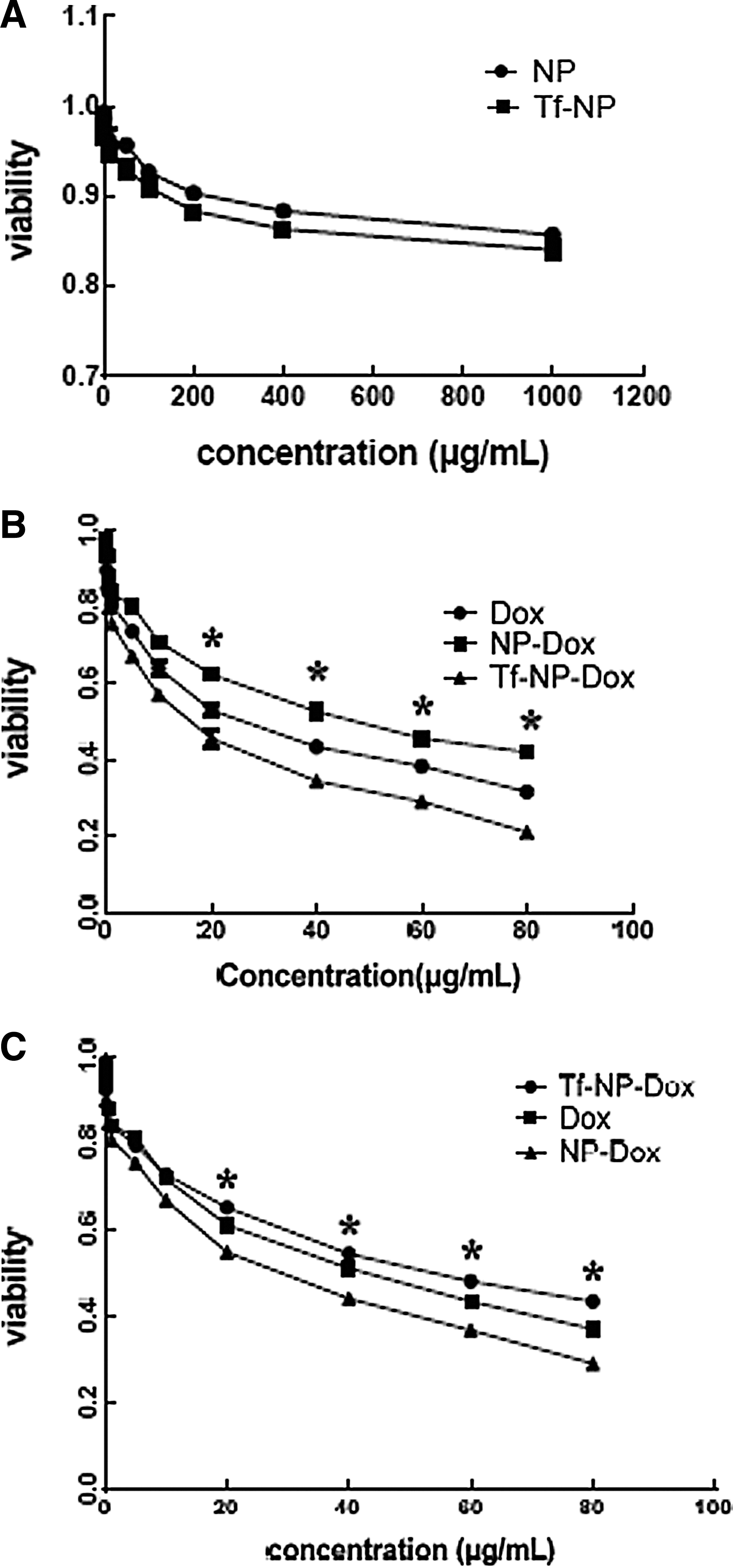
In vitro cytotoxicity of NPs. The cytotoxicity of NPs on C6 glioma cells was evaluated by CCK-8 assay.
Uptake of Dox by C6 glioma cells in vitro
HPLC assay showed that the intracellular Dox concentration in C6 glioma cells treated with Dox, NP-Dox and Tf-NP-Dox increased in a time dependent manner (Fig. 3). After 12-h incubation, the intracellular Dox concentration in Tf-NP-Dox group was 2.07-folds higher compare with NP-Dox group (p<0.05). Within the initial 2 hours, intracellular Dox concentration in free Dox group was higher compared with other groups. However, after 4 hours the uptake of Tf-NP-Dox was accelerated, and Dox concentration in Tf-NP-Dox group was higher compared with other groups.
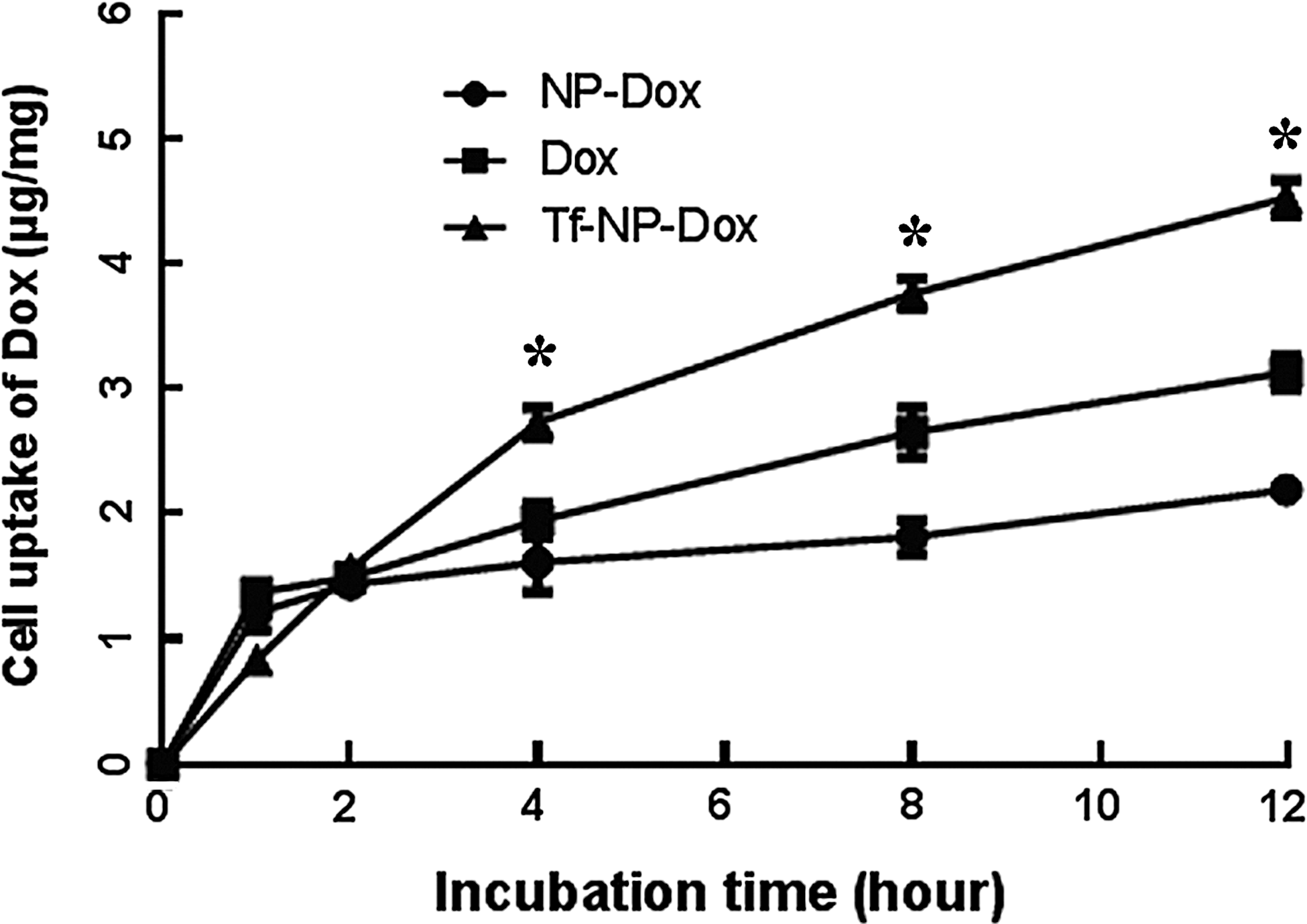
Uptake of Dox by C6 glioma cells in vitro. C6 glioma cells were treated with Dox, NP-Dox, and Tf-NP-Dox (Dox concentration 4 μg/mL) at 37°C from 1 to 12 hours and the intracellular Dox concentration was detected by HPLC analysis. The uptake index was expressed as Dox (μg)/cellular protein (mg). *p<0.05 Tf-NP-Dox versus NP-Dox or free Dox.
Biodistribution of Dox in the brain of glioma-bearing rats
To evaluate the therapeutic efficacy of Tf-NP-Dox for glioma in vivo, the glioma-bearing rats were treated with the different agents via systemic administration, on the 10th days postintracranial implantation. Dox concentration in left caudate nucleus (normal brain tissue) and right caudate nucleus (tumor tissue) were analyzed by HPLC. The results showed that Dox concentration was much higher in right caudate nucleus than the contra-lateral part in Tf-NP-Dox group (p<0.05, Fig. 4). Moreover, Dox concentration of right caudate nucleus in Tf-NP-Dox group was much higher compared with other groups (p<0.01, Fig. 4).
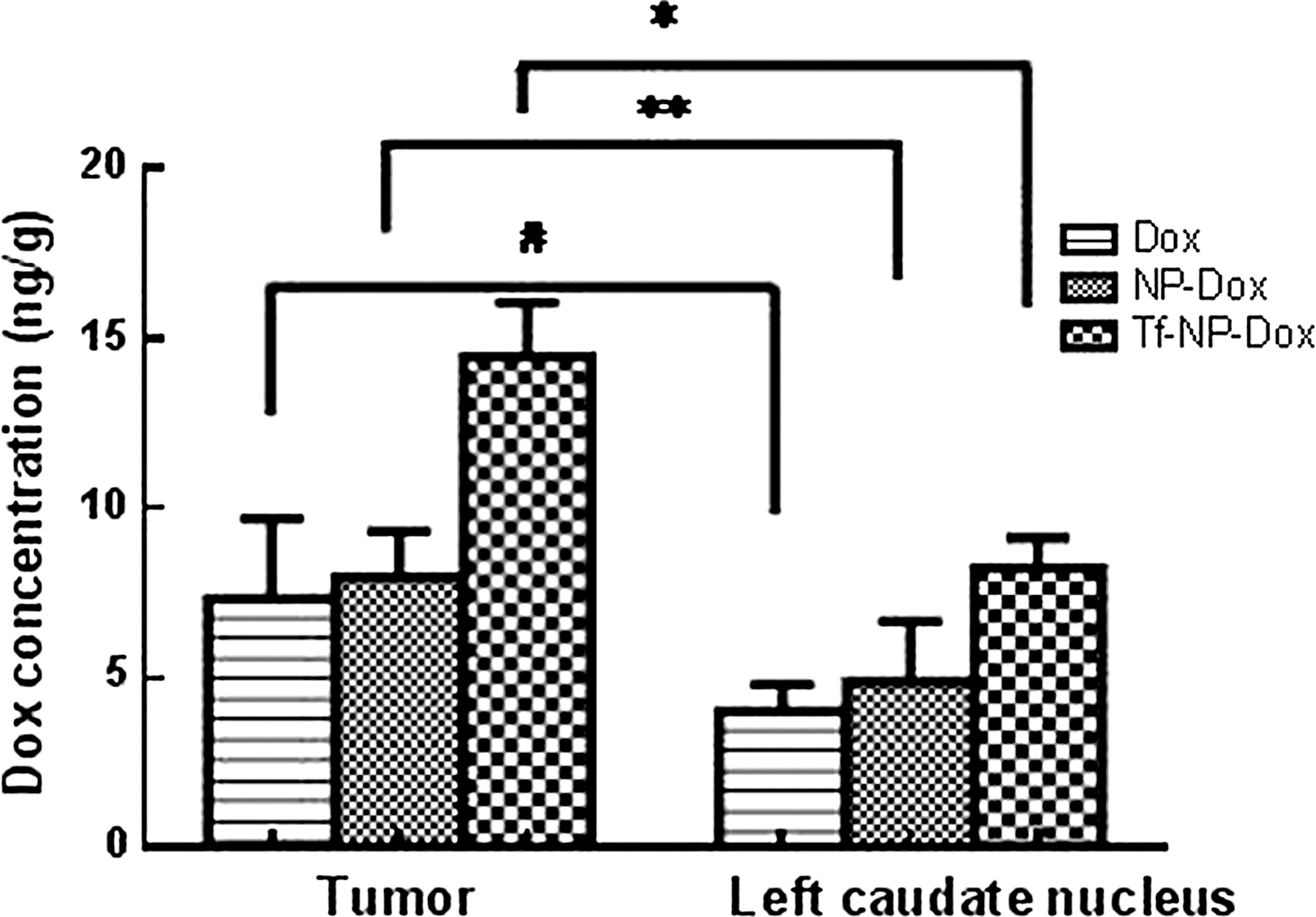
Biodistribution of Dox in the brain of glioma-bearing rats. The C6 glioma cells-bearing rats received i.v. of Dox, NP-Dox, and Tf-NP-Dox and Dox concentration in the tumor tissue and left caudate nucleus was detected by HPLC analysis (n=3). *p<0.01, **p<0.05, # p<0.05.
Tf-NP-Dox effectively inhibits glioma growth in vivo
After treatment with different formulations, the inhibitory ratio on xenograft glioma tumor by Dox, NP-Dox, and Tf-NP-Dox were 13%, 19%, and 36%, respectively on day 15 postintracranial implantation (Fig. 5A), suggesting that Tf-NP-Dox exhibited stronger inhibitory effect on glioma growth than Dox and NP-Dox (p<0.01).

Tf-NP-Dox effectively inhibits glioma growth in vivo.
The survival range of glioma bearing rats from saline, free Dox, NP-Dox, and Tf-NP-Dox group was 14–23, 15–27, 17–31, and 21–37 days, respectively (Fig. 5B). The rank of median survival time was Tf-NP-Dox (27 days)>NP-Dox (23 days)>Dox (20 days)>saline (18 days). As shown in Table 1, by log-rank test the median survival time of Tf-NP-Dox group was significantly prolonged compared with that of saline control (p<0.01), free Dox (p<0.01) or NP-Dox group (p<0.05).
The dosage of Dox was 4×1.5 mg/kg. Log-rank test versus control. The increases in survival (%) was compared to saline (ISTC), to free Dox solution (ISTS) or to NP-Dox (ISTG).
p<0.01 versus saline.
p<0.01 versus free Dox.
p<0.05 versus NP-Dox.
Dox, doxorubicin; NP, nanoparticle; Tf, transferrin.
Discussion
The antitumor effects on brain glioma mainly depends on the transport of chemotherapeutic agent across BBB to reach the glioma site. To achieve this, various approaches have been exploited, such as chemical, biological delivery vectors and molecular Trojan horses. In this study, we developed a functionalized NP modified by Tf with antitumor agents encapsulated inside. Tf is a monomeric glycoprotein that can transport one (monoferric Tf) or two (diferric Tf) iron atoms. 14 TfR is overexpressed on the brain capillary endothelium and the surface of glioma cells but its expression is low on normal tissues. 7,15 Tf-mediated transcytosis has been demonstrated to transport across BBB. 16,17 Consequently, Tf coupled nanocarrier has the potential for glioma targeting therapy. To prepare NPs that target glioma, PEGlation is essential because the bulky PEG head group inhibits the rapid uptake of reticuloendothelial system (RES). 18 PLA is biodegradable and biocompatible, but rapidly removed from blood circulation after intravenous injection by RES. Coating the NP surface with PEG has been shown to confer long circulation properties to PLA. PLA-PEG NPs have been extensively investigated for their potential as controlled and targeted drug carriers in the last decade. 19,20 In this study, we found that MPEG-PLA and maleimide-PEG-PLA assembled into NP and the vesicles were generally round with a diameter around 100 nm as examined by TEM micrographs. The size of Tf-NP-Dox was a little larger than NP-Dox, which indicated Tf conjugation to NP surface. The mean Tf molecules on each Tf-NP-Dox surface were around 32 as detected by ELISA Kit, confirming the successful conjugation of Tf onto the NP surface. The results of in vitro release study in different media demonstrated slow-releasing property.
C6 glioma cells were chosen as the model brain tumor cells in the present study. To exclude the possible effects of blank NP and Tf-NP on the growth of C6 glioma cells, the cytotoxicity of NP and Tf-NP was estimated and no significant toxicity was found. Among the free Dox, NP-Dox, and Tf-NP-Dox, Tf-NP-Dox exhibited stronger cytotoxicity to C6 cells via CCK-8 assay. IC50 of Dox, NP-Dox and Tf-NP-Dox was 21.8, 45.2, and 9.3 μg/mL, respectively, demonstrating a 4.86-fold increase of cell inhibition by Tf-NP-Dox compared to NP-Dox and a significantly higher cytotoxicity mediated by Tf. The cytotoxity of NP-Dox was a little lower compared with free DOX, which might attribute to low-releasing aspect of NP-Dox. In Tf-competitive inhibition assay, Tf pretreatment decreased markedly the cytotoxicity of Tf-NP-Dox, but had little effect on the cytotoxicity of NP-Dox or Dox. Taken together, these results confirm that the conjugation of Tf to NP increases the cytotoxicity of Tf-NP-Dox to C6 glioma cells, consistent with recent studies. 21,22
The targeting effect of Tf-NP-Dox to C6 glioma was quantitatively measured via HPLC assay. The intracellular Dox dosage of all group increased in a time-dependent manner. Tf-NP-Dox could transport Dox into C6 glioma cells at a faster rate compared to NP-Dox or Dox, demonstrating that the endocytosis by the C6 glioma cells was increased by the Tf modification. This could be explained by the fact that the increased endocytosis is mediated by the specific binding between Tf and TfR followed by an increase in the internalization of C6 glioma cells. The results agreed well with the cytotoxicity assay, suggesting that Tf mediates a stronger targeting endocytosis by C6 glioma cells.
To evaluate the efficacy of Tf-NP-Dox in vivo, brain glioma-bearing rat model was applied. For the chemotherapy of glioma in the brain, the ability of Tf-NP-Dox to penetrate BBB and enter the glioma tissue is essential. The capacity of Tf-NP-Dox targeting to glioma in vivo was assessed by intratumoral Dox concentration after systemic administration. We found that Dox concentration was much higher than the contra-lateral part, indicating that Tf-NP-Dox was able to enter the brain and further concentrate at the tumor sites. Furthermore, the analysis of survival curves and tumor volume showed that Tf-NP-Dox significantly inhibited the rapid growth of glioma, compared to Dox and NP-Dox. These in vivo data complement the in vitro studies and provide strong evidence for the potential of Tf-NP-Dox for glioma therapy.
In summary, PEG-PLA NP was conceived via emulsification-solvent evaporation method with surface modified by Tf targeting to glioma. Tf-NP-Dox demonstrated effective antitumor effect on C6 glioma cells in vitro. Moreover, in glioma bearing rats, Tf-NP-Dox could transport Dox into the tumor sites, leading to better therapeutic efficacy manifested by effective inhibition of tumor growth and prolonged survival. Although the pharmacokinetics of Tf-NP-Dox should be further studied before its clinical application, our results suggest that Tf-NP-Dox is a potential nanoscale drug delivery system for glioma chemotherapy.
Footnotes
Acknowledgments
This work was supported by the University Graduate Students' Scientific Research Innovative Program of Jiangsu Province, China (CX10B-0502) and the National Science Foundation of People's Republic of China (81272799, 81271554).
Disclosure Statement
There are no financial conflicts.