
Abstract
Purpose:
This study was to demonstrate the utility of 99mTc-3P-RGD2 micro-single-photon emission computed tomography/computed tomography (SPECT/CT) for the integrin αvβ3 expression quantification in NCI-H446 and A549 lung cancer xenografts.
Materials and Methods:
99mTc-3P-RGD2 was prepared with high radiochemical purity (97%±2%) and showing high in vitro stability. The in vitro affinities of 99mTc-3P-RGD2 to NCI-H446 and A549 tumor cells were analyzed with γ-counter, while the in vivo uptakes in NCI-H446 and A549 xenografts were evaluated with micro-SPECT/CT. The region of interest was drawn over the tumor site and contralateral muscle on the SPECT/CT image, and the tumor to nontumor (T/NT) ratio was calculated to estimate αvβ3 expression and tumor uptake. The expressions of integrin αvβ3 in vitro and in vivo were analyzed using a flow cytometer and immunofluorescence.
Results:
Micro-SPECT/CT demonstrated focal uptake in the tumors. T/NT ratio in NCI-H446 xenografts was significantly higher compared with the A549 tumor model, as 5.92±0.82 and 3.62±0.91, respectively, with p<0.05. In addition, integrin αvβ3 expression in NCI-H446 cells was significantly higher compared with the A549 cells, which was consistent with the imaging data. A linear relationship was observed between 99mTc-3P-RGD2 uptake and αvβ3 expression (R2 =0.7667, p<0.001).
Conclusion:
99mTc-3P-RGD2 SPECT/CT could be used to quantify integrin αvβ3 expression within tumors, providing a rational basis for integrin αvβ3-targeted cancer therapy.
Introduction
Lung cancer is one of the leading causes of death worldwide, 1 and early diagnosis and appropriate treatments are critical in improving lung cancer patients' quality of life. Radiology greatly contributes to lung cancer diagnosis. Computed tomography (CT) and magnetic resonance imaging are generally used for lung cancer screening and cerebral metastasis detection; however, there are some limitations in solitary pulmonary nodule characterization and distant metastasis detection. 2,3 [18F]-fluorodeoxyglucose (FDG) position emission tomography (PET)/CT has played an increasingly important role in lung cancer diagnosis and staging. However, there are limitations for [18F] FDG with respect to detecting tumors with a low metabolic activity and evaluating efficiency of antivascular therapies.
Integrin plays a crucial role in facilitating angiogenesis, which is a vital process for tumor growth and metastasis. 4,5 The RGD peptide (Arg-Gly-Asp), which selectively targets integrin αvβ3, is a biomolecule that accelerates the binding of radionuclide particles, including 64Cu, 111In, 18F, 99mTc, and 68Ga, 6 –8 to integrin αvβ3, thereby enabling imaging of αvβ3-positive tumors with PET and single-photon emission computed tomography (SPECT) with high accuracy. 6 The 99mTc-3P-RGD2 tracer, which is characterized by rapid blood clearance and urinary system excretion, 9 has been demonstrated to be more effective than 99mTc-RGD2, 99mTc-P-RGD2, 99mTc-2P-RGD2, or 99mTc-3G-RGD2. 10 –12
Besides its diagnostic role, RGD is an important agent in cancer treatment. In recent years, the use of anti-angiogenesis therapy, anti-integrin αvβ3 therapy, RGD-related chemotherapy, and gene therapy has rapidly developed. 13 –18 However, the expression level of integrin αvβ3 varies in different tumors, and the limited knowledge of αvβ3 expression in patient tumors has restricted the broad application of anti-αvβ3 therapy in clinical settings. Noninvasive imaging of αvβ3-positive tumors and quantification of αvβ3 expression levels are important not only for diagnosis, but also for choosing appropriate treatment strategies and monitoring the response to therapy.
In this study, the efficacy of 99mTc-3P-RGD2 for the detection of small-cell lung carcinoma NCI-H446 xenografts and nonsmall-cell lung carcinoma A549 xenografts was analyzed, and the correlation between αvβ3 expressions and tumor uptake rates was evaluated.
Materials and Methods
Chemicals and antibodies
The 99mTc-3P-RGD2 compound represents [99mTc (HYNIC-3P-RGD2) (tricine) (TPPTS)], where HYNIC=6:22 hydrazinonicotinyl, 3P-RGD2=PEG4-E[PEG4-c(RGDfK)]2 (PEG4=15-amino-4,7,10,13-tetraoxapentadecanoic acid), and TPPTS=trisodiumtriphenylphosphine-3,3′,3′′-trisulfonate (Fig. S1).
Kit formation of the 3P-RGD2 compound was kindly provided by Professor Shuang Liu of the School of Health Science, Purdue University (West Lafayette, IN). Na99mTcO4 was purchased from China Isotope Corporation (Nanjing, China). The anti-αvβ3 antibody was purchased from Abbiotec (San Diego, CA). Secondary antibodies AlexaFluor488-conjugated goat anti-rabbit antibody, Alexa594-conjugated goat anti-rabbit IgG, and nucleus-staining 4′,6-diamidino-2-phenylindole (DAPI) were purchased from Life Technologies (Carlsbad, CA). The anti-CD31 antibody was acquired from BD (Heidelberg, Germany).
Cell culture
NCI-H446 and A549 cells were purchased from KeyGen Biology Company (Nanjing, China). They were cultured in Dulbecco's modified Eagle's medium (Hyclone, Logan, UT), supplemented with 10% fetal bovine serum (Hyclone) and 100 IU/mL penicillin–streptomycin (Invitrogen, Burlington, ON, Canada), and grown at 37°C in 5% CO2.
Animal model
The NCI-H446 and A549 lung carcinoma cell line-bearing athymic nude mice were purchased from KeyGen Biology Company. Mice were housed under environmentally controlled conditions (22°C; 12-hour light/12-hour/dark cycle with the light cycle from 6:00 to 18:00 and the dark cycle from 18:00 to 6:00) and provided with pathogen-free food and water.
Animal care and use were performed strictly in accordance with the ethical guidelines of the Nanjing Medical University Animal Care and Use Committee, and the study protocol was approved by the local institutional review board.
99mTc-3P-RGD2 has been proven to be a safe and well-tolerated radiotracer in nonhuman primates. It shows high tumor uptake, rapid blood and renal clearance, and radiotracer accumulation in the bladder. 9
Preparation of 99mTc-3P-RGD2
99mTc-3P-RGD2 was prepared according to previously reported methods 19 using a lyophilized kit formulation containing 20-μg HYNIC-3P-RGD2, 5-mg TPPTS, 6.5-mg tricine, 40-mg mannitol, 38.5-mg disodium succinate hexahydrate, and 12.7-mg succinic acid. 99mTc-labeling was accomplished by adding 1–1.5 mL of Na99mTcO4 solution (1110–1850 MBq). The reconstituted mixture was incubated at 100°C for 20 minutes, and the radiochemical purity was determined using radio-high-performance liquid chromatography (HPLC).
Radio-HPLC analysis
Radio-HPLC was conducted on an LC-20AT system (Shimadzu, Japan). The flow rate was 0.5 mL/min. The gradient mobile phase was set up with solvent A (0.1% trifluoroacetic acid in Millipore water) and solvent B (0.1% trifluoroacetic acid in acetonitrile), 0–5 minutes 5% solvent B, 5–15 minutes from 5% to 70% solvent B, and 15–30 minutes from 70% to 5% solvent B. The ultraviolet/visible detector was set at λ=220 nm, and a Zorbax-Rx C18 HPLC column (4.6×250 mm, 100 pore size) was used. The radiosensitivity was 200 K at room temperature.
Stability in vitro
The stability of 99mTc-3P-RGD2 in newborn calf serum was determined after incubating the radiolabeled compound (37 MBq) in 2 mL newborn calf serum at 37°C. Every 20 μL mixture was injected directly into the radio-HPLC to analyze the radiochemistry purity, which was followed by radiolabeling efficiency analysis at 0, 3, 4, and 6 hours.
Immunofluorescence analysis for the expression of integrin αvβ3 in vitro
NCI-H446 and A549 cells (106 cells/mL) were washed with phosphate-buffered saline (PBS), fixed with 4% paraformaldehyde (Sigma-Aldrich, St. Louis, MO) for 1 hour, and blocked for 1 hour with 3% bovine serum albumin. Then, the cells were incubated with the anti-αvβ3 antibody (Abbiotec) (diluted at 1:200) overnight at 4°C. After three washes with PBS, cells were incubated with the secondary antibody, Alexa Fluor488-conjugated goat anti-rabbit antibody (green) (Life Technologies), at 1:200 dilution for 1 hour at room temperature, followed by nuclei staining with DAPI (Life Technologies). The fluorescence was visualized under a fluorescence microscope (Axioplan2; Zeiss, Jena, Germany) under 400× magnification, with the same exposure time, equal brightness, and contrast adjustments used for all samples.
Flow cytometry assessment for integrin αvβ3 expression in vitro and in vivo
To analyze the expression of integrin αvβ3, cultured cells or cell suspensions from tumor tissues were incubated with the fluorescein isothiocyanate anti-αvβ3 antibody (Abbiotec) at the desired concentration for 30 minutes at room temperature. After three washes, the cells were analyzed using flow cytometry (FACScalibur; Becton Dickinson, Franklin Lanes, NJ).
In vitro binding of 99mTc-3P-RGD2 to NCI-H446 and A549 cells
The binding affinities of 99mTc-3P-RGD2 to H446 and A549 cells were determined using a γ-counter. Briefly, NCI-H446 and A549 cells were seeded in 24-well plates. After a 12-hour incubation, 3.7 kBq Na99mTcO4 or 99mTc-3P-RGD2 was added to the control well or to RGD wells at intervals of 10, 60, 120, and 240 minutes. Then, the cells were washed twice with PBS and the bound radionuclide was removed from each well with 1 mol/L sodium hydroxide solution. Finally, all cells were transferred to tubes and analyzed using a γ-counter. The 99mTc-3P-RGD2 uptake rate of cells was calculated using the following formula:
Binding rate=bound part/(bound part+unbound part)×100%.
Whole-body micro-SPECT/CT imaging
SPECT/CT scans and images were obtained with a Micro-SPECT/CT PLUS system (Bioscan, Washington, DC) equipped with a 0.74-mm nine-pinhole collimator: SPECT: 140 keV, 30 seconds/frame, 256×256 matrix, 20% window; CT scanner: 55 kVp, exposed time 1000 ms, 180° plane. Static scans of 12 tumor-bearing mice (6 NCI-H446, 6 A549) were obtained 60 minutes after tail vein injection of ∼37–55.5 MBq of 99mTc-3P-RGD2 in 0.1 mL saline. All 12 mice were anesthetized with 1.5% isoflurane for micro-SPECT/CT and throughout imaging. It took 30 minutes to complete the whole-body SPECT scan and 15 minutes to complete the whole-body CT scan. SPECT and CT data were reconstructed using InvivoScope software (Bioscan). The volumes of interest were drawn manually to cover the entire tumor. Based on the view in the CT image, the soft nontumor tissue reference (in the same transaxial plane, muscle) was also marked, and the tumor to nontumor (T/NT) ratios were calculated.
The correlation between the tumor uptake rates (measured by micro-SPECT/CT) and the integrin αvβ3 expressions (measured by flow cytometry) was analyzed.
Blocking experiments in vitro and in vivo
An in vitro blocking study was performed in NCI-H446 cells, 125I-c(RGDyK) was prepared from Na125I (purchased from China Isotope Corporation) and c(RGDyK) (kindly provided by Dr. Shuang Liu of the School of Health Science, Purdue University, West Lafayette, IN). The radiochemical purity was 3.7×10¹³ Bq/mmol. Two microtiter 24-well vinyl assay plates were coated with 100 μL/well of a solution of NCI-H446 cell suspension (5×10⁴/well) in PBS in a coating buffer (500 mmol/L Tris-HCl, pH 7.4; 150 mmol/L NaCl; 2 mmol/L CaCl2; 1 mmol/L MgCl2; 1 mmol/L MnCl2; and 0.1% bovine serum albumin 300 μL) for 24 hours at 4°C. The plates were washed twice with PBS. Then, 100 μL PBS containing 10 kBq of 125I-c(RGDyK) and appropriate dilutions (0–1000 nmol/L) of 3P-RGD2 in PBS were incubated in the wells at 4°C for 2 hours. After incubation, the plates were washed thrice with PBS. The wells were cut out and counted in a gamma counter. The IC50 value was calculated by nonlinear regression using a GraphPad Prism (GraphPad Prism 4.0; GraphPad Software, San Diego, CA). Each data point is the average of three determinations.99mTc-3P-RGD2 was used as a radiotracer and 3P-RGD2 as the blocking agent in the NCI-H446 lung cancer models; the same procedure was proceeded as in whole-body micro-SPECT/CT imaging, but 3P-RGD2 (140 nmol, 100 μg/100 μL) was coinjected via tail vein as the blocking agent.
Immunostaining of tumor tissue sections
Tumor tissue sections (5-μm thick) were prepared according to standard protocols and then assessed for integrin αvβ3 expression. The sections were fixed in acetone and stained as described above. The primary antibodies used were rabbit anti-integrin αvβ3 and rat anti-CD31 (BD Biosciences, San Jose, CA). Secondary reagents used were Alexa488-conjugated goat anti-rabbit antibody (green) (Life Technologies) and Alexa594-conjugated goat anti-rabbit IgG (red) (Life Technologies). Sections were blocked for 1 hour using 4% goat serum and then labeled with primary antibodies followed by the secondary antibodies. Nuclei DNA was stained using DAPI (Life Technologies). Images were then visualized by Carl Zeiss Axioplan 2 microscopy (Zeiss, Welwyn Garden City, United Kingdom).
Statistical analyses
Data are expressed as the mean±SD. Statistical analyses were carried out using the Student's t-test. All data were analyzed using SPSS software. For all analyses, p<0.05 was considered statistically significant.
Results
The efficiency of HYNIC-3P-RGD2 peptide labeling
The radiochemical purity of 99mTc-3P-RGD2, determined by radio-HPLC, was 97%±2% (n=10), and the retention time was 14.15 minutes (Fig. 1; Fig. S2).

99mTc-3P-RGD2 labeling efficiency was showing high radiochemical purity (over 95%), analyzed by radio-high-performance liquid chromatography (HPLC); the retention time was 14.15 minutes.
99mTc-3P-RGD2 stability in vitro
Stability analysis of 99mTc-3P-RGD2 in newborn calf serum revealed that the labeled HYNIC-3P-RGD2 prepared from the kit was stable. The radiochemical purity of 99mTc-3P-RGD2 in newborn calf serum was >95% at 0, 3, and 4 hours, and even after 6 hours of incubation (Fig. 2). The retention times were ∼14.5 minutes.
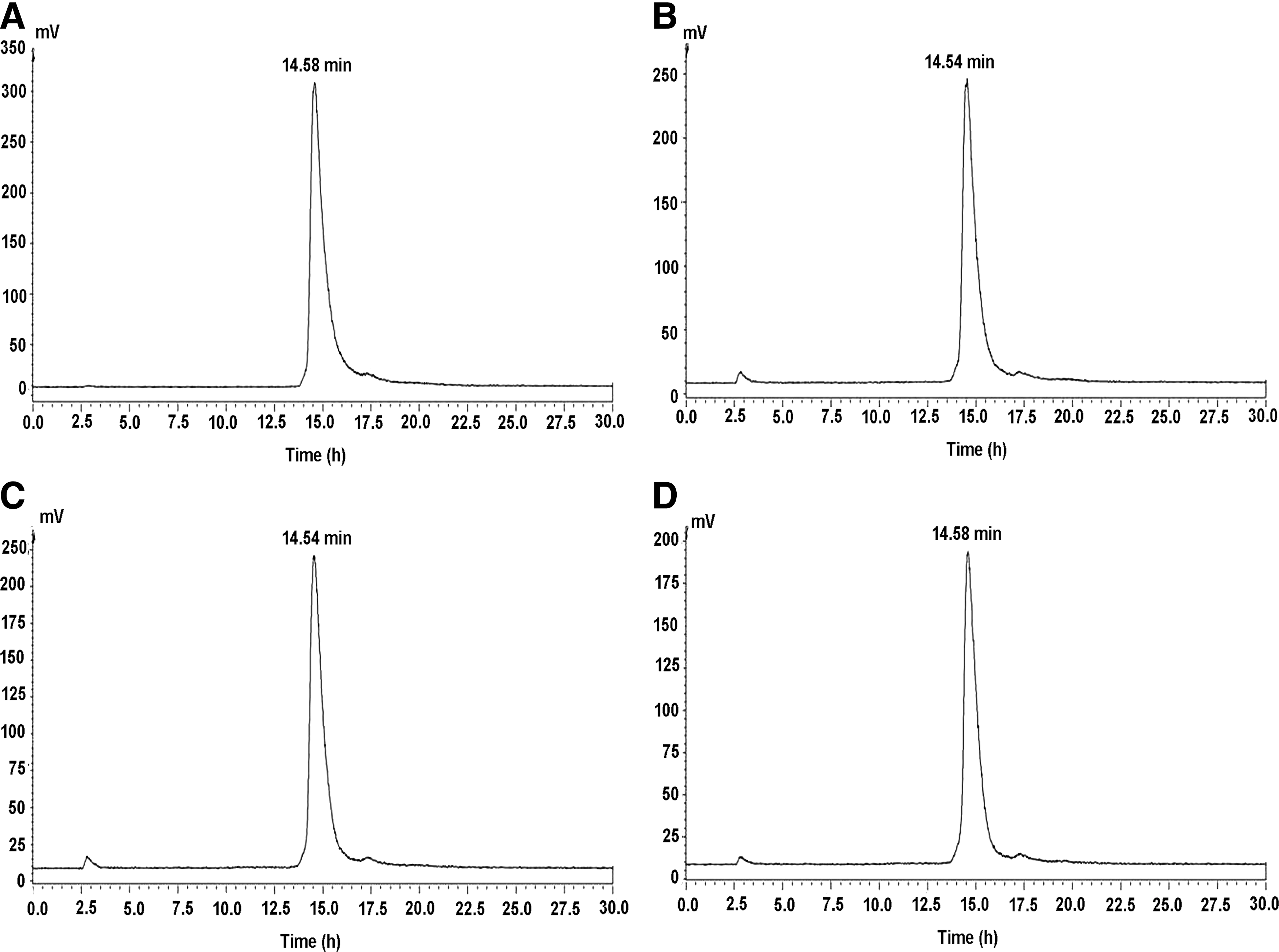
99mTc-3P-RGD2 presented high radiochemical purity (over 95%) incubated after labeling in newborn calf serum at 0
Binding studies
The binding abilities of Na99mTcO4 or 99mTc-3P-RGD2 to NCI-H446 or A549 cells at different time points are shown in Figure 3 and Table 1. There was no overall difference between the uptake of Na99mTcO4 in NCI-H446 and A549 cells; however, the overall uptake of 99mTc-3P-RGD2 was significantly higher in NCI-H446 cells than in A549 cells. The difference between Na99mTcO4 and 99mTc-3P-RGD2 uptake levels was significant at 4 hours in A549 cells (t=−4.079, p=0.015) and NCI-H446 cells (t=−11.818, p<0.001). Furthermore, the difference in 99mTc-3P-RGD2 uptakes between A549 and NCI-H446 cells was also significant at 4 hours (t=−4.005, p=0.016).

Mean±standard deviation percent.
t=11.818, p<0.001.
t=−4.079, p=0.015.
t=−4.005, p=0.016.
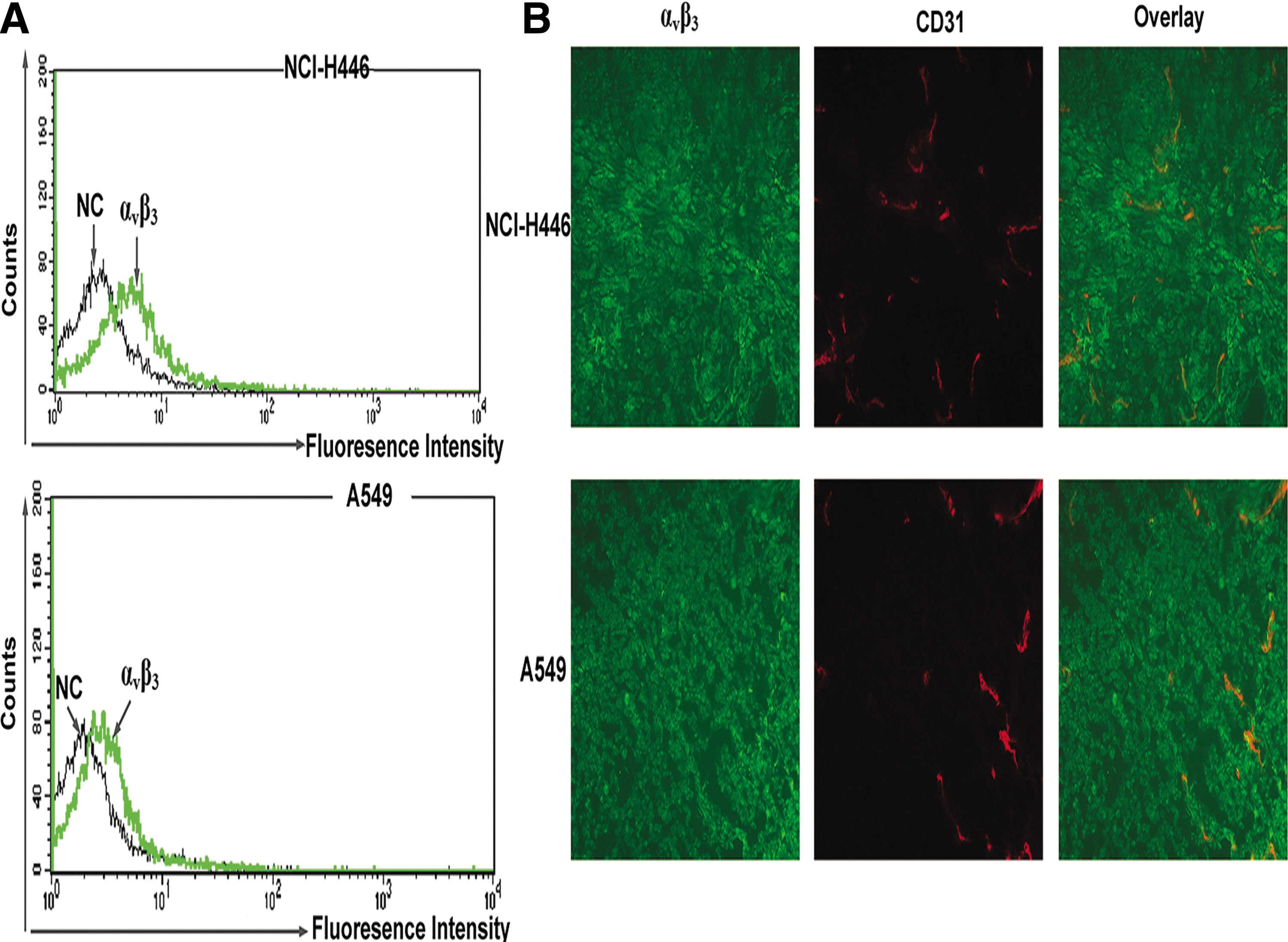
Expression levels of integrin αvβ3 in NCI-H446 and A549 cells
The expression levels of integrin αvβ3 in NCI-H446 and A549 cells are shown in Figure 3. Integrin αvβ3 was expressed at a higher level in NCI-H446 cells than in A549 cells, as analyzed by flow cytometry.
Uptakes of 99mTc-3P-RGD2 in NCI-H446 and A549 xenografts
Representative whole-body scans of NCI-H446 and A549 tumor-bearing mice at 3 hours after intravenous administration of 99mTc-3P-RGD2 are shown in Figure 4. In the NCI-H446 tumor models, the tumors appeared clear with high contrast to the contralateral background at 60 minutes postinjection, and the average N/NT ratio at 3 hours was 5.92±0.82. By contrast, in the A549 tumor models, the tumor could be visualized with moderate tumor-to-background contrast at 60 minutes postinjection with an average T/NT ratio at 3 hours of 3.62±0.91. The NCI-H446 tumor uptakes of 99mTc-3P-RGD2 was significantly higher compared with A549 (t=4.557, p=0.001).

Left: The 3D and transverse views of representative micro-single-photon emission computed tomography/computed tomography images of nude mice bearing NCI-H446 tumors (∼0.27 cm3; mean tumor to nontumor [T/NT] ratio=5.92±0.82) 3 hours after intravenous injection of 99mTc-3P-RGD2; Right: nude mice bearing A549 tumors (∼0.15 cm3; mean T/NT ratio=3. 62±0.91) 3 hours after intravenous injection of 99mTc-3P-RGD2. Significant difference exists, t=4.557, p=0.001.
Blocking experiments in vitro and in vivo
The authors determined the integrin αvβ3 binding affinity of 3P-RGD2 by displacement of 125I-c(RGDyK) bound to NCI-H446 cells. The IC50 value was calculated to be 8.759 nmol/L. An in vivo blocking study was performed in NCI-H446 tumor models, unlabeled (cold) 3P-RGD2 was injected (140 nmol, 100 μg/100 μL) via the tail vein. The NCI-H446 tumor uptake in the control and blocking image was compared at 60 minutes postinjection. In blocking studies, the radioactivity uptake was significantly decreased (T/NT=1.93±0.36), compared with controls (5.97±0.42) (Fig. S3).
Integrin αvβ3 expression in NCI-H446 and A549 tumor tissues
Immunofluorescence staining was performed to examine the integrin αvβ3 and CD31 (blood vessel marker) expression levels in tumor tissues of different types of lung carcinoma. As shown in Figure 5, integrin αvβ3 expression was higher in NCI-H446 tumor tissues than in A549 tumor tissues.
Correlation of 99mTc-3P-RGD2 in vivo uptake and integrin αvβ3 expression
As shown in Figure 6, NCI-H446 and A549 tumor uptake was positively correlated with integrin αvβ3 expression (R2 =0.7667, r=0.876, y=5.3146x+2.9934, p<0.001).
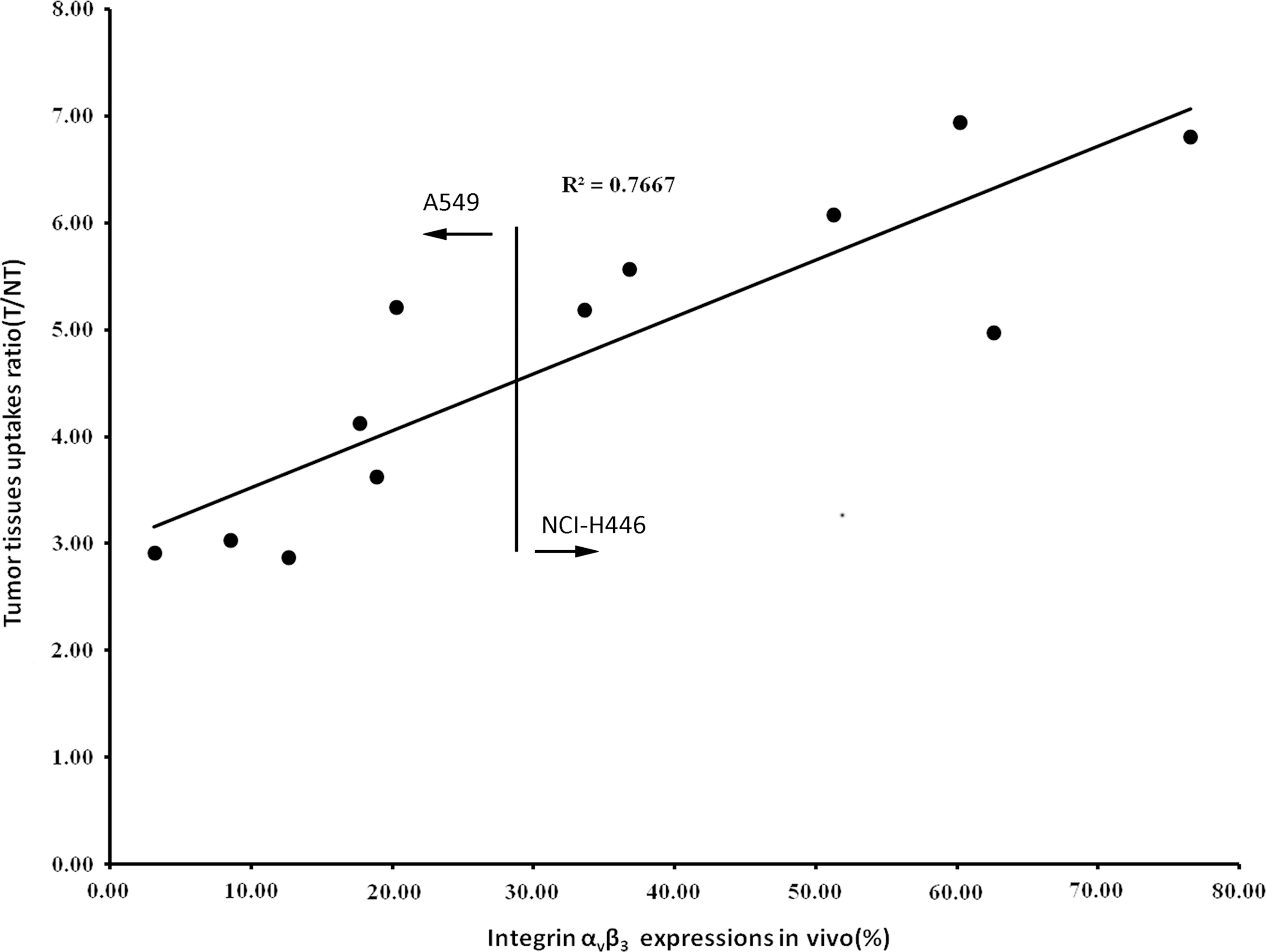
NCI-H446 and A549 tumor uptakes (n=12) of 99mTc-3P-RGD2 at 3 hours postinjection correlated well with αvβ3 expressions obtained from flow cytometry analysis in a liner manner. y=5.3146x+2.9934, R2 =0.7667, p<0.001.
Discussion
RGD (Arg-Gly-Asp) is a tripeptide sequence that has been used as a vehicle for various chemotherapeutic agents, nanoparticles, and gene therapy targets 17,20 –22 due to its high affinity with integrin αvβ3. Integrin αvβ3 plays a crucial role in angiogenesis and in facilitating tumor cell metastasis and invasion and has been studied intensively as a molecular target for both tumor image tracers and tumor therapy. 23,24
99mTc-3P-RGD2 is a promising radiotracer for αvβ3-positive tumors. Besides 99mTc-NC100692 and [18F]Galacto-RGD, recent clinical studies showed 99mTc-3P-RGD2 and SPECT had high sensitivity and accuracy in the diagnosis of lung cancer and thyroid cancer. In vitro and in vivo studies also showed 99mTc-3P-RGD2 with high affinity to glioma and breast cancer. 15,25 –27 Dimeric cyclic RGD peptides and 3PEG4 increases the uptake of 99mTc-3P-RGD2 compared to 99mTc-NC100692. 11 In addition, its relatively simple one-step preparation, high yields, and better biodistribution make 99mTc-3P-RGD2 a superior radiotracer to [18F]Galacto-RGD.
In this study, 99mTc-3P-RGD2 was labeled in kit formulation with high radiolabeling yield and confident stability (Figs. 1 and 2). It showed good sensitivity for detecting A549 and NCI-H446 xenograft foci. Nevertheless, the affinity to A549 and NCI-H446 differed both in vitro and in vivo, which correlated with αvβ3 expression level variation (Figs. 3 and 4). Correlation was present between the uptake of 99mTc-3P-RGD2 and integrin αvβ3 expression, represented by the following linear equation: y=5.3146x+2.9934, R2 =0.7667. This indicates that 99mTc-3P-RGD2 could be used not only to distinguish but also to quantify the expression level of integrin αvβ3 in a noninvasive manner. In general, the expression levels of integrin αvβ3 differ among different tumor cells or among different patients for the same tumor type, which presents a challenge for clinicians when faced with the decision of whether or not to adopt anti-αvβ3 therapy. The extra time required for testing different treatment options until finding the most efficient therapy means that patients are likely to miss the best window of treatment time for anti-αvβ3 therapy. 99mTc-3P-RGD2 micro-SPECT/CT provided quantitative data of integrin αvβ3 in tumors, which forms a basis for its use in αvβ3-targeted therapy. 13 –17 Further studies are warranted.
Conclusions
99mTc-3P-RGD2 images showed focal uptake in tumor sites in both NCI-H446 and A549 xenografts. The linear relationship between the tumor radiouptake and integrin αvβ3 expression in NCI-H446 and A549 tumors provides a significant foundation to justify a clinician's choice in using anti-integrin αvβ3 or RGD-related therapies. Further studies are warranted, as this study is expected to pave the way for increased development of radioisotope-labeled RGD analogue therapies such as 177Lu-labeled RGD and 90Y-labeled RGD.
Footnotes
Acknowledgments
The authors thank Zhang Jianping for technical assistance in image acquisition. All of the Micro-SPECT/CT scans were performed at the Department of Nuclear Medicine, Fudan University Shanghai Cancer Center. This research was supported by the Jiangsu Provincial Nature Science Foundation [BL2012037, BK2011104]; the Chinese National Nature Sciences Foundation [81271604]; and a key grant of the Nanjing Health Bureau [ZKX1001].
Disclosure Statement
The authors declare no potential conflicts of interest.
References
Supplementary Material
Please find the following supplemental material available below.
For Open Access articles published under a Creative Commons License, all supplemental material carries the same license as the article it is associated with.
For non-Open Access articles published, all supplemental material carries a non-exclusive license, and permission requests for re-use of supplemental material or any part of supplemental material shall be sent directly to the copyright owner as specified in the copyright notice associated with the article.