
Abstract
Aim:
DNA methylation plays important roles in various kinds of carcinogenesis. Vitamin C could induce Tet-dependent DNA demethylation in embryonic stem cells. Therefore, the antagonizing activity of vitamin C on ultraviolet (UV)-induced apoptosis was investigated in this study.
Methods:
Apoptosis of human epidermoid carcinoma A431 cells and p16-knockout (KO) or p21-KO fibroblasts was assessed by a fluorescence-activated cell sorter. Real-time PCR and western blot were used to determine the relative expression levels of p12, p21, and Tet1/2/3 genes. The global DNA methylation levels were determined using MethylFlash Methylated DNA Quantification Kit in A431 cells with or without vitamin C treatment. To examine the DNA demethylation activity of vitamin C, DNA immunoprecipitation (DIP)-qPCR was performed to determine the relative levels of 5-methylcytosine (5mC) or 5-hydroxymethylcytosine (5hmC) in p16 and p21 promoter regions containing cytosine-phosphorothiolated guanine (CpG) islands.
Results:
The increasing apoptosis of A431 cells under prolonged UV irradiation was remarkably decreased by the combination of vitamin C treatment, suggesting that vitamin C protects against UV-induced apoptosis. Concurrently, vitamin C induced a significant reduction of global DNA methylation in a time- and dose-dependent manner in A431 cells. Vitamin C also reactivated the expression of p16 and p21 at mRNA and protein levels. Mechanistically, about 27% 5hmC-positive cells were observed in vitamin C-treated A431 cells, and the 5hmC enrichment at p16 and p21 promoter regions was also largely increased by vitamin C. Moreover, the expression of p16 and p21 was decreased in Tet1/2 double-knockdown cells, in which the inhibitory effect of vitamin C on UV-induced apoptosis was dismissed. Furthermore, the inhibition of UV-induced apoptosis on vitamin C treatment nearly disappeared in p16- or p21-knockout primary cultured fibroblasts.
Conclusion:
These results demonstrate that vitamin C effectively antagonizes UV-induced apoptosis through regulation of Tet activity, DNA demethylation, and subsequent tumor suppressor gene activation in skin cancer cells.
Introduction
DNA hypermethylation is a common epigenetic abnormality in various cancers and may serve as a useful clinical marker for cancer diagnosis. The hypermethylation of specific cytosine residues in DNA represses transcription of tumor suppressor genes, leading to gene silencing. 1,2 Epigenetic alterations in multiple genes are believed to play crucial roles in skin carcinogenesis, and it involves cytosine-phosphorothiolated guanine (CpG) methylation in sunlight-induced skin cancer. 3,4 Exploring the fundamental mechanism of the enzymology and control of DNA methylation might assist in understanding the generation of genetic heterogeneity in skin cancer progression.
Ultraviolet (UV) radiation is a very prominent environmental toxic carcinogen for squamous cell carcinoma of the skin. 5 UV-induced gene mutations, DNA damage, and methylation alterations in promoter regions of tumor suppressors such as CDKN2A (p16) could potentially disrupt skin cell proliferation and apoptosis, and finally result in skin carcinogenesis. 6 –9 Induction of apoptosis after UV exposure appears to be a protective mechanism to get rid of damaged cells that bear the risk of malignant transformation. The ideal destination is to find a way to antagonize UV-induced apoptosis as well as malignant transformation. 10 The approach to protect cells undergoing epigenetic alterations is a possible way for skin cancer precaution and therapy. 11,12
Due to the reversibility of DNA methylation, some chemical drugs such as 5′-azacytidine (5-Aza-dc), decitabine, and histone deacetylase inhibitors are being used for tumor treatment. Novel demethylating agents such as antisense DNA methyltransferase and small interference RNA are being developed. 1 Recent investigations demonstrate that some dietary phytochemicals may prevent cancers through modulation of epigenetic modification processes. Tea catechins are found to modify epigenetic events, including DNA methylation and histone acetylation, to regulate the transcription of tumor suppressor genes in skin cancer cells. 6 Recently, vitamin C is revealed to induce Tet-dependent demethylation in embryonic stem (ES) cells and reprogramming. 13,14 However, it remains unknown whether vitamin C could induce methylation alterations in skin cancer cells to inhibit skin carcinogenesis.
The aim of the current study was to investigate the functions of vitamin C and the underlying molecular mechanism in skin cancer cells. Here, we demonstrated that vitamin C treatment protected against UV-induced apoptosis of skin cancer cells and reduced the global DNA methylation level. Specifically, vitamin C reactivated the expression of tumor suppressors p16 and CDKN1A (p21) through promotion of 5-hydroxymethylcytosine (5hmC) level in a Tet-dependent manner. These results suggest an epigenetic mechanism for skin cancer chemoprevention by the dietary phytochemical vitamin C.
Materials and Methods
Cell culture and treatment
Human skin cancer A431 cells were originally obtained from ATCC and cultured as monolayers in DMEM supplemented with 10% fetal bovine serum and 100 μg/mL penicillin/streptomycin (Invitrogen) at 37°C in 5% CO2. The normal human epidermal keratinocytes (NHEK) cells were purchased from Clonetics and cultured according to the instructions (keratinocyte growth medium supplemented with 5 ng/L human recombinant EGF and 0.05 mg/mL bovine pituitary extract (Invitrogen). p16 knockout (KO) and p21 KO mice were originally obtained from Jackson Lab. Wild-type (WT), p16 KO, and p21 KO mice at E13.5 were subject for primary culture of fibroblasts from the skin tissues according to the regular primary culture methods. These cells were cultured in the same conditions with A431 cells. Cells were seeded at a density of 1×10⁶ cells per dish and allowed to attach for 24 hours before treatment with chemicals. Cells were exposed to 30 J/m2 UV light (254 nm, UV-C) using a solar simulator (SUV100; Sigma) that was equipped with an energy output control. The subconfluent cells were treated with either various concentrations of vitamin C (L-ascorbic acid 2-phosphate) (10–200 μg/mL, Sigma) or a single dose of 5-aza-2-deoxycytidine (5-aza-dc) (5 μM) for different time intervals according to the experimental requirements.
Apoptosis analysis
Cells were stained by the fluorescent dye propidium iodide 15 and assessed by a fluorescence-activated cell sorter as previously described. 16 The percentages of apoptotic cells were presented as the state of cell apoptosis.
RNA extraction and Quantitative real-time PCR
Total RNA was extracted from the cells with different treatment using Trizol Reagents Kit (Invitrogen), and cDNA was synthesized through the reverse transcription reaction reagents (Superscript III; Invitrogen). Gene expression level was determined using SYBR Green/Fluorescein PCR Master Mix. cDNA was amplified using real-time PCR with a Bio-Rad thermocycler and SYBR Green detection system (Bio-Rad Laboratories). The relative expression levels of genes were normalized to the expression level of the housekeeping gene β-actin. The 2−ΔΔCt methods were used to quantify the relative expression of genes. Primer sequences were as follows:
5hmC immunostaining
A431 cells were fixed in 4% paraformaldehyde for 2 hours at room temperature and then processed for 5hmC cytostaining. Briefly, cells were permeabilized with 0.3% Triton X-100 in PBS buffer, and washed with PBS for 10 minutes. The cells were then blocked with 5% preimmune goat serum in PBS for 30 minutes, followed by incubation with 3% H2O2 for 20 minutes to quench endogenous peroxidase. After washing the cells with PBS, cells were incubated with anti-5hmC antibody (1:1000; Millipore) for 2 hours, followed by sequential incubation of cells with the FITC-conjugated secondary antibody. The nuclei were stained with DAPI (1:1000). The data shown were captured by fluorescence microscopy (20×).
Global DNA methylation level determination
The total genomic DNA was extracted from the cells, which were treated with vitamin C or 5-aza-dc using the Qiagen DNA Mini Kit (Qiagen) following the manufacturer's instructions. The global DNA methylation levels were determined using the MethylFlash Methylated DNA Quantification Kit (Colorimetric) according to the manufacturer's instructions. They provide the levels of global DNA methylation, and are not specific to any particular gene.
DNA-immunoprecipitation-qPCR
DNA-immunoprecipitation (DIP) utilizes naked genomic DNA as a binding substrate for 5mC or 5hmC antibodies (1:1000; Millipore). Whole genomic DNA was extracted from the cells using the Qiagen DNA Mini Kit (Qiagen). Then, the genomic DNA was subjected to DIP analysis as previously described. 17 qPCR (real-time PCR) was performed to determine the relative levels of 5mC or 5hmC immunoprecipitated DNA in p16 and p21 promoter regions containing CpG islands.
Western blot
Protein level analysis was performed using western blot. Cell lysates from different treatment groups were prepared for loading samples, which were electrophoresed on premade 10% Tris–glycine gels and then transferred onto nitrocellulose membranes. After blocking with 5% nonfat dry milk at room temperature for 1 hour, the membranes were incubated with primary antibodies against p16 (1:500; Santa Cruz Biotechnology), p21 (1:500; PharMingen), Tet1 (1:400, Abcam), Tet2 (1:1000, Abcam), and β-actin (1:10,000; Abcam) at 4°C overnight followed by a peroxidase-conjugated secondary antibody (1:1000; Santa Cruz Biotechnology). Then, they were developed using an enhanced chemiluminescence kit (Pierce). β-actin served as a loading control.
Cell transfection and gene knockdown
A431 cells were transfected using Lipofectamine 2000 reagents (Life Technologies) following the manufacturer's instructions. Tet1 and Tet2 were knocked down by their specific shRNAs, which were constructed into pSuper vectors. Tet1 shRNA targeted sequence was CGATGCAAGCCATCCTTTCGA, Tet2 shRNA targeted sequence was GCCAAGTCATTATTTGACCAT, and control shRNA sequence was GCTACGAAGCACCTCTCTTAG
Statistical analysis
The data for global DNA methylation levels are expressed as percentages with the basal levels in NHEK cells taken as 100%. Student's t-test was used to determine the statistical difference between different groups. All results were repeated at least thrice. The results in this study are expressed as the means±standard deviation. The data were considered significant if p<0.05.
Results
Vitamin C protects against UV-induced apoptosis and induces the reduction of global DNA methylation in skin cancer cells
Several dietary phytochemicals were reported to prevent cancers through modulation of epigenetic modification in cancer development. 18,19 Epidemiologic evidence and some studies reveal that vitamin C is a protective effector for nonhormone-dependent cancers. 20 –22 However, it remains unclear whether vitamin C plays a role in skin carcinogenesis prevention, and the molecular mechanism of vitamin C in cancer prevention is also not clear.
UV light is a strong apoptotic trigger that induces caspase-dependent biochemical changes in skin cells. Therefore, the effect of vitamin C in UV-induced apoptosis was detected in human skin cancer A431 cells. Interestingly, the proportion of apoptosis in vitamin C co-cultured A431 cells is much lower than the control group (Fig. 1A), suggesting that vitamin C could protect against UV irradiation-induced apoptosis in skin cancer cells. It provides a functional in vitro model for us to explore the molecular mechanism of cancer prevention of vitamin C. A recent study reported that vitamin C could induce Tet-dependent DNA demethylation. 13 We hypothesized that vitamin C might induce DNA demethylation in skin cancer cells, in which aberrant methylation frequently exists. To test this hypothesis, the global DNA methylation level was determined in vitamin C-treated cells. Consistently, the global DNA methylation level was downregulated by vitamin C in a dose-dependent manner. As a control, the DNA methylation level of normal human epidermal keratinocytes NHEK cells and the methylation inhibitor 5-Aza-dc-treated A431 cells was maintained at a very low level (Fig. 1B). To test whether the effect of vitamin C acts in a time-dependent manner, the global DNA methylation level of vitamin C-treated cells was examined at different time points. Similar to 5-Aza-dc effects, vitamin C induced the reduction of DNA methylation in a time-dependent manner. Together, vitamin C acts as a protector of UV-induced apoptosis and it could induce demethylation in human skin cancer cells.

Vitamin C induces the reduction of global DNA methylation in human A431 skin cancer cells.
Vitamin C reactivates the expression of silenced tumor suppressor p16 and p21
Based on the earlier results, we postulated that vitamin C protects against UV-induced apoptosis depending on the induction of DNA demethylation. It is well known that DNA demethylation could reactivate some silenced genes. In various cancers, the tumor suppressors p16 and p21 were frequently methylated in CpG island accumulated promoter regions. 23,24 The possibility of reactivation of p16 and p21 by vitamin C was checked. As expected, the silenced p16 and p21 expression was reactivated by vitamin C at mRNA level and protein level in a dose-dependent manner (Fig. 2). Methylation inhibitor 5-Aza-dc could also upregulate p16 and p21 expression in A431 cells (Fig. 2A, B), suggesting that reduction of DNA methylation, by either vitamin C or 5-Aza-dC, could, indeed, reactivate tumor suppressor gene expression.
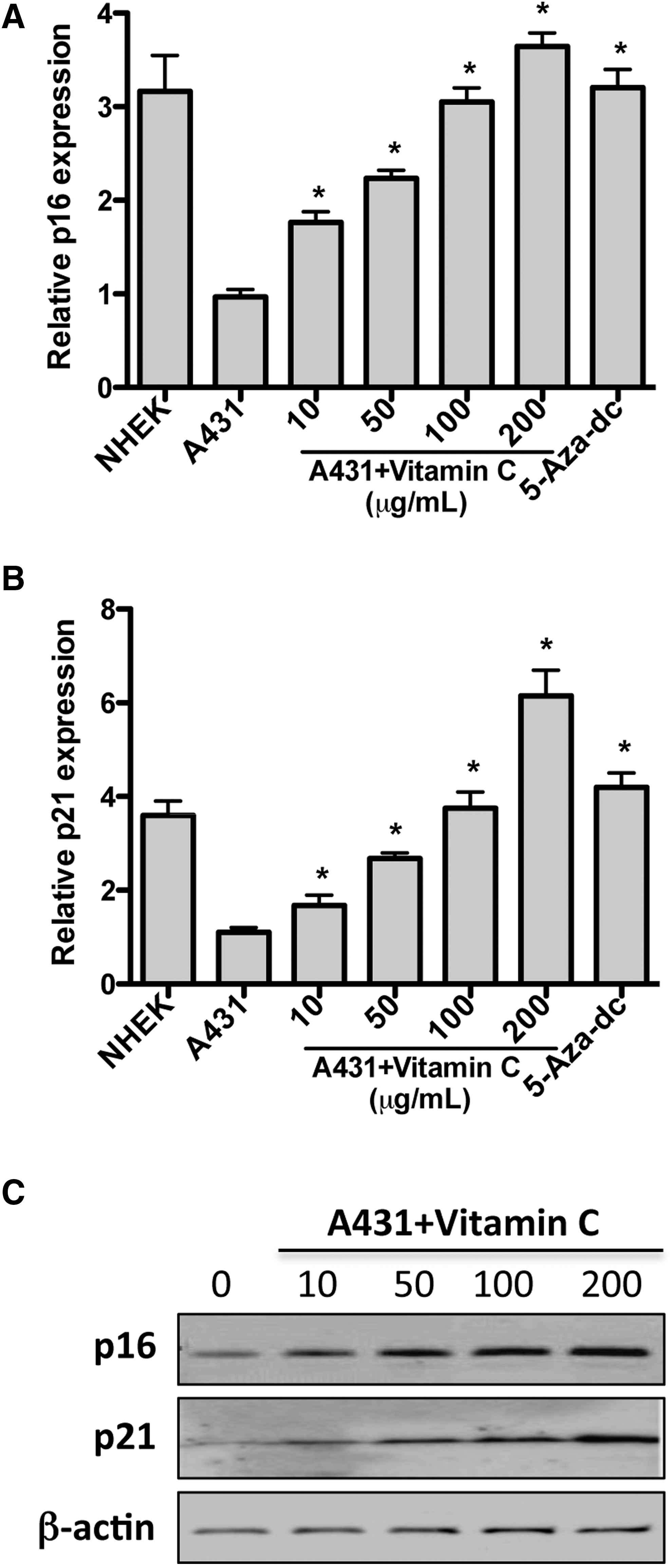
Treatment of cells with vitamin C reactivates silenced tumor suppressor p16 and p21.
Vitamin C induces the loss of 5mC at tumor suppressor gene promoters through increasing 5hmC level in skin cancer cells
Given that vitamin C reactivates p16 and p21 expression through induction of DNA demethylation, the methylation level in the two tumor suppressor gene promoters should be downregulated. 5-hydroxymethylcytosine (5hmC), an intermediate state of complete DNA demethylation, is present in many biological processes. 25 The 5hmC level was determined by immunostaining in this study after A431 cells were treated with vitamin C for 3 days. The result in Figure 3A showed that 5hmC-positive cells were significantly increased compared with the control group, and the statistical analysis further verified this view (Fig. 3B). To analyze the methylation or hydroxymethylation alterations in specific gene (p16 and p21) promoters, DIP-qPCR was performed using anti-5mC and anti-5hmC antibodies. Specific primers flanking the reported CpG islands in p16 and p21 promoter regions 23,24 were used to determine the 5mC and 5hmC levels in these two genes. Our results revealed that 5mC levels at p16 and p21 gene promoters were decreased in vitamin C-treated cells (Fig. 3C). Correspondingly, the 5hmC levels at the same regions were increased by vitamin C (Fig. 3D). Collectively, vitamin C induces the reduction of 5mC level at tumor suppressor gene promoters through increasing their 5hmC level, which then reactivates the transcription of silenced genes such as p16 and p21.

Vitamin C induces loss of 5mC at tumor suppressor gene promoters through increasing 5hmC level.
Vitamin C antagonizes UV-induced apoptosis through upregulation of tumor suppressors in a Tet-dependent manner
A previous study reports that vitamin C induces Tet1/2-dependent DNA demethylation in ES cells 13 and Tet proteins were the main executors to convert 5-mC to 5hmC. 26 In this study, we found that vitamin C could protect against UV-induced apoptosis and it upregulates p16/p21 expression through promotion of 5mC conversion to 5hmC. We asked whether the effect of vitamin C in antagonizing UV-induced apoptosis is dependent on Tet-p16/p21 regulatory machinery. We first checked the relative expression of the three members of Tet family (Tet1, Tet2, and Tet3), and Tet1 and Tet2 showed a relatively higher abundance in A431 cells (Fig. 4A). Then, Tet1 and Tet2 were knocked down simultaneously in A431 cells and both proteins were knocked down efficiently (Fig. 4B). The expression of p16 and p21 was examined in Tet1/2 knockdown cells and both of them were significantly downregulated (Fig. 4C), suggesting the regulation relationship between Tet1/2 and p16/p21 tumor suppressors. To explore the functional correlation between Tet proteins and UV-induced apoptosis, Tet1/2 knockdown cells were subjected to UV irradiation. Interestingly, Tet1/2 knockdown cells showed much higher sensitivity to UV irradiation, and the protection effects of vitamin C were impaired in Tet1/2 knockdown cells (Fig. 4D). Notably, the vitamin C-induced reduction of global DNA methylation was also partially blocked by Tet1/2 knockdown (Fig. 4E). These data suggest that vitamin C antagonizes UV-induced apoptosis, is dependent on Tet1/2 and that Tet1/2 might regulate p16 and p21 expression through modulating DNA demethylation. To further study p16/p21 functions in prevention of UV-induced apoptosis by vitamin C, cell apoptosis was analyzed in p16 knockout (KO) (Fig. 4F) and p21 KO (Fig. 4G) fibroblasts, respectively. Similar to Tet1/2 knockdown cells, the proportion of apoptotic cells in p16 or p21 KO groups is obviously higher than in wild-type (WT) groups, and the protection effects of vitamin C are very weak in knockout groups compared with WT cells, demonstrating that the functions of vitamin C are dependent on tumor suppressors. Taken together, vitamin C protects against UV-induced apoptosis depending on activation of Tet-p16/p21 signaling cascade.

The effects of vitamin C acts by Tet-dependent tumor suppressor upregulation in skin cancer cells.
Discussion
In mammalian cells, exposure to UV irradiation leads to cellular damage, subsequent induction of genes, and activation of a variety of signaling pathways. 27,28 The UV-damaged cells, which cannot be repaired, are likely to undergo apoptosis. Moreover, UV irradiation is an important exogenous carcinogen for skin carcinogenesis, which accompanies a manifestation of both genetic and epigenetic modifications, including DNA methylation and histone acetylation changes. 29 In recent years, it is believed that epigenetic events play essential roles in cancer development and epigenetic cancer therapy is a promising approach for disease treatment. 30,31
Dietary phytochemicals, including vitamins and green tea components, have been shown to be involved in epigenetic modifications to regulate cellular function and to modify the risk of cancer. 6,18,20 Especially, effects of vitamin C on cell growth, viability, and DNA synthesis in prostate cancer cells have been clearly studied. 20,32 Vitamin C is marketed as a dietary supplement partly because of its antioxidant properties and vitamin C deficiency will result in behavioral change, developmental defects, as well as other diseases. 33,34 Epidemiological evidence shows that vitamin C intake is effective in cancer prevention. 21 Vitamin C has been applied in clinical incidence in gastric cancer and urothelial cell carcinoma. 33,35,36 However, less attention has been paid to the epigenetic modifications by vitamin C in prevention of skin cancer risk and the mechanism of cancer prevention of vitamin C remains unclear.
In this study, the effect of vitamin C in UV-induced apoptosis of skin cancer cells was tested using the A431 human epidermoid carcinoma cell line as an in vitro model system. We first found that vitamin C could prevent against UV irradiation-induced apoptosis (Fig. 1). The inhibitory activity of vitamin C on apoptosis is consistent with its effect on multiple kinds of cells. 37 –39 Vitamin C is an antioxidant that maintains the activity of dioxygenases which act in function in reprogramming and epigenetic regulation. 40 Moreover, vitamin C could improve induced pluripotent stem cell quality and induced Tet-dependent DNA demethylation in ES cells. 13,14 Abnormal DNA methylation is a hallmark of cancer and leads to silencing of tumor suppressor genes and cancer progression. Here, we found that vitamin C treatment could lead to the obvious reduction of global level and increase of 5hmC level (Fig. 3). Specific to particular genes, the DNA methylation level of CpG island regions in tumor suppressor genes p16 and p21 was downregulated and the corresponding 5hmC level was increased after co-culture with vitamin C (Fig. 3). Furthermore, vitamin C elicited the reactivation of the expression of tumor suppressors, which lost the restriction of DNA methylation (Figs. 2, 3). A similar effect of vitamin C was also found in another human SCC-13 cell line (data not shown), which confirmed our findings. It demonstrates that vitamin C might act through modifying epigenetic state in skin cancer cells and correct the harmful modification alterations during carcinogenesis.
DNA hydroxylation is an intermediate state of active DNA demethylation, which was mainly completed by Tet family proteins. 41 –43 We found that Tet1/2 is responsible for p16/p21 re-expression, 5hmC upregulation, and global methylation decrease by vitamin C in this study (Fig. 4). Moreover, functional assay revealed that protection of UV-induced apoptosis by vitamin C was dependent on Tet-p16/p21 regulatory cascade (Fig. 4). More importantly, vitamin C could specifically enhance the activity of Tet1 in a biochemical assay. 13 It is predictable that vitamin C could also promote Tet activity in skin cancer cells. Tet1 and Tet2 were identified in this study, but we cannot exclude the involvement of Tet3 in UV protection. (-)-Epigallocatechin-3-gallate could also reactivate p16 and p21 expression through reducing DNA methylation and histone acetylation. 6 The reactivation of tumor suppressors by this chemical might also depend on regulation of Tet activity. Notably, the aspect of vitamin C on histone acetylation, another important epigenetic modification, might play important roles in gene regulation, which will be investigated in our future study.
In summary, vitamin C is revealed to protect against UV-induced apoptosis in A431 cells and vitamin C-induced DNA demethylation in tumor suppressor gene promoters leads to their expression. Mechanistically, vitamin C acts through modulation of Tet-p16/p21 regulatory cascade. Our data suggest that epigenetic regulation of tumor suppressor expression by vitamin C might provide a possibility to rectify the cells undergoing cancerization and to protect normal cells from epigenetic deregulation under chemotherapy or radiation therapy. These findings are of importance for understanding the anticancer mechanisms and clinical applications of dietary pigment, such as vitamins, green tea polyphenols, and curcumin, an anti-carcinogenic agent that could also inhibit UV irradiation-induced oxidative stress and apoptosis. 44 This gives us a hint that the anti-tumor activity of dietary agents is very complicated, and the underlying mechanism needs further studies.
Disclosure Statement
The authors declare no conflict of interest.