
Abstract
Lutetium-177 is an assured therapeutic radionuclide with favorable half-life and suitable β− energy. Radiolabeled 177Lu-EDTMP (Ethylenediamine tetramethylene phosphonic acid) is by and large used for bone pain palliation in cancer patients. In vitro cell studies are carried out in osteosarcoma cells MG-63 to evaluate the combined effect of anticancer drug camptothecin (CPT) and 177Lu-EDTMP. Two concentrations of 177Lu-EDTMP (3.7 and 37 MBq) were incubated with MG63 cell line for 48 hours with and without pretreatment of CPT (10 nM) for 1 hour. After completion of incubation, the cells were harvested and cellular toxicity was estimated by LDH, MTT, and trypan blue dye. Apoptotic DNA fragmentation was estimated by ELISA kit. The expression of proteins such as bcl2, PARP, and MAPK (mitogen-activated protein kinase) that were related to apoptotic signaling pathways was assessed by western blotting. The results indicated that cellular toxicity and apoptosis were relatively higher in MG63 cells that were treated with CPT prior to treating with 177Lu-EDTMP in comparison with the corresponding individual controls.
Introduction
Almost 65% of patients suffering from prostate or breast cancer and 35% of those with advanced stage of the lung, thyroid, and kidney cancer are likely to have skeletal bone metastases.
1
Bone metastases, a frequent complication of cancer causes excruciating pain. The treatment typically include agents such as analgesics, biphosphonates, external beam radiation therapy, hormone therapy, chemotherapy, and/or radiolabeled bone pain palliative agents.
2
Several phosphonates labeled with β
Materials and Methods
Materials
Chemicals and kits for assays were purchased from Sigma Chemical, Inc. unless otherwise stated. In situ Cell Death Detection ELISA Kit was purchased from Roche Diagnostics GmbH. Lu-177 and EDTMP ligand used were produced in-house in Isotope Applications & Radiopharmaceuticals Division, Bhabha Atomic Research Centre, Mumbai, India.
Cell culture
Human osteosarcoma cells MG63 and noncancerous cells Chinese Hamster Ovary (CHO) were obtained from the National Center for Cell Sciences (NCCS) Pune, India. MG63 cells and CHO cells were cultured in MEM and RPMI 1640 respectively, supplemented with 10% serum (Invitrogen) and antibiotic solution. Cells were grown in humidified 5% CO2 atmosphere in incubator at 37°C and were passaged every alternate day.
Radiolabeling of EDTMP with 177Lu
Radiolabeling of 177Lu-EDTMP was carried out according to the modified protocol described by Kumar et al. 14 In brief, 177Lu-EDTMP complex, suitable for present studies, were prepared as follows. One hundred microliter of a stock solution EDTMP (2 mg/mL) in 0.5 M NaHCO3 buffer, pH 9 was mixed with 100 μL of 177LuCl3 (20 mCi, 740 MBq). The pH of the reaction mixture was adjusted to ∼7 and incubated at 50°C for a period of 30 minutes. The preparation containing the complex was allowed to attain room temperature and used for all subsequent studies. The radiochemical purity of 177Lu-EDTMP was determined by paper chromatography technique using ammonia:ethanol:water (1:10:20 v/v) as the eluting solvent.
Determination of IC50 of CPT in MG63 cell line by MTT assay
To find the IC50 value of CPT, 1×10³ MG63 cells were plated in 96-well plates and different amount of CPT (5–1250 nM) were added and incubated for 48 hours. IC50 was determined by MTT assay. Briefly, after completion of incubation, 10 μL of MTT solution (5 mg/mL) was added and incubated for 2 hours. Formazan crystal was solubilized by addition of 100 μL of solubilizing buffer (20% SDS in 50% DMF). Absorbance was measured at 570 nm with reference to 630 nm in BioTek Universal Microplate Reader (BioTek, Inc.). Percent cell proliferation was calculated as ratio of OD of treated to control cells multiplied by 100. IC50 value was manually determined from three independent reactions. IC50 value of CPT was calculated from the straight line graph after plotting percent cell proliferation with respect to logarithmic value of concentration.
Treatment of tumor cells with 177Lu-EDTMP
MG63 (2×10⁵) cells were plated overnight in six-well plates. The cells were treated with 10 nM of CPT for 1 hour followed by addition of both 3.7 and 37 MBq of 177Lu-EDTMP. The cells were incubated for 48 hours at 37°C. After the incubation, cells were harvested and different assays were performed to estimate the extent of cell damage and its underlying mechanism of cell death. Similar experiments were carried out using noncancerous CHO cells for comparative evaluation of cell toxicity.
Study of cell toxicity
The MG63 and CHO cells were treated with CPT, 177Lu-EDTMP, and its combination, and incubated for 48 hours in complete medium. Cell toxicity study was carried out with LDH (lactate dehydrogenase), trypan blue dye uptake, and MTT assay.
Study of cell membrane integrity by LDH assay
After completion of incubation, the cells' supernatant were harvested and centrifuged at 1400 g for 5 minutes. Supernatant was collected to estimate the release of LDH. The LDH assay was carried out according to the protocol described in the kit (Sigma). In brief, LDH assay mixture was prepared by mixing equal volume of LDH assay substrate, cofactor, and dye prepared freshly before use. The LDH assay mixture was mixed with supernatant media (2:1, v/v) in 96-well plates. They were mixed well and incubated for 25 minutes at room temperature and protected from light. The reaction was terminated by adding 1/10 volume of 1 N HCl and the absorbance was recorded at 490 nm. The percent release of LDH was calculated as ratio of OD (optical density) of treated and control samples multiplied by 100.
Study of cell death assay by trypan blue dye uptake assay
Cells were harvested after treatment by trypsinization and the viability was determined by counting cells in equal volume of cells and 0.4% trypan blue dye. Living cells excludes dye while dead cells take up the dye, which were counted in haemocytometer. Percent cell death was calculated as the ratio of dead to total cells multiplied by 100.
Study of cell proliferation by MTT assay
To find cell proliferation, cells were harvested after treatment by trypsinization, 1×10³ cells were plated in 96-well plates and 10 μL of MTT solution (5 mg/mL) was added and incubated for 2 hours. MTT assay was carried out as described earlier.
Study of apoptosis by estimation of DNA fragmentation by ELISA method
For determination of magnitude of apoptosis, DNA fragmentation study was carried out according to protocol described in In Situ Cell Death Detection ELISA Kit. MG63 cells (1×10⁵ cells) were harvested after a different treatment, lysed in lysis buffer for 30 minutes, and centrifuged to 10,000 g for 10 minutes. Supernatant was carefully transferred to new tubes. ELISA plate was coated overnight at 4°C with anti-histone antibody followed by incubation of 10 μL of cell lysates for 90 minutes. Thereafter, the wells were washed thrice with buffer provided with kit and incubated with anti DNA-horse radish peroxidase for 90 minutes. The wells were again washed thrice with buffer and substrate solution was added and incubated for 20 minutes. The color developed was quantified at 405 nm. DNA fragmentation was expressed as enrichment factor, which is the ratio of OD of treated and control cells.
Study of proteins expression related to apoptotic cell death
MG63 cells were harvested after desired treatment and lysed in cell lysis buffer (10 mM Tris pH 7.4, 100 mM NaCl, 1 mM EDTA, 1 mM NaF, 20 mM Na4P2O7, 20 mM Na3VO4, 1% Triton X-100, 10% glycerol, 0.1% SDS, 0.5% deoxycholate, and 1 mM PMSF) and protease inhibitor cocktail. Protein concentration in samples was estimated by Bio-Rad Protein assay. Protein (40 μg) was loaded on 10% and 15% SDS PAGE gel for electrophoresis. Protein bands were transferred onto nitrocellulose membrane by electroblotting. The membrane was blocked using 5% nonfat milk protein followed by incubation with primary antibodies for PARP, bcl2, and MAPK (mitogen-activated protein kinase) such as p38 and phospho p38 and β-actin for 1.5 hours. The membrane was washed with Tris buffer saline containing Tween-20 (0.1%) followed by treatment with secondary antibody. The membrane was incubated with ECL reagents (Cell Signaling Tech.), followed by exposure to X-ray film (Hyper film; Amersham). X-ray film was processed and developed by Kodak developer and fixer solution. Protein blot densitometry was carried out using UVIBand Map software (UVItech Limited).
Statistical analysis
Unless mentioned, the results are mean±SD of at least three independent experiments where p-value of 0.05 or less is considered significant.
Results
Determination of radiolabeling yield
A ligand to metal ratio of 5:1 was used for the complexation, wherein >95% labeling yield was obtained. Further decrease in ligand concentration resulted in low complexation yield. Hence, the ratio was maintained to obtain a radiochemical purity as determined by paper chromatography to be >95% consistently in repeated experiments.
Determination of IC50 of CPT
The IC50 value of CPT is 37.17±0.67 nM for MG63 cells incubated for 48 hours (Fig. 1). To study the synergistic/additive effect of drug on 177Lu-EDTMP-treated MG63 cells, 10 nM (almost one fourth of IC50) of CPT had been chosen for carrying out further experiments.
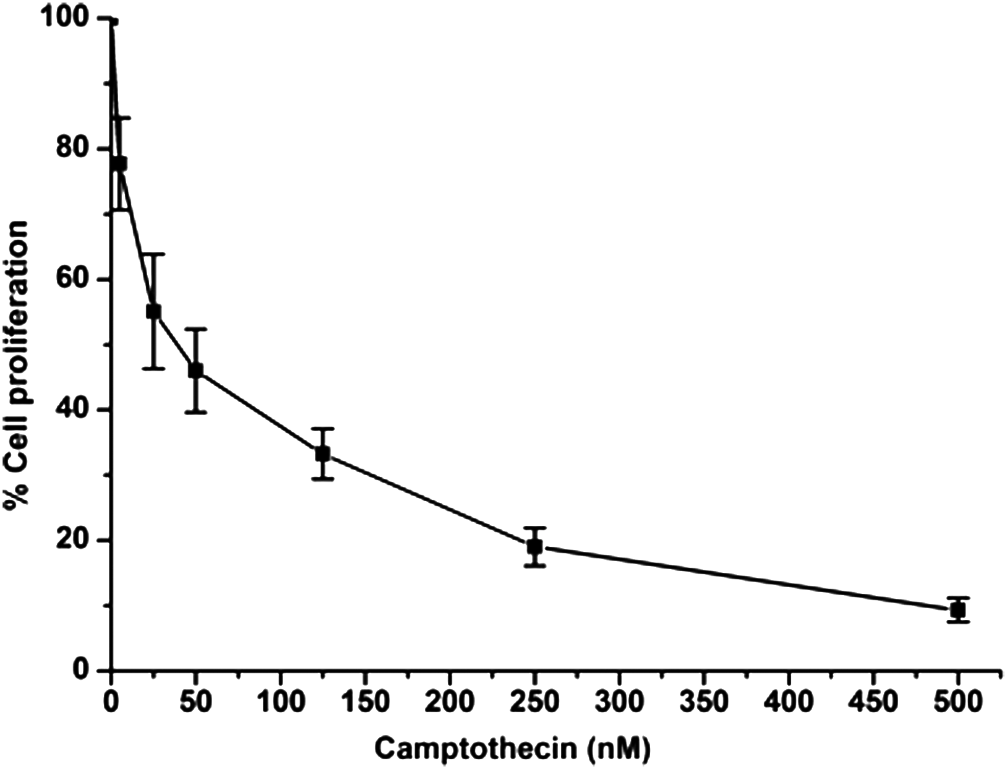
Percent cell proliferation of MG63 cells treated with CPT for 48 hours.
Effect of 177Lu-EDTMP and CPT on toxicity of MG63 and CHO cells
Estimation of lactate dehydrogenase
LDH released into the media by the cells when the cell membrane is damaged is a marker of cell toxicity. To measure the release, LDH assay was carried out in MG63 and CHO cells treated with 3.7 and 37 MBq activity of 177Lu-EDTMP and CPT over 48 hours of incubation (Table 1). It was observed that MG63 cells treated with CPT followed by either 3.7 or 37 MBq of 177Lu-EDTMP incubation showed increase in LDH release, which was significantly (p<0.05) higher than the corresponding controls. Although similar results are obtained in the CHO cells, the extent of LDH release was lower compared with MG63. The results also showed that there is no significant difference between LDH release from MG63 and CHO cells treated with CPT, 3.7 and 37 MBq 177Lu-EDTMP under same conditions. It was found that the CPT (111.89±3.24) is more toxic than the 3.7 MBq (104.04%±1.19%) of 177Lu-EDTMP and equally toxic to 37 MBq (107.1%±0.62%) of 177Lu-EDTMP in terms of membrane damage that reflects on the LDH release in MG63 cells, while there is no significant difference observed in the CHO cells treated with CPT, 3.7 and 37 MBq 177Lu-EDTMP. Thus, the LDH assay indicates that the combined treatment in case of MG63 cells shows higher toxicity than in the case of CHO cells.
CHO, Chinese hamster ovary.
Estimation of cell death by trypan blue dye uptake
Similar to the observation in the LDH assay, it was also found that in trypan blue dye uptake assay, CPT is more toxic than 3.7 and 37 MBq of 177Lu-EDTMP (Table 1) in MG63 cells. It was observed that MG63 cells treated with CPT followed by 3.7 and 37 MBq of 177Lu-EDTMP incubation showed increase in cell death, which was significantly (p<0.05) higher than the corresponding controls (Table 1). Similar trend was observed in CHO cells but to a smaller extent. There is also significant difference (p<0.05) observed between the combinatorial treatment of CPT with 3.7 (26.96%±2.13%) and 37 MBq (34.42%±1.65%) of 177Lu-EDTMP in MG63 cells, which is comparatively higher than the CHO cells. Cell death in CHO cells are 9.85%±1.32% and 18.14%±3.46% in the combined treatment with 3.7 and 37 MBq respectively,
Cell proliferation by MTT assay
Similar to the observation in LDH and cell death assay, it was found that CPT is more effective in inhibiting MG63 cell proliferation than either 3.7 or 37 MBq of 177Lu-EDTMP (Table 1). It was also observed that cells treated with CPT followed by 3.7 and 37 MBq of 177Lu-EDTMP incubation showed decrease in cell proliferation, which was significantly (p<0.05) lower than the corresponding controls (Table 1) in MG63 cells. Although similar results are obtained in the CHO cells, the extent of cell proliferation was higher compared with MG63. There is also significant difference (p<0.05) observed between the combined treatment of CPT with 3.7 (56.46%±2.98%) and with 37 MBq (46.94%±2.85%) of 177Lu-EDTMP in MG63 cells, however, there is no significant difference observed between the combined treatment of CPT with 3.7 (87.21%±1. 82%) and with 37 MBq (84.13%±2.93%) of 177Lu-EDTMP-treated CHO cells. The overall cell death observed in CHO cells was significantly lesser than that of MG63 cells treated with same concentrations.
Effect of 177Lu-EDTMP and CPT on induction of apoptotic cell death in MG63 cells
To elucidate the mode of cell death induced by 177Lu-EDTMP in combination with CPT in MG63 cells, magnitude of apoptosis was determined by In Situ Cell Death Detection ELISA method (Fig. 2). This method is based on the fact that during apoptosis the fragmented nuclear DNA and histone exudes to cytoplasm, which can be detected by ELISA method and expressed as enrichment factor. Our results showed that the enrichment factor of CPT (2.98±0.21) was higher compared to the 3.7 (1.2%±0.16%) and 37 MBq (1.82±0.15) of 177Lu-EDTMP (Fig. 2). It was also observed that cells treated with CPT in combination with 3.7 and 37 MBq of 177Lu-EDTMP incubation showed increase in enrichment factor, which was significantly (p<0.05) higher than the corresponding controls (Fig. 2). There is also significant difference (p<0.05) observed between the combinatorial treatment of CPT with 3.7 (6.31±0.16) and 37 MBq (8.57±0.11) of 177Lu-EDTMP.
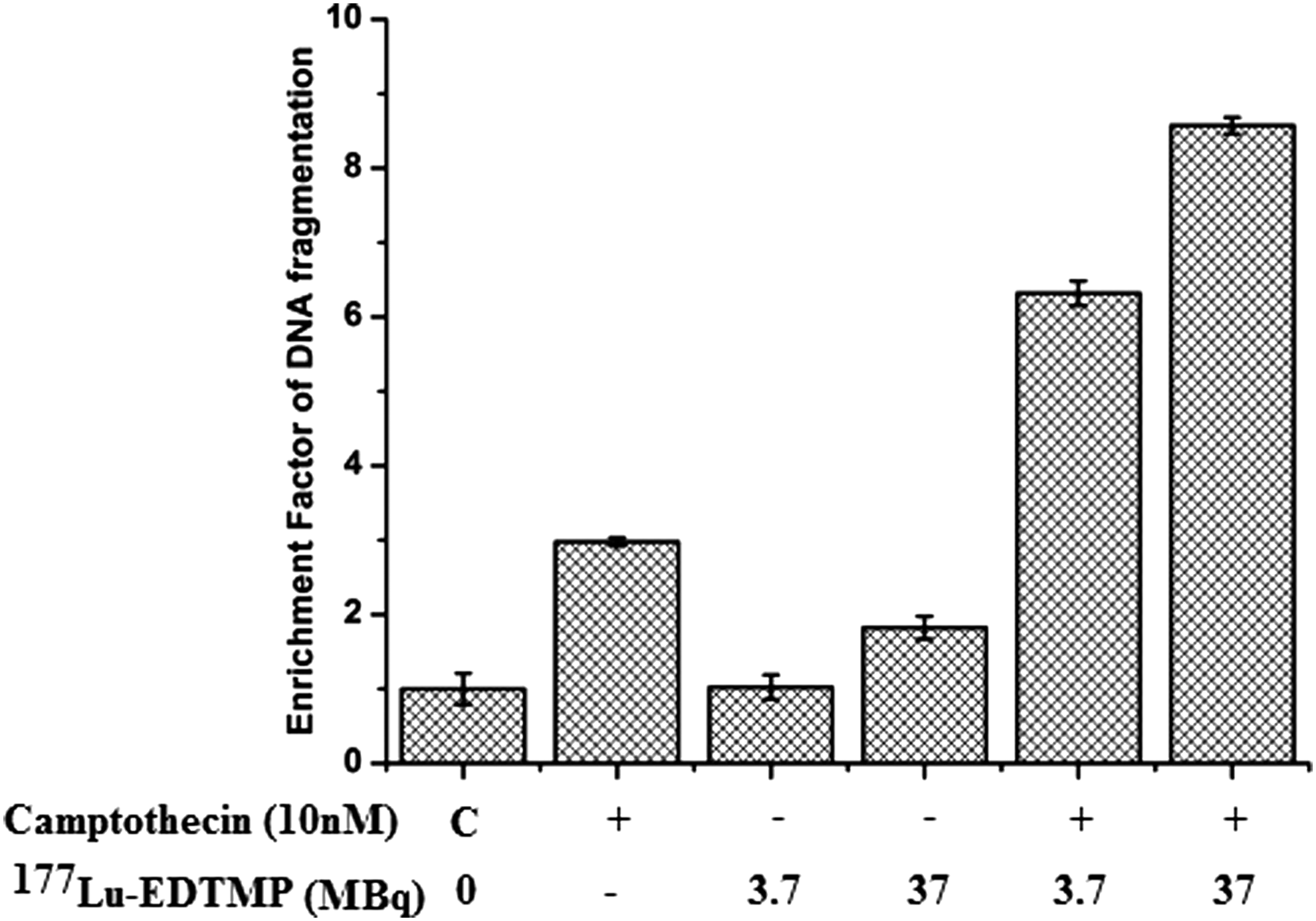
Estimation of magnitudes of apoptosis by DNA fragmentation in MG63 cells treated with CPT and 177Lu-EDTMP for 48 hours (n=3, p≤0.05 compared with their corresponding control).
Effect of 177Lu-EDTMP and CPT on expression of proteins related to apoptosis in MG63 cells
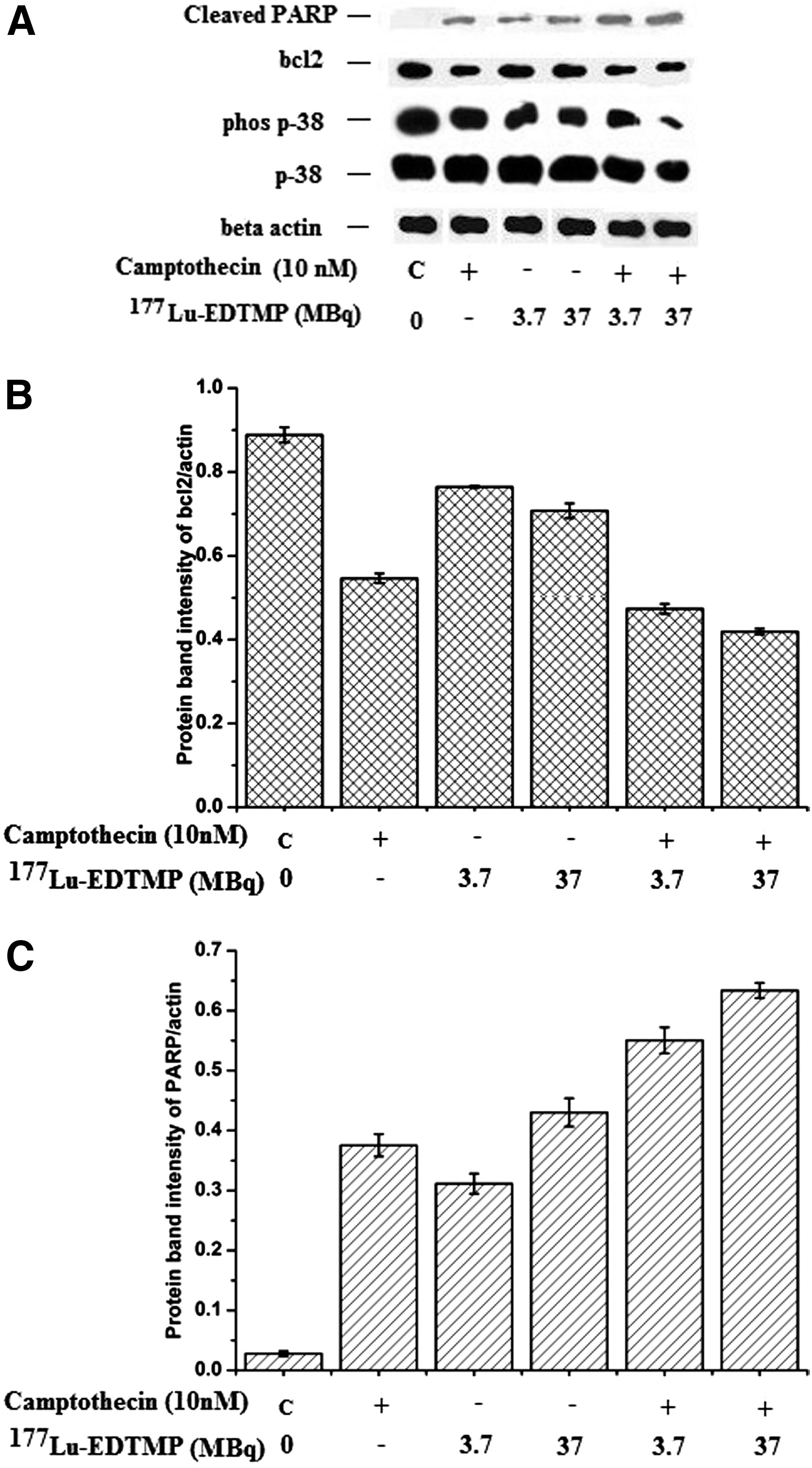
To investigate the underlying mechanism of cell death induced by CPT in combination with 177Lu-EDTMP, expression of apoptotic proteins in MG63 cells was studied by western blotting (Fig. 3A). Densitometry of bcl2 and cleaved PARP proteins from western blot was achieved and expressed as a ratio of protein of interest to the corresponding β-actin protein as a loading control (Fig. 3B, C). It was found that the bcl2 expression significantly decreased (p<0.05) in the combined treatment compared with the individual controls (Fig. 3B). Unlike bcl2, cleavage of PARP protein was significantly increased (p<0.05) in the combined treatment compared the individual controls, which was yet again the highest in the combined treatment of 37 MBq of 177Lu-EDTMP with CPT in MG63 cells (Fig. 3A, C). Western blotting was also carried out for MAPK pathway proteins p38 and their phosphorylated form in control and treated cells. An increase in expression of p38 proteins was observed in cells treated with 177Lu-EDTMP or CPT compared to the combined treatments (Fig. 3A). However, strong phosphorylation of p38 was observed in cells independently treated with 177Lu-EDTMP and CPT while the weakest phosphorylation of p38 was observed in cells treated with 37 MBq of 177Lu-EDTMP in combination with CPT (Fig. 3A).
Discussion
There are various therapeutic modalities for bone pain palliation including chemotherapy and radiotherapy by radiolabeled bone seeking agents such as biophosphonates, cyclic/acyclic polyaminophosphonic acid derivatives. 1–2 The average clinical response time for radiopharmaceuticals based on bone pain palliation is normally between 1 and 3 weeks, however, effectiveness may be seen as late as 4 weeks after treatment. 1 Furthermore, several clinically available bone pain palliative agents cause bone marrow suppression due to high energy of emitted β irradiation. 1–2 To overcome this situation, radionuclides of moderate energy such as 177LuCl3 became the isotope of choice for bone pain palliation. 3 The main objective of this study was to understand the underlying mechanism of the therapeutic effect in a combined treatment of 177Lu-EDTMP and CPT in MG63 cells. To evaluate the enhancement of the therapeutic gain in vitro, bone osteosarcoma cells (MG63) were irradiated with 177Lu-EDTMP alongwith the chemotherapeutic drug (CPT). In our previous study, we have found that 177Lu-EDTMP bound to the mineralized bone cells and induced cell toxicity by apoptosis. 14 In this study, we observed enhanced cell toxicity and apoptosis in the combined treatment of CPT with both 3.7 and 37 MBq of 177Lu-EDTMP compared with the individual control, which might be due to the combined effect of toxicity on MG63 cells. CPT bind to the topoisomerase I-DNA complex, leading to DNA strand breaks upon replication 13 and DNA strand break might be further amplified due to the β irradiation from 177Lu-EDTMP, resulting in enhanced cell toxicity of tumor cells. In this article, cell toxicity was assessed by three different methods and their values differ quantitatively due to sensitivity of techniques. 15,16 However, the patterns of toxicity observed by the three methods were quite similar. The same studies were carried out using noncancerous CHO cells. The extent of cell death observed was significantly lesser than MG63 cells. DNA fragmentation is used as marker of apoptosis by authors, 17 which are in agreement with the result of DNA fragmentation obtained from the combined treatment of CPT with 3.7 and 37 MBq of 177Lu-EDTMP.
The survival or death of a cell is largely determined by the antiapoptotic and proapoptotic regulator proteins. During apoptotic cell death, antiapoptotic proteins such as bcl2, bclxl, mcl, and others get downregulated, 18 which is in agreement with the observation where downregulation of bcl2 protein was observed (Fig. 3A, B) in the combinatorial treatment of CPT with 3.7 and 37 MBq of 177Lu-EDTMP. The native PARP proteins (113 kDa) cleaved into 89 and 24 kDa protein fragments during the apoptosis 19 and corresponding increase in PARP cleavage compared to the controls confirm the apoptotic cell death in combined treatment of CPT with 3.7 and 37 MBq of 177Lu-EDTMP.
Protein p38 are poorly activated by mitogens but strongly activated by inflammatory cytokines and wide variety of cellular stress inducers and its activation occurs through phosphorylation. After activation, these cytosolic proteins translocated to the nucleus and activate numerous proteins and/or transcription factors. 20,21 The marginal decrease in expression of p38 protein in the combined treatment might be due to the chemo/radiation stress influencing the expression of protein. Protein p38 are key molecular players in MAPK signaling and phosphorylation of these proteins is required in cell survival and proliferation. This significant decrease in phosphorylation of p38 in the combined treatment may be linked to activation of antisurvival signaling in cells, which leads to downregulation of bcl2 and ultimately apoptotic death (Fig. 3A). 22 –24 It has been reported that radiolabeled bone pain palliating agents accumulated 5%–25% more in metastatic regions than the normal osteoblast cells. 1 The CPT accumulation in cells, arrest DNA replication fork and induced DNA double-strand break, while β radiation emitted from 177Lu-EDTMP has the potential to kill the abnormal cells followed by apoptotic cell signaling pathways. This results in synergistic cytotoxic effect observed in vitro on MG63 cells treated with combination of CPT and 177Lu EDTMP.
Conclusion
CPT being a chemotherapeutic agent binds to DNA and induce DNA strand break followed by cell death, while the higher affinity of radiolabeled polyaminophosphonic acid such as 177Lu-EDTMP toward abnormal osteoblast and osteolytic cells results in increased cellular toxicity and apoptotic cell death. By combining both modalities, cellular toxicity and apoptotic cell death reported were higher than the individual controls indicating that CPT sensitizes 177Lu-EDTMP induced apoptotic cell death in osteosarcoma cells.
Footnotes
Acknowledgments
The authors are thankful to Dr. Sharmila Banerjee, Head Radiopharmaceuticals Chemistry Section, Isotope Applications and Radiopharmaceuticals Division (IA&RPhD), Bhabha Atomic Research Centre (BARC) and Dr. Tapas Das for providing the in-house EDTMP for this work. The authors gratefully acknowledge Dr. A. Dash, Head Radiochemicals and Radiation Sources Section and his colleagues for providing the in-house 177LuCl3. The authors acknowledge the support provided by Dr. Gursharan Singh, Associate Director (I), Radiochemistry and Isotope Group and Head IA & RPhD, BARC, India during this work. The authors are thankful to Dr. M.R.A. Pillai, Former Head, Radiopharmaceutical Division, BARC, India for giving the direction and suggestions for carrying out this work.
Disclosure Statement
No competing financial interests exist.