




Abstract
In this study, a novel way of distinguishing the intrinsic relationship between ανβ3 integrin targeting and detection of tumor growth by using a radiolabeled tracer based on a cyclic Arg-Gly-Asp (RGD) peptide was provided. The potential of the in vivo scintigraphic imaging of the developing vasculature from the early stage of tumor growth was evaluated. Alongside with the scintigraphic images, biodistribution studies were performed at distinct time points to validate this noninvasive imaging approach. The ability to noninvasively assess the tumor growth of ανβ3 integrin-positive glioblastoma tumors provides a method to better understand tumor angiogenesis in vivo and allows for a direct assessment of anti-integrin treatment efficacy.
Introduction
Angiogenesis, the sprouting of new capillaries from existing microvasculature, is a crucial process in the pathogenesis of a large number of nonrelated diseases, such as tumor growth, rheumatoid arthritis, ocular diseases, and psoriasis. 1 –3 In cancer, early angiogenesis is essential for tumor growth and provides important targets for diagnosis and therapy. 4,5 Specifically, the formation of new blood vessels is a key requirement for solid tumor growth beyond the size of 2–3 mm3, since diffusion is no longer sufficient to supply the tissue with oxygen and nutrients. 4
A number of molecular imaging approaches have been applied to visualize angiogenesis such as radiotracer-based imaging (single-photon emission computed tomography [SPECT] or positron emission tomography [PET]), magnetic resonance imaging (MRI), ultrasound, and optical imaging. 6 –8 Among the most widely used molecules for angiogenesis imaging studies are vascular endothelial growth factor (VEGF) and its receptors (VEGFRs) and integrin receptors. 9
Peptides based on the Arg–Gly–Asp (RGD) sequence have been identified as an essential binding motif to some integrin receptors that mediate cell adhesion between cells and proteins of the extracellular matrix. 10,11 They are widely used as specific ligands for the ανβ3 integrin receptors, which are abundantly expressed on several tumors, including osteosarcomas, melanomas, lung carcinomas, breast cancer, and glioblastoma multiforme (GBM). 12 –14 GBM is among the most highly vascularized tumors. It is also the most common primary brain tumor and one of the most lethal cancers. 15,16 Current standard treatment of GBM consists of surgery and combined radiation therapy and chemotherapy. 17,18
The relatively high sensitivity of radiotracer-based imaging systems SPECT and PET provides an advantage in the practical application of molecular imaging techniques for the evaluation of angiogenesis. 6,19,20 In the past decade, many RGD peptides and nonpeptide RGD mimetics have been labeled with different radioisotopes to develop angiogenesis-targeting radiocompounds for both diagnosis and therapy (99mTc, 125I, 111In, 90Y, 18F, 177Lu, 89Zr, 64Cu, and 68Ga), and some of them have already been introduced into clinical trials. 21 –31
Recently, the authors focused on the biological evaluation of an RGD derivative, cRGDfK-Orn3-CGG, with promising tumor uptake and retention in a GBM tumor mouse model. 31 They designed this novel RGD peptide sequence, relying on the previous studies of this group with high prostate cancer uptake in subcutaneous PC-3 tumor-bearing mice, in which the chelating moiety, comprising the amino acids Gly-Gly-Cys, 32 –34 was N-terminally conjugated to a gastrin-releasing peptide analogue, bombesin, through aminoacid spacer groups. 32,34 In relation to the tri-peptide chelating ligand, of the so-called N3S type, it has been shown that it forms a well-defined and stable complex with [Tc(V)O3]+, which is a widely studied metal core. 35 As far as the tri-amino acid spacer of ornithines might be concerned, by maximizing the effect of the charge with the introduction of this polar linker, the hydrophilic character of the final compound influenced the targeting efficiency of ανβ3 integrin receptors and the overall biodistribution profile. More specifically, c(RGDfK)-(Orn)3-[CGG-99mTc] showed high and stable labeling with Tc-99m, high tumor uptake, and rapid blood clearance, while the main route of its excretion was through the urinary pathway. Moreover, this new RGD derivative was also conjugated with magnetic nanoparticles with promising theranostic applications. 36
These findings gave rise to the possibility of extending applications of cRGDfK-Orn3-[CGG-99mTc] from targeted imaging of ανβ3 expression to noninvasively monitoring of tumor growth over a prolonged period of time (35 days) after subcutaneous inoculation of GBM cells (U87MG) at the left front flank of severe combined immunodeficient (SCID) mice. Along with the scintigraphic images, biodistribution studies were performed to validate this noninvasive imaging approach.
To the best of knowledge, only two studies based on the noninvasive nuclear imaging techniques for visualizing the angiogenic activity during tumor growth are found in literature. The first one refers to a monomeric cyclic RGD derivative labeled with F-18 [ 18 F-FB-c(RGDyK)] that was applied for the microPET imaging of brain tumor growth in an orthotopic U87MG glioblastoma xenograft model. 24 The second published work deals with a dimeric 99mTc-HYNIC-labeled c(RGDfK) with three PEG4 chain spacer groups (99mTc-3P-RGD2) that was used as an imaging SPECT/CT platform to monitor αvβ3 expression and tumor necrosis during glioma growth in a subcutaneous U87MG athymic nu/nu mouse model. 37 The same group has also used the 99mTc-3P-RGD2 derivative for the monitoring progression of breast cancer lung metastases. 38
The novelty of this study relies on the use of a 99mTc-labeled monomeric cyclic RGD derivative, c(RGDfK)-(Orn)3-[CGG-99mTc], which even if it disposes lower tumor accumulation in relation to multimeric 99mTc-RGD peptides 39 –43 was able to clearly detect integrin expression variations during glioblastoma tumor growth. Nevertheless, apart from dimerization that contributes to a higher integrin αvβ3 binding affinity, a direct comparison of tumor uptake among 99mTc-labeled RGD derivatives is somehow difficult due to the wide variety of (1) the linkers and chelating groups 44 –46 and (2) the tumor cell lines with different ανβ3 receptor density levels that are used. 6,47 –49
Materials and Methods
Materials
The RGD derivative, cRGDfK-Orn3-CGG, was designed by this group 31 and was consequently obtained from Caslo Laboratory at the Technical University of Denmark. All other chemicals were of reagent grade and used without further purification. Technetium–99m, in the form of 99mNaTcO4 in saline, was eluted from a commercial 99Mo–99mTc generator (Drytec; GE Healthcare). High-glucose Dulbecco's modified Eagle Medium (DMEM) was purchased from Sigma-Aldrich. Trypsin–EDTA, L-glutamine, penicillin–streptomycin solution, and heat-inactivated fetal bovine serum (FBS) were obtained from Biochrom KG.
Methods
The 99mTc-complexes were identified by comparative RP-HPLC analysis on a Waters 600 chromatography system coupled to both a Waters 2487 Dual λ absorbance detector and a Gabi γ detector from Raytest. Radiochromatographic analysis was performed on a C-18 RP (25.4×2.5 cm, 5 μm porosity) column eluted with a binary gradient system at a 1 mL/min flow rate.
Scintigraphic images were acquired on a small mouse-sized camera manufactured by this team. 50 The detection device is based on two Position Sensitive Photomultiplier Tubes (H85000; Hamamatsu), a parallel hole collimator, and a NaI(Tl) pixelated scintillator. The spatial resolution of the systems is ∼1.5 mm at 0 mm distance from the collimator's surface.
Formation of the cRGDfK-Orn3-[CGG-99mTc(V)] complex
The RGD derivative, cRGDfK-Orn3-CGG radiolabeled with 99mTc, was previously evaluated as a tumor-imaging agent with regard to its radiochemical, radiobiological, and imaging characteristics. 31 Radiolabeling of cRGDfK-Orn3-CGG was performed according to an already published method through the 99mTc-gluconate precursor. Briefly, sodium gluconate is used as an intermediate exchange ligand for 99mTc, and stannous chloride is the reducing agent. Radiochemical analysis as well as in vitro stability and metabolic studies showed high radiochemical yield and stability of the radiolabeled derivative.
Cell culture
Human glioblastoma cancer cells (U87MG) were maintained in DMEM high glucose supplemented with 10% FBS, 1% L-glutamine, 1% penicillin/streptomycin, and 1% GlutaMAX. The cells were incubated in a controlled humidified atmosphere containing 5% CO2 at 37°C and were subcultured weekly. They were detached from the flask surface by using a trypsin/EDTA solution (0.25%).
Animal models
SCID mice from the same colony weighing 20–25 g were obtained from the breeding facilities of the Institute of Biology NCSR “Demokritos.” Under sterile conditions, mice were subcutaneously inoculated with 100 μL of cell suspension (∼107 U87MG cells/animal) just above the left anterior leg. Tumors overexpressing the ανβ3 receptors were allowed to grow up to 35 days postinoculation. Mice did not demonstrate any abnormal behavior (hyperactivity, weight loss, dehydration of skin, fur standing up, or hunched posture) that would urge us to sacrifice them earlier than day 35. All in vivo studies were performed in compliance with European legislation, while this Institution has approved of all animal protocols presented in the current study.
Quantitative evaluation of integrin expression
The authors performed a quantitative evaluation of the ανβ3-mediated tumor expression during the early stages of U87MG glioblastoma differentiation using the radiolabeled RGD peptide, cRGDfk-Orm3-[CGG-99mTc]. They evaluated the in vivo profile of tumor angiogenesis on days 0, 3, 9, 21, and 35 after cancer cell inoculation. Taking into consideration the findings of a similar study by Shao et al., 37 where the glioma growth rate was slow up to the 3rd week of cell inoculation, and by correlating them with tumor measurements taken by a caliper, the authors excluded the time window 9–21 from the imaging study, since tumor dimensions were not high enough to be detected. Dynamic imaging studies were performed in different groups of SCID mice (n=3) bearing U87MG tumors at each of the above-mentioned time points. Biodistribution studies were also performed on these mice after the imaging studies were completed. SCID mice were injected through the tail vein with 100 μL (∼3.70 MBq/animal) of cRGDfK-Orn3-[CGG-99mTc] diluted in saline (pH 7.0), and the spatiotemporal localization of the 99mTc-labeled RGD was calculated.
Biodistribution studies
Biodistribution studies were performed in combination with scintigraphic imaging of radiolabeled RGD peptides. When dynamic scintigraphic imaging was completed at 1 hour postinjection (p.i.), the tumor-bearing SCID mice were sacrificed by ether anesthesia. Groups of SCID mice (n=3–5) were sacrificed on each day designated for assessment of tumor growth. The main organs were subsequently removed and, together with samples of muscles and urine, were weighed and counted in a NaI well counter. Stomach and intestines were not emptied before the measurements. Uptake of the radiotracer in each tissue was calculated and expressed as the percentage of injected dose per gram of the tissue (% ID/g). The % ID in whole blood was estimated assuming a whole-blood volume of 6.5% of the total body weight. Biodistribution data are reported as % ID/g and are presented as mean±SD (n=3–5).
Imaging studies
Determination of the spatiotemporal localization of cRGDfK-Orn3-[CGG-99mTc] was performed in different groups of SCID mice bearing U87MG tumors by dynamic scintigraphic imaging on different days of angiogenesis. After injecting 100 μL (∼3.70 MBq) of radiolabeled peptide intravenously into the tail vein, the animals were anesthetized. Immediately after radiotracer injection, anesthesia was induced by intraperitoneal administration of 100 μL/10 g body weight of a stock solution containing 0.5 mL ketamine–hydrochloride (100 mg/mL), 0.25 mL xylazine–hydrochloride (20 mg/mL), and 4.25 mL NaCl 0.9%. Dynamic images of the injected mice were obtained from 4 to 10 minutes up to 1 hour p.i.
Data were continuously acquired and sequential images of 2-minutes frames were stored for all animals. All images were planar, scintigraphic, and no tomographic reconstruction was performed. The raw data obtained by the imaging system have a size of 50×100 pixels, which corresponds to an area of 50×100 mm. The images were stored in raw format and processed with ImageJ open source software (version 1.47c; NIH). Then, they were linearly interpolated in ImageJ, to provide a smoother final image with 250×500 pixels. No smoothing algorithm was used. ImageJ is also used to select the colormap and enhance image contrast at a certain level where the organs/structures of interest can be distinguished. All successive 2-minute frames were summed to achieve an image with high statistics, on which regions of interest (ROIs) were drawn. Then, those ROIs were applied to the 2-minute frames to provide semiquantitative time–activity curves. The percentage of the injected dose per organ (%ID/org) was calculated. Accordingly, the %ID/g in tumors was estimated based on the weight of the organs.
Statistical analyses
The biodistribution data are presented as mean %ID/g±SD (n=3–5) and were statistically analyzed with Student's t-test. Analyses were two-tailed, with a p-value <0.05 being considered statistically significant and p=0.1 as not significant.
Results and Discussion
Evaluation of angiogenesis with biodistribution and imaging techniques
The new RGD derivative was efficiently labeled with 99mTc through the CGG chelating moiety with a high-specific activity of 5.55 GBq/μmol. The assessment of GBM tumor growth as a function of time in a U87MG GBM tumor-bearing mouse model was studied by using the radiolabeled peptide cRGDfK-Orn3-[CGG-99mTc] from the day of inoculation of cancer cells (day 0) until tumors reached a volume of ∼3 cm3 (day 35). In Table 1, the mean values of animal weight and tumor weight on various angiogenesis stages during tumor development are presented. Figure 1 shows the time course profile of glioblastoma growth in U87MG tumor-bearing mice for the experimental period of 35 days.
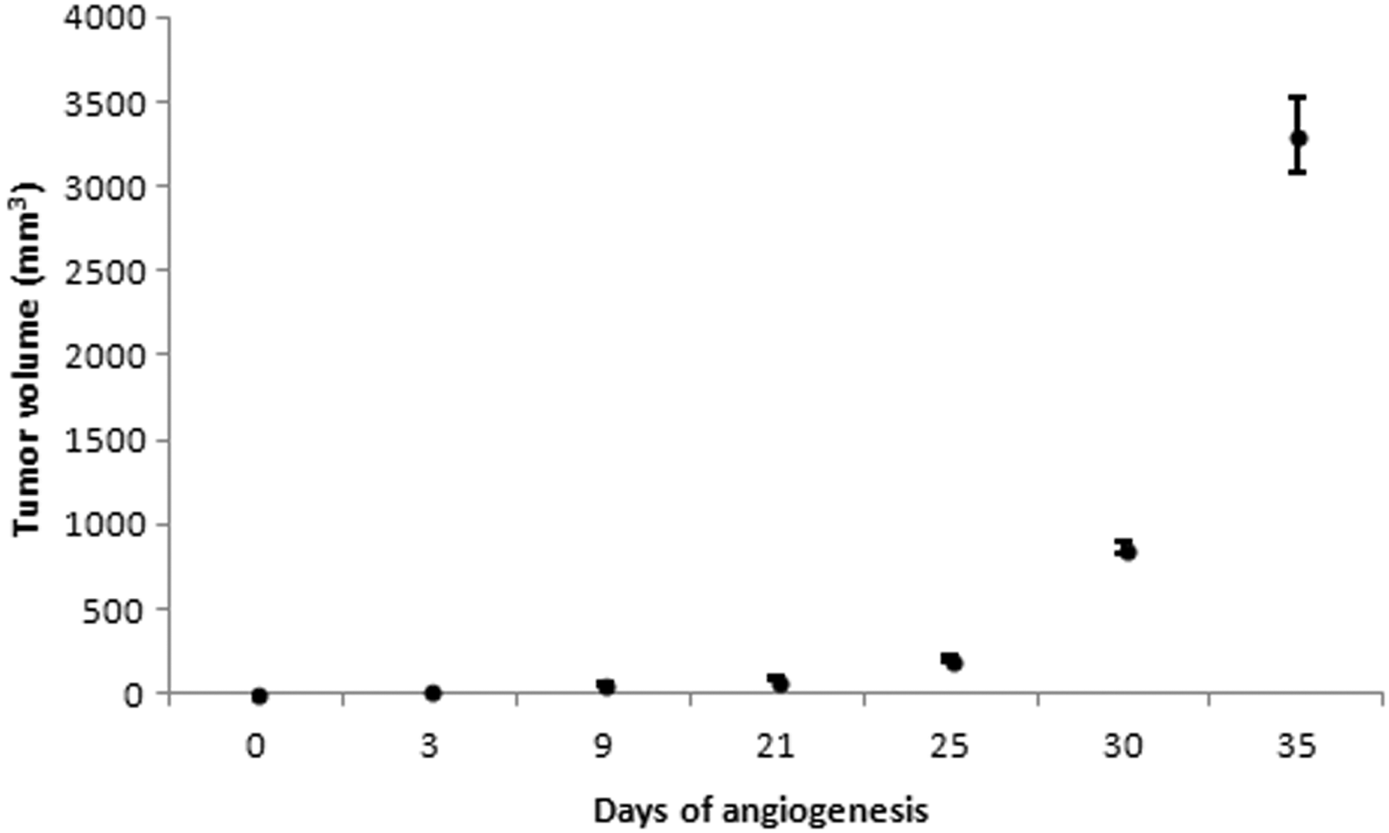
Time course profile of GBM growth in U87MG tumor-bearing mice (n=3/day) during the 35-day experimental period. GBM, glioblastoma multiforme.
In this study, biodistribution studies (Fig. 2) were performed on different days of U87MG tumor growth. The significantly high tumor uptake of the radiolabeled RGD derivative at 1 hour p.i. on day 21 of angiogenesis (11.60%±2.05% ID/g) can be attributed to the peak of angiogenesis. 34,51,52 Significantly, (p<0.05) low tumor uptake on days 3 and 9 of the study in relation to day 21 can be attributed to the early vascular stage of the tumor. 51 –53 On the other hand, reduced tumor uptake on days 29 (p=0.1) and 35 (p<0.05) relative to day 21 can be attributed to the presence of hypoxic/necrotic regions in the tumor resulting in reduced expression of ανβ3 receptors as well as to the maturation of the vascular bed network, elimination, and other factors affecting late angiogenesis development. High kidney values were observed during the whole study, reflecting the elimination route of the radiolabeled compound. Uptake in all other organs such as the liver, stomach, intestines, and muscles was insignificant.

Biodistribution results of cRGDfK-Orn3-[CGG-99mTc] in U87MG tumor-bearing mice (n=3–5) at 1 hour p.i. (% ID/g), on different days of tumor growth. ID/g, injected dose per gram; p.i., postinjection.
Relative tracer uptake expressed as tumor-to-blood, tumor-to-muscle, tumor-to-intestines, tumor-to-heart, and tumor-to-liver ratios at 1 hour p.i. was plotted versus days of tumor growth (Fig. 3). The high tumor-to-background ratios were mainly attributed to high tumor uptake in combination with low background. More specifically on day 21 of the study, the tumor-to-muscle and tumor-to intestines contrast ratios were very high with values of 10.5 and 6.2, respectively.

Graphical presentation of tumor-to-tissue ratios, at 1 hour p.i. of cRGDfK-Orn3-[CGG-99mTc], on different days of tumor growth (n=3–5).
Dynamic imaging studies of anesthetized U87MG tumor-bearing mice were performed on a high-resolution, dedicated small animal γ-camera, up to 1 hour p.i. versus several days of tumor development (Fig. 4). The experimental tumor at the left anterior leg was much more clearly delineated between 21 and 35 days of the under-study tumor growth period. ROI analysis was performed on the collected planar images to obtain semiquantitative information and the relative ID/g (%) tumor values were calculated. Imaging and biodistribution studies were in good agreement and indicated that the highest tumor uptake of cRGDfK-Orn3-[CGG-99mTc] in U87MG tumor-bearing mice was observed ∼3 weeks after inoculation (day 21) (Table 2).
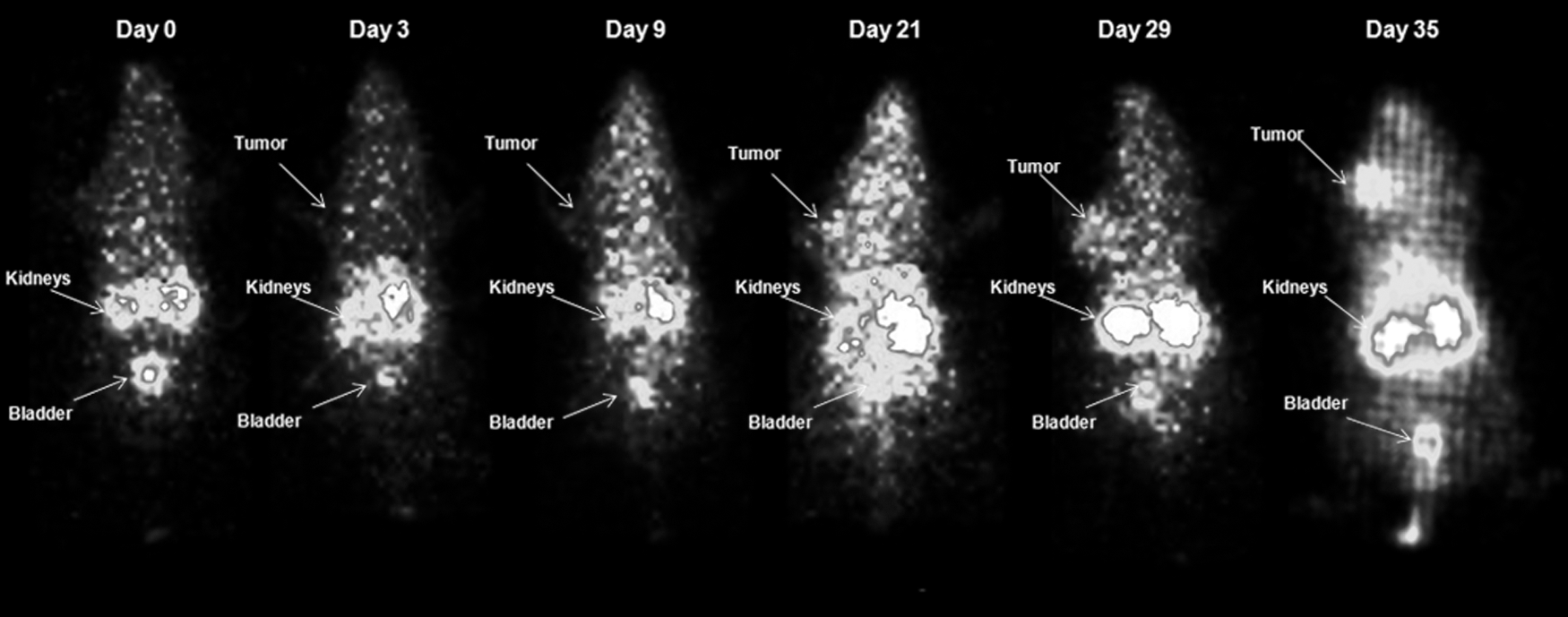
Representative planar γ images of U87MG tumor-bearing mice injected with 3.70 MBq of cRGDfK-Orn3-[CGG-99mTc] at 1 hour p.i. on different days of tumor growth (n=3). The arrows indicate the presence of the tumor, kidneys, and bladder.
Data obtained from imaging (n=3) and biodistribution (n=3–5) studies.
ID/g, injected dose per gram.
According to the in vivo imaging and biodistribution studies, the predominant excretion route of the cRGDfK-Orn3-[CGG-99mTc] was through the urinary system, which is in agreement with the initial study of the radiopeptide. 31 This hydrophilic RGD derivative had been appropriately designed to improve excretion kinetics and consequently to reduce accumulation of radioactivity at the upper abdominal area.
A method of understanding GBM tumor growth and its relationship with ανβ3 integrin expression by using a high-resolution γ-camera has been demonstrated. Tumor growth assessment by this kind of noninvasive imaging could be useful in directing physicians to continue or modify the therapeutic protocol of choice. Experimentally, the application of this radiotracer for tracking the evolution of cancer in different types of malignant tumors remains to be explored without necessitating animal euthanization as was confirmed by these findings. Nevertheless, a correlation of imaging with histochemical analysis of tumor tissues for distinguishing the vascular growth from integrin expression or for examining the heterogeneity during tumor growth would be undoubtedly very useful as a complementary tool.
The novel RGD derivative, cRGDfK-Orn3-CGG, is considered to be appropriately designed for radiolabeling with 99mTc for monitoring of ανβ3 integrin-positive glioblastoma tumor growth in vivo. Furthermore, a work on the conjugation of this peptide to magnetic nanoparticles, which will lead to a dual-modality SPECT/MRI contrast agent that can also act as a drug nanocarrier as well as a hypothermia therapeutic agent, has been recently accepted for publication. 54 –56
Conclusions
A novel way of distinguishing the intrinsic relationship between ανβ3 integrin targeting and detection of tumor growth by using a radiolabeled tracer based on a cyclic RGD peptide was provided. The potential of the in vivo scintigraphic imaging of the developing vasculature from the early stage of tumor growth was evaluated. The ability to noninvasively assess the tumor growth of ανβ3 integrin-positive glioblastoma tumors provides a method to better understand tumor angiogenesis in vivo and allows for a direct assessment of anti-integrin treatment efficacy.
Footnotes
Acknowledgments
This research has been cofinanced by the European Union (European Social Fund—ESF) and Greek national funds through the Operational Program “Education and Lifelong Learning” of the National Strategic Reference Framework (NSRF)—Research Funding Program: Heracleitus II. Investing in knowledge society through the European Social Fund.
Disclosure Statement
No conflicting financial interests exist.