
Abstract
Dendrimers are synthetic nanomolecules with well-defined chemical structures. Different strategies have been used for radiolabeling dendrimers with different radioisotopes. In this study, the aim was to conjugate dendrimers with 177Lu, to observe the in vivo behavior of the labeled compound and to measure the elementary changes in tumor tissue that could be caused by ionizing radiation. PAMAM G4 dendrimers conjugated with DOTA were labeled with 177Lu. The radiolabeled compound was characterized and its stability was evaluated by reverse phase high performance liquid chromatography. Radiolabeling yield was >98% and stable for 24 hours. Biodistribution studies of 177Lu-DOTA-dendrimers in C57BL/6 melanoma-bearing mice showed blood clearance with hepatic and renal depuration and tumor uptake. The concentrations of Br, Ca, Cl, Fe, K, Mg, Na, Rb, S, and Zn were determined in tumor tissues of C57BL/6 mice treated with 177Lu-DOTA-dendrimers and in untreated mice. The results showed decreased concentrations of Br (62%), Ca (24%), Cl (51%), K (12%) and Na (60%) and increased concentrations of Fe (8%), Mg (28%), Rb (100%), S (6%) and Zn (4%) in tumor tissues of mice treated with 177Lu-DOTA-dendrimers. These data may be useful to evaluate changes in tumor tissues as indicators of damage that could be caused by ionizing radiation.
Introduction
Dendrimers are nanomolecules, which have a molecular structure similar to a highly branched tree. 1 –4 They are symmetric molecules with a well-defined structure, high water solubility, biocompatibility, and high functionality, providing an ideal structure for biomedical applications. 2,5,6 Dendrimer structure can be divided into three parts: core, interior (branches), and surface group (periphery). The surface group can be modified to attach molecules such as peptides, antibodies, drugs, or radionuclides. 4 Branching units are organized in layers called generations. The increase in the generation corresponds to the increase in multiplied number of branches. 1
PAMAM dendrimers have large numbers of cationic surface groups and internal amine groups; conjugated with bifunctional chelator they allow stable binding of radionuclides. Bifunctional chelating agents are molecules that bind avidly to metal ions. They have ability to be appended covalently to biologically active target vectors. 7 DOTA-NHS ester (2,2′,2′′-(10-(2-((2,5-dioxopyrrolidin-1-yl)oxy)-2-oxoethyl)-1,4,7,10-tetraazacyclododecane-1,4,7-triyl)triacetic acid) is a bifunctional chelator. It has a remarkable scope for complexing metal ions with +2 and +3 charges. 1
Metal complexes with PAMAM dendrimers have found a broad range of applications in chemistry, biology, and medicine. 8 –10 Among metals, Lutetium-177 (177Lu) is an isotope used for diagnostic and therapy of small tumors. 11 –14
The half-life time of 177Lu is about 1 week (6.65 days). It can be produced by a simple reaction (176Lu (n, γ) 177Lu) with a high specific activity and decays to stable 177Hf.15 177Lu is a β-emitter with Emax = 497 keV (78.6%), 384 keV (9.1%), and 176 keV (12.2%) with a maximum tissue penetration of 2 mm, which is adequate for therapy. 14 177Lu also emits low-energy γ of 113 keV (6.4%) and 208 keV (11%) for imaging. 14,15
It is well known that many metals and ions play essential roles in metabolic and physiological processes, such as maintenance and regulation of cell function, activation, or inhibition of enzymatic reactions and regulation of membrane function. These processes can act directly or indirectly in the tumor microenvironment. 16 –18 Tumor tissue is characterized by an abnormal architecture, by poorly differentiated and unregulated multiplication of cells, and by disordered growth. Some studies show substantial differences in certain metal and ion concentrations between normal tissues and tumor tissues. 18 –21 It is also observed that concentrations of several trace elements are enhanced, whereas those of other are depressed, indicating their role in the growth of tumor. 16,21
In this study, the aim was to radiolabel PAMAM-G4 dendrimers with 177Lu, study its biodistribution, and measure concentrations of metals and ions in tumor tissue of mice treated with this compound. These data may be useful to analyze changes in tumor tissues as an indicator of damage that could be caused by ionizing radiation.
Materials and Methods
Preparation of conjugate
To perform conjugation, 4 mg of DOTA-NHS ester (Macrocyclics) was dissolved in 400 μL of phosphate buffer 0.1 M pH 8.5 and was added to 10 mg of PAMAM G4 dendrimers (MW = 14.215 Da; Sigma-Aldrich). Methanol of the stock dendrimers solution was extracted previously with gentle flowing of N2 gas. This solution was incubated for 1 hour at room temperature. Afterward, the solution was purified by size exclusion chromatography using a PD-10 desalting column containing Sephadex G25 (GE Healthcare) with sodium acetate buffer 0.15 M (pH 7.0) as a mobile phase. Buffers were prepared with Milli-Q water to prevent metal contamination.
Radiolabeling
Radiolabeling of 1 mg of DOTA-dendrimers was performed with gentisic acid (10 mg/mL) and 37 MBq (1 mCi) of [177Lu]LuCl3 (MURR, Columbia, MO). The solution was incubated for 30 minutes at 50°C. [177Lu]-DOTA-dendrimers were purified by size exclusion chromatography (PD-10 column). Sodium acetate buffer (0.15 M, pH: 7.5) was used as eluent. Samples were analyzed by reverse phase HPLC.
High performance liquid chromatography
Reversed-phase high performance liquid chromatography (HPLC) was performed on a C18 column (Ultra C18, 10 μm, 4.6 × 250 mm; Restek) using an Agilent 1200 system with an in-line radioactivity (Raytest) and ultraviolet detector (UV) 214 nm detectors. The gradient system was run at a flow rate of 1 mL/min using the following conditions: eluent A (water 0.01% TFA); and eluent B (acetonitrile 0.01% TFA). Gradient: 0 minute 100% A–0% B, 20 minutes 0% A–100% B.
Cell culture
The B16-F10 murine melanoma cell line was obtained from American Type Culture Collection. It was maintained in Dulbecco modified Eagle medium (PAA Laboratories GmbH) supplemented with 10% fetal bovine serum (PAA Laboratories GmbH) and glutamine 2 mM (AppliChem GmbH). To be inoculated in mice, B16-F10 murine melanoma cells were suspended in phosphate saline buffer 0.9%.
Animal and tumor growth evaluation
All animal studies were performed in compliance with the national laws for the conduct of the animal experimentation. Six weeks-old male C57BL/6 mice (URBE, Uruguay) were used. They received a subcutaneous inoculation of 2.5 × 10⁵ cell/0.1 mL in their right flanks, and by 2 weeks after inoculation, the tumors were palpable and were used for biodistribution studies.
Biodistribution
Biodistribution studies were performed after the intravenous (IV) injection of 0.2 mg of dendrimers labeled with 177Lu. 7.4 MBq (0.2 mCi) of 177Lu-DOTA-dendrimers were injected in C57BL/6 melanoma-bearing mice, weighing 25 ± 3 g. Groups of three mice were sacrificed 1, 4 and 24 hours postinjection. Tumor, tissues and organs of interest were harvested, weighed, and radioactivity quantified in a solid scintillation counter (ORTEC). Results were expressed as percentage of injected dose per gram (% ID/g).
Collection and preparation of the samples
177Lu-DOTA-dendrimers 7.4 MBq (0.2 mCi), were injected through the tail vein in three C57BL/6 melanoma-bearing mice. Animals were sacrificed by cervical dislocation after 24 hours postinjection; tumors were removed, weighed, freeze-dried and homogenized. For control group tumor tissues of mice untreated with 177Lu-DOTA-dendrimer (n = 7) were analyzed.
Instrumentation, methods and certified reference materials
Element concentrations in tumor tissues were measured by neutron activation analysis (NAA) technique. 22,23 Bovine Liver Powder (NIST 1557b) was used as certified reference material and for analytical quality control.
Both samples and certified reference material were irradiated for 300 seconds with a thermal neutron flux of ∼4.85 × 10¹² n·cm2·second−1, in a pneumatic station at the nuclear reactor (IEA-R1, 3–4.5 MW, pool type), IPEN/CNEN-SP, Brazil. After a decay time of 60 seconds, a γ counting of 1800 seconds was used for determining 80Br (T1/2 = 18 minutes, Eγ = 616 keV), 49Ca (T1/2 = 9 minutes, Eγ = 3084 keV), 38Cl (T1/2 = 37 minutes, Eγ = 1642 keV), 42K (T1/2 = 12 hours, Eγ = 1525 keV), 27 Mg (T1/2 = 9 minutes, Eγ = 844 and 1014 keV), 24 Na (T1/2 = 15 hours, Eγ = 1369 keV), and 37S (T1/2 = 5 minutes, Eγ = 3104 keV). For determining 60Co (T1/2 = 5.27 a, Eγ = 1173 keV), 59Fe (T1/2 = 44 days, Eγ = 1099 keV), 86Rb (T1/2 = 18.7 days, Eγ = 1077 keV) and 65Zn (T1/2 = 244 days, Eγ = 1116 keV), samples and certified reference material were irradiated with a thermal neutron flux of 8 × 10¹² n·cm2·second−1 for 4 hours. After a decay time of 7 days and a γ counting time of 6 hours, the element concentrations were determined. All analyses were done in duplicate.
A γ-spectrometer system composed of a ORTEC HPGe detector (Model GEM-60195, FWHM = 1.87 keV for 1.33 MeV of 60Co), calibrated for energy through the measurements of standard sources of 56Co, 137 Cs, and 152Eu, coupled to an MCA ORTEC Model 919E and a PC, were used to measure the induced γ-ray activity. The concentration of each element was calculated using in house software. 24
Results
Radiolabeling
The labeling efficiency for [177Lu]-DOTA-dendrimers was >98% in 1 hour. Figure 1 presents HPLC radiochromatograms of [177Lu]-DOTA-dendrimers. The retention time was 8.58 minutes for γ detector (ChA), which corresponds with the peaks of the ultraviolet detector (UV) (λ = 214 nm).

Reverse phase high performance liquid chromatography of [177Lu]-DOTA-dendrimers after 30 minutes of incubation. Ultraviolet detector (UV) and γ detector (ChA).
Biodistribution
The results of experiments are expressed as % ID/g (n = 3) and are presented in Figure 2. Biodistribution results indicate a high uptake in liver (about 20% ID/g), and spleen (18% ID/g), rapid clearance of the compound from blood (activity decreased by half in 4 hours), and renal elimination. The uptake by tumor was 5% ID/g and remained constant over 24 hours.
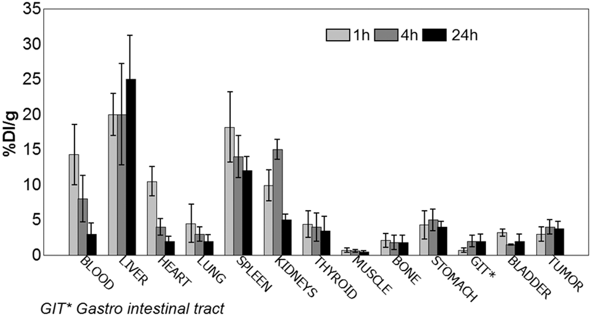
Biodistribution of 177Lu-DOTA-dendrimers at different times (1, 4, and 24 hours) in melanoma-bearing mice (n = 3 for each time). Data are expressed as percentage of injected dose per gram (% ID/g).
Concentrations of metals and ions in tumor tissue
The concentrations of metals and ions in tumor tissues of mice treated with 177Lu-DOTA-dendrimers and in tumor tissues of mice untreated with this compound were determined by NAA technique and are summarized in Figure 3. These results show a decrease in the levels of Br (62%), Ca (24%), Cl (51%), Co (7%), K (12%) and Na (60%) and an increase in the levels of Fe (8%), Mg (28%), Rb (100%), S (6%) and Zn (4%) in tumor tissues of mice treated with 177Lu-DOTA-dendrimers.
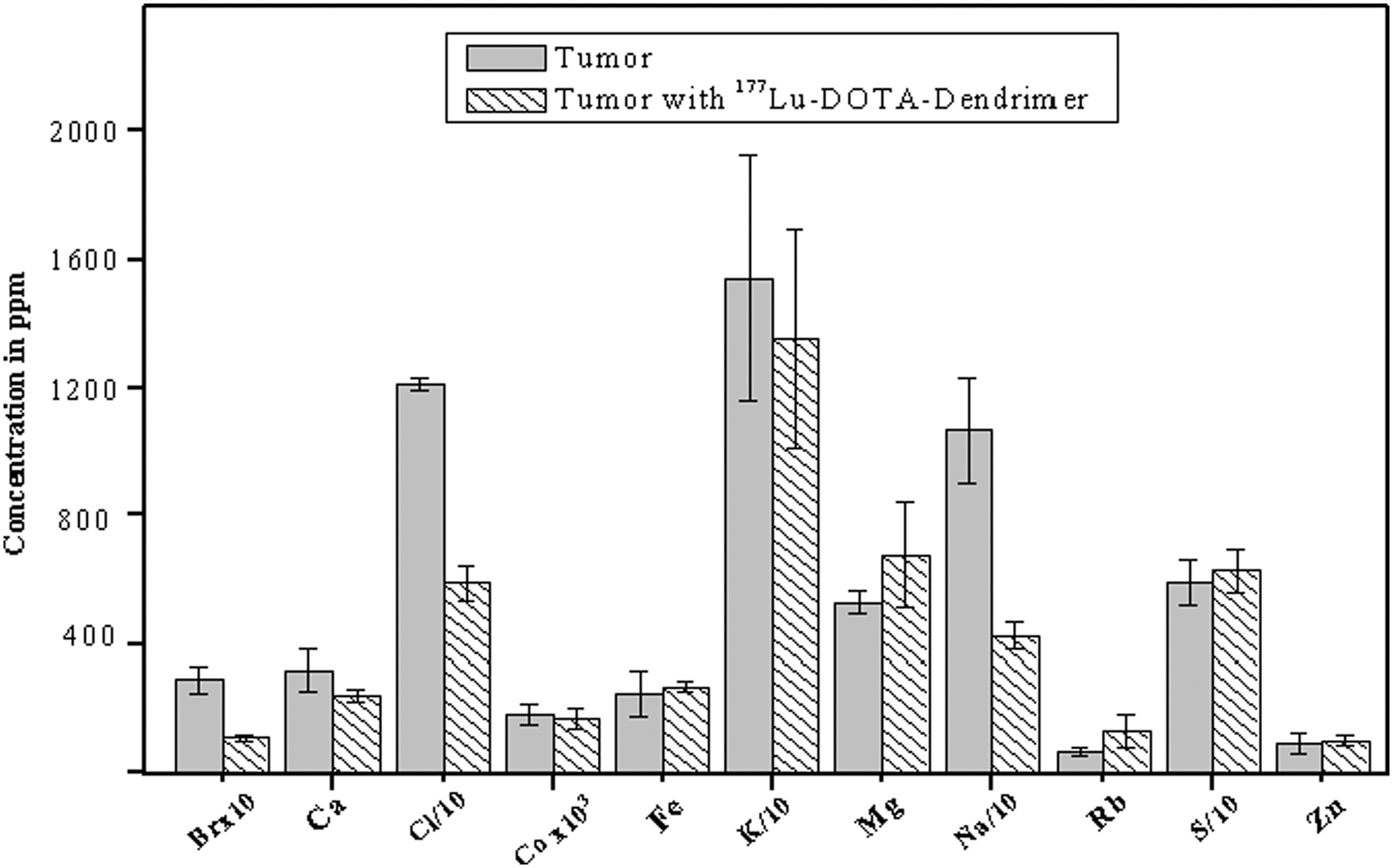
Concentrations of Br, Ca, Cl, Co, Fe, K, Mg, Na, Rb, S, and Zn in tumor tissue and tumor tissue treated with 177Lu-DOTA-dendrimers.
The differences between levels of Br, Ca, Cl, Na, and Rb in tumor tissues of mice treated with 177Lu-DOTA-dendrimers and tumor tissues of mice untreated with this compound were statistically significant (p < 0.05).
Discussion
In this study, it was found that synthetic nanomolecular dendrimers can be labeled with 177Lu via DOTA, with very good efficiency and stability.
A Melanoma model was used in this study because previous work has shown the ability of this model to accumulate nanoparticles with relative ease (well-watered solid tumor). 10 Furthermore, melanoma is highly metastatic and is associated with a poor prognosis; it is an interesting model from medical point of view.
Biodistribution results showed blood clearance with hepatic and renal depuration. This is probably the result of the molecular weight of the dendrimers (14 kDa), which can be metabolized by hepatocyte cells, and that are below the level of renal cutoff (40–60 kDa). Tumor uptake in 177Lu-DOTA-dendrimers was significant (5% ID/g). This tumor uptake could be triggered by increased vascularity of tumor tissues, which can lead to an improvement of elemental and molecular concentrations and enhanced permeability and retention effects.
There is considerable interest in the relationship between metals and ions and tumors. 17 In this study, the concentration of some elements in tumor tissues of mice treated with 177Lu-DOTA-dendrimers and tumor tissues of mice untreated with this compound were analyzed. The total amounts of metals and ions in tumor tissues were evaluated without considering the elements that could possibly reach the tumor through circulating blood and lymph from other highly irradiated organs such as liver, spleen, or blood itself.
The results demonstrate statistically significant differences in concentrations of Br, Ca, Cl, Na and Rb. These elements play important roles in cellular processes. Na and Cl are essential for regulation of cell membrane permeability. Ca is involved in processes like secretion, adhesion, motility, growth, and cellular differentiation. 25 Br is a cofactor for peroxidasin-catalyzed formation of sulfilimine crosslinks, a post-translational modification important for tissue organization and development found within the collagen IV scaffold of basement membranes. 26 Collagen IV scaffolding is also found during pathogenesis, it is accumulated in the tumor interstitium and it is associated with tumor fibrosis. 27 Furthermore, basement membranes are essential components of all blood vessels or capillaries and have important roles as regulators of angiogenesis and tumor growth. 27
Another element with significant difference was Rb. It presented an increase of 100% in tumor tissues of mice treated with 177Lu-DOTA-dendrimer. There is little information about the effects of Rb in the human body. It is found in the form of monovalent cation, which is accumulated intracellularly. Rb is transferred through membranes by the Na+ K+-ATPase pump and supports some functions in immune response, probably by supporting cell differentiation. 28
Although the role of Rb is largely unknown, there are some studies that show an anticancer effect of Rb. Brewer 29 established that the optimal pH range for cancer cells is 6.2–7.0. Rb increases the pH value of tumor cells up to 8 or more; with pH values above 8 cell mitosis ceases and the lifetime of the cell is reduced. On the other hand some studies suggest that Rb may be related to the development and progression of cancer cells. 30,31 The exact effects of Rb in tumor tissues are unclear and need to be investigated.
Conclusion
Dendrimers have become an important tool for use in the field of medicine. In particular, their ability to interact with charged functional groups is important. The results of this study confirmed that PAMAM G4 dendrimers can be labeled with the β and γ emitter 177Lu via DOTA, with very good efficiency and stability.
Although it is difficult to determine the probable effects of 177Lu-DOTA-dendrimers on essential elements of tumor metabolism, this study demonstrated altered element concentrations in tumor tissues of mice treated with 177Lu-DOTA-dendrimers. We would like to point out that this study represents only a small part of an important research field. Further studies are necessary to examine the effect of β emitters such as 177Lu-DOTA-dendrimers in tumor tissues and finally their potential use in the treatment of cancer.
Footnotes
Acknowledgment
The authors would like to acknowledge the financial support of the Coordenação de Aperfeiçoamento de Pessoal de Nível Superior (CAPES).
Disclosure Statement
No conflicting financial interests exist.