
Abstract
The present article describes the preparation, characterization, and biological evaluation of Thulium-170 (170Tm) [T 1/2=128.4 days; E β(max)=968 keV; E γ=84 keV (3.26%)] labeled tin oxide microparticles for its possible use in radiation synovectomy (RSV) of medium-sized joints. 170Tm was produced by irradiation of natural thulium oxide target. 170Tm-labeled microparticles were synthesized with high yield and radionuclidic purity (>99%) along with excellent in vitro stability by following a simple process. Particle sizes and morphology of the radiolabeled particles were examined by light microscope, dynamic light scattering, and transmission electron microscope and found to be of stable spherical morphology within the range of 1.4–3.2 μm. The preparation was injected into the knee joints of healthy Beagle dogs intraarticularly for biological studies. Serial whole-body and regional images were taken by single-photon-emission computed tomography (SPECT) and SPECT-CT cameras up to 9 months postadministration, which showed very low leakage (<8% of I.D.) of the instilled particles. The majority of leaked radiocolloid particles were found in inguinal lymph nodes during the 9 months of follow-up. All the animals tolerated the treatment well; the compound did not show any possible radiotoxicological effect. These preliminary studies showed that 170Tm-labeled microparticles could be a promising nontoxic and effective radiopharmaceutical for RSV applications or later local antitumor therapy.
Introduction
Rheumatoid arthritis is an inflammatory disorder of synovial joints that affects more than 1% of the adult population around the world. It is manifested in severe pain and restricted joint mobility. 1 –4 This inflammatory disorder is characterized by inflammation of synovium that results in the damage of articular cartilage and in the advanced stage to the underlying bone surface. The treatment strategy involves the destruction of the diseased pannus and inflamed synovium with the expectation that the regenerated synovium will be free of disease. The major outcomes of the treatment are the alleviation of the pain, improvement in mobility, and normalization of joint functions, which all contribute to significant improvement in quality of life. Intraarticular administration of β− emitting radionuclides of suitable decay properties to relieve pain and inflammation, referred as “radiation synovectomy (RSV)” or “radiosynoviorthesis,” has emerged as an effective treatment option and a viable alternative to the use of analgesics, chemicals, and surgery for the patients suffering from acute and chronic inflammatory joint disorders. 2 –12 In this modality, the β− emitting radionuclide, either in the form of colloid or radiolabeled particulate (preferably of 1–10-μm-size range), is injected into the articular cavity in which they are phagocytized by the outermost synovial lining cells and deliver local radiation dose to the synovium. 2,3,5 –7 The effective cytotoxic dose to be delivered to the joints is determined by the size of the joint, synovial thickness, synovial structures, and others. RSV is enticing because it is generally an outpatient procedure, succinct, cost effective, and associated with low rate of side-effects, and it is applicable to all joints, especially to the small ones. First introduced by Fellinger and Schmidt in 1952, 13 RSV had been extensively used in Europe and Asia for the last six decades.
Selection of an appropriate β− emitting radionuclide of optimum tissue penetration range along with other desirable decay properties is arguably the first and foremost consideration in designing a suitable agent for RSV. The β− radiation energy should be sufficient to penetrate and ablate proliferating layer of the inflamed synovium with minimum radiation-induced damage to the underlying articular cartilage or adjacent bone underneath. 5,14 –16 This is achieved by the choice of suitable radionuclide having β− emission of optimum tissue penetration range depending on the volume of the joint to be treated. A host of therapeutic radionuclides, namely, 90Y, 153Sm, 165Dy, 166Ho, 169Er177Lu, 186Re, 188Re, and 198Au has been clinically used for radionuclide therapy and especially RSV. 2,7,10 –12,14,15,17 –23 Beside the selection of appropriate radionuclide, the other major challenge in the RSV procedure is to minimize the leakage of administered radioactivity from the synovial cavity resulting in unwanted dose to the nontarget organs, such as liver, spleen, and lymph nodes. 5,10,20,24 The leakage usually results from either the very small size of the particulates or due to the poor in vivo stability of the preparation leading to the dissociation of activity from the particle. In general, the radionuclides are used in the form of colloids or macroaggregates whereby citrate, silicate, sulfide, ferric hydroxide, and others have been used as the carrier agents. 5,7 –9,11,12,14,17,19 –21 Alternatively, the radionuclides are labeled with preformed microparticulates, such as hydroxyapatite. 2,7,10,24 –27 The primary aspect is that the prepared radiopharmaceuticals have to be nontoxic, biocompatible, and biodegradable particulate systems of an appropriate colloid particle size. The combination of radiometal and the carrier agent is to be chosen such that it leads to high in vivo stability of the radiolabeled colloidal particulates. Moreover, the biological half-life of the radiolabeled agent should be significantly longer than the physical half-life of the radionuclide used.
In the present study, the authors propose the use of the lanthanide Thulium-170 (170Tm) as a viable radionuclide for use in RSV. 170Tm decays to stable 170Yb by emission of medium-energy β− particles having maximum energy of 968 keV following a half-life of 128.6 days. 28 The energy of β− emission is suitable for treatment of arthritis of medium-sized joints (tissue penetration=5 mm). 29 In this respect, 170Tm could be an alternative to 186Re. 170Tm also emits diagnostically useful γ photon of 84 keV (3.26%), 28 which can be utilized to follow the in vivo localization and pharmacokinetics of 170Tm-labeled particulates/colloids by scintigraphy or SPECT-CT and also to carry out simultaneous dosimetry studies. In addition, the relatively low abundance (3.26%) of the γ photon would ensure minimum increase in radiation dose burden on patients. Moreover, 170Tm can be produced by a relatively easy route involving thermal neutron bombardment on natural Tm2O3 (100% 169Tm) in medium-flux research reactors. The requirement for an enriched target does not arise and radionuclidic impurities are not formed by radiative capture during neutron activation. The high thermal neutron capture cross-section of 169Tm (σ=103 b) makes it possible to produce 170Tm with adequate quantity and sufficient specific activity using medium-flux research reactors. 29,30
Das et al. 29 have earlier proposed the use of 170Tm as a promising alternative to 89SrCl2 (Metastron®) for palliation of pain due to osseous metastases. Apart from this, 170Tm has also been used in interstitial brachytherapy in the form of encapsulated sources. 31,32 However, to the best of our knowledge, the use of 170Tm in RSV is proposed for the first time in the present article. It is pertinent to point out that the physical half-life of 170Tm is significantly higher compared with all other radionuclides used in RSV. One of the reasons for the lack of wide practice of RSV is logistical issues associated with the shipping of the radiopharmaceuticals based on short-lived radionuclides. Hence, 170Tm-based RSV could be a benefit for countries who do not have access to reactor. To develop an RSV agent, a carrier microparticle capable of giving robust and irreversible binding with 170Tm is essential to be identified. Working toward this, we report herein the preparation, characterization, and preliminary biological evaluation of 170Tm-labeled tin-dioxide microparticles 33 of particle size between 1 and 10 μm for its possible use in RSV of medium-sized joints.
Materials and Methods
Thulium oxide (spectroscopic grade, >99.999% chemically pure), used as the target for production of 170Tm, was obtained from E. Merck. Radionuclidic purity of 170Tm was determined by using HPGe detector coupled to a 4K multichannel analyzer (MCA) system. Eu-152 reference source, obtained from Amersham, Inc., was used for both energy and efficiency calibrations of the detector. All other radioactivity measurements were carried out using Capintec CRC-15R dose calibrator while radioactive counting was done by using well-type NaI(Tl) scintillation counter (NK-350; Gamma), keeping the baseline at 50 keV and a window of 100 keV for detecting the 84-keV γ radiation of 170Tm.
Light microscope (LM) and transmission electron microscope (TEM) (JEOL) were used for studying the particle size distribution and morphology of the particles. Thin-layer chromatography (TLC) was done using silica gel (SG)–impregnated glass fiber plates, procured from Gelman Sciences, Inc. Detailed distribution of radioactivity in developed ITLC-SG plates was done by using TLC scanner device obtained from Raytest.
In vivo biological evaluation of the radiolabeled microparticles was performed in healthy Beagle dogs. Fused single-photon-emission computed tomography (SPECT)–computed tomography (CT) images were recorded using “Anyscan” SPECT-CT machine obtained from Mediso, Inc. Serial whole-body SPECT images were recorded using “Nucline X-ring” SPECT camera procured from Mediso, Inc. The animals were kept and treated in compliance with all applicable sections of the Hungarian Laws No. XXVIII/1998 and LXVII/2002 on the protection and welfare of animals and animal welfare directions and regulations of the European Union. The study was also approved by the Governmental Ethical Committee (permission No. 22.1/609/001/2010).
Production of 170Tm nuclide
170Tm was produced by thermal neutron bombardment on natural Tm2O3 (169Tm is 100% abundant) target at a flux of 7×10¹³ n·cm2·s−1 for 60 days in the “DHRUVA” reactor of the Bhabha Atomic Research Centre (Mumbai, India). In a typical batch, 10 mg of the target material was weighed, flame sealed in a quartz ampoule, and irradiated after placing the ampoule inside an aluminum container in the reactor. The irradiated powder was dissolved in 1 M HCl by gentle warming and the resulting solution was evaporated to near dryness. The nearly dried solution was reconstituted in double-distilled water whereby 170TmCl3 was obtained and this radiochemical form was used for the subsequent studies. Radioactivity assay involving the total activity produced and radiochemical purity of the 170Tm was checked by recording the γ-ray spectra using an HPGe detector coupled to a 4K MCA system.
Preparation of 170Tm-carrier microparticles
170Tm-labeled microparticles were prepared by incorporating 170Tm into tin dioxide microparticles following the procedure mentioned here. A solution of SnCl2 having a concentration of 120 mg/mL was prepared by dissolving SnCl2 in 0.1 M HCl and 100 μL aliquot of this solution was dispensed in each evacuated vial, which was subsequently stored at 4°C until further use.
Prior to radiolabeling, the vials were allowed to attain ambient temperature. Radiolabeling was achieved by incubating the vials at 100°C for 90 minutes after the addition of 300 μL of 170TmCl3 (214 MBq) and 200 μL of 0.4 M sodium phosphate buffer (pH 7.4). The final concentration of the suspensions was 20 mg/mL.
170Tm-labeled microparticles were produced under sterile conditions in a GLP-certified laboratory (Medi-Radiopharma Co., Ltd.) using sterile components, reagents, and sterile laminar box for the preparations and radiolabelings. Produced radiocolloids were not checked for sterility after the preparations.
In vitro characterization of 170Tm-carrier microparticles
Particle size distribution and examination of particle morphology were accomplished by LM, TEM, and dynamic light scattering (DLS) before and after radiolabeling process. A Malvern Zetasizer Nano ZS instrument was used for DLS analyses. The labeling efficiency and radiochemical stability of the 170Tm-labeled microparticles were checked by employing TLC technique, carried out in SG-impregnated glass fiber plates using 0.1 M sodium citrate as the eluting solvent. In brief, an aliquot of the reaction mixtures (∼5 μL) was spotted 1.5 cm above from the one end of the TLC strips (12×1 cm2). The spots were dried and the strips were eluted in 0.1 M sodium citrate. After 15 minutes, the developed strips were dried and cut into 1-cm segments and the activity associated with each segment was determined by using a well-type NaI(Tl) counter with an automatic sample changer. Results of labeling efficiency were verified by repeated TLC analyses (the first TLC analysis was repeated three times after labeling method) and by parallel measurements of unbound Tm-170-spotted samples. Raytest MiniGita TLC scanner device (Mini Gamma Isotope Thin Layer Analyzer) was also applied to determine the detailed distribution of radioactivity in developed TLC plates. In ITLC-SG, 170Tm-labeled microparticles were observed to be retained near the point of spotting (R f=0), while the possible unexpected degradation products (R f=0.1…1) and unbound 170TmCl3 (R f=1) were migrated toward the solvent front under identical conditions.
To find out the in vitro stability of the radiolabeled microparticles, the product was stored at room temperature up to 9 months postpreparation and its radiolabeling efficiency and particle size distribution were determined at different time intervals (1 hour, 3 hours, 24 hours, 2 days, 7 days, 2 weeks, 4 weeks, 5 weeks, 9 weeks, 14 weeks, and 9 months) following the quality control procedure mentioned earlier. In our preliminary studies, the serum stability of the produced radiocolloid was evaluated in vitro in canine blood serum. A 50-μL aliquot of labeled compound (20 MBq) was added to 500 μL of serum. The sample was incubated at 37°C and labeling efficiency was measured at different times (1 hour, 3 hours, 24 hours, 2 days, 7 days, 2 weeks, and 4 weeks). Percentage of radioactivity in the colloidal form was determined with the same ITLC-SG method.
In vivo biological studies
In vivo biological studies were performed by injecting the radiolabeled microparticles intraarticularly to the right knee joints of three healthy, male Beagle dogs. In three animals, injected volumes of the preparation were maintained at 150, 300, and 500 μL and the corresponding 170Tm activities were 54, 107, and 178 MBq, respectively. Control three-dimensional SPECT-CT mapping examination was performed 4 hours after administration of microparticles to detect the potential early activity leakage from the joints and so the potential degradation of radiocolloid before the proper time. SPECT-CT examinations were performed by a human SPECT-CT camera (AnyScan; Mediso, Inc.). Serial whole-body and regional images were taken with a human SPECT camera up to 9 months postadministration. Images were acquired with 60-second-time (and with 300 seconds at late measurements) prerequisites using a 1024×1024×16 matrix size. Scans were evaluated visually and by region of interest/volume of interest (ROI/VOI) analysis. ROI calculations were performed by outlining the knee joints and detecting active regions using Interview Software developed by Mediso Ltd. Serial blood, urine, and feces samplings were carried out for radioactivity measurements in order to find out the possible excretion of the injected activity through these pathways. Blood samples were collected pre- and postinjection of the radiolabeled particulates for carrying out hematological and biochemical studies as well as for performing comet assays to check the possible radiotoxicological effects.
Dosimetric estimations
The dosimetric estimations of the 9-month treatment and follow-up were based on methods developed for radiation dose calculations by the Medical Internal Radiation Dose (MIRD) Committee of the Society of Nuclear Medicine.
34
–37
Dose calculations are made using the simple expression
Results and Discussion
Production of 170Tm
170Tm was produced with a specific activity of 6.50 TBq·g−1 when natural thulium oxide was irradiated at a thermal neutron flux of 7×10¹³ n·cm2·s−1 for 60 days. γ-Ray spectrometric analysis carried out just after radiochemical processing using the appropriately diluted sample showed the presence of a major γ peak at 84 keV, which is attributed as photo-peak of 170Tm, along with two minor peaks at 54 and 59 keV, which are X-rays associated with the decay of 170Tm. No other photo-peaks were visible in the γ-ray spectrum. However, it is documented in the literature that 171Tm (T ½=1.92 years), which could be formed via the double-neutron capture on 169Tm, may be present as a radionuclidic impurity in the processed 170Tm. 38 Although it is difficult to experimentally determine the extent of 171Tm formed, theoretical calculation shows that 170Tm is expected to be obtained with a radionuclide purity of ∼99.6% at EOB.
In vitro characterization of 170Tm carrier microparticles
The characteristics of the microparticles and particle size distribution were studied by LM, TEM, and DLS. The size and shape of the particles were visually observed through LM and TEM and it was found that particles are almost spherical in shape and all are having diameter <10 μm. Figure 1 depicts the particle morphology and particle size distribution of the 170Tm-tin-dioxide microparticles recorded using TEM. DLS results confirmed that the hydrodynamical mean diameter was 1852 nm one hour postpreparation with a relatively low polydispersity value (PdI=0.470). Apart from clearly showing the morphology of the particles, the image provides evidence in favor of the fact that the whole fraction of the particles of radiocolloid have the diameters in the range of 1–10 μm. Moreover, results of TEM and DLS examinations of the product had correlation to earlier described tin-dioxide microparticle products with respect to the particle size distribution. 33,39
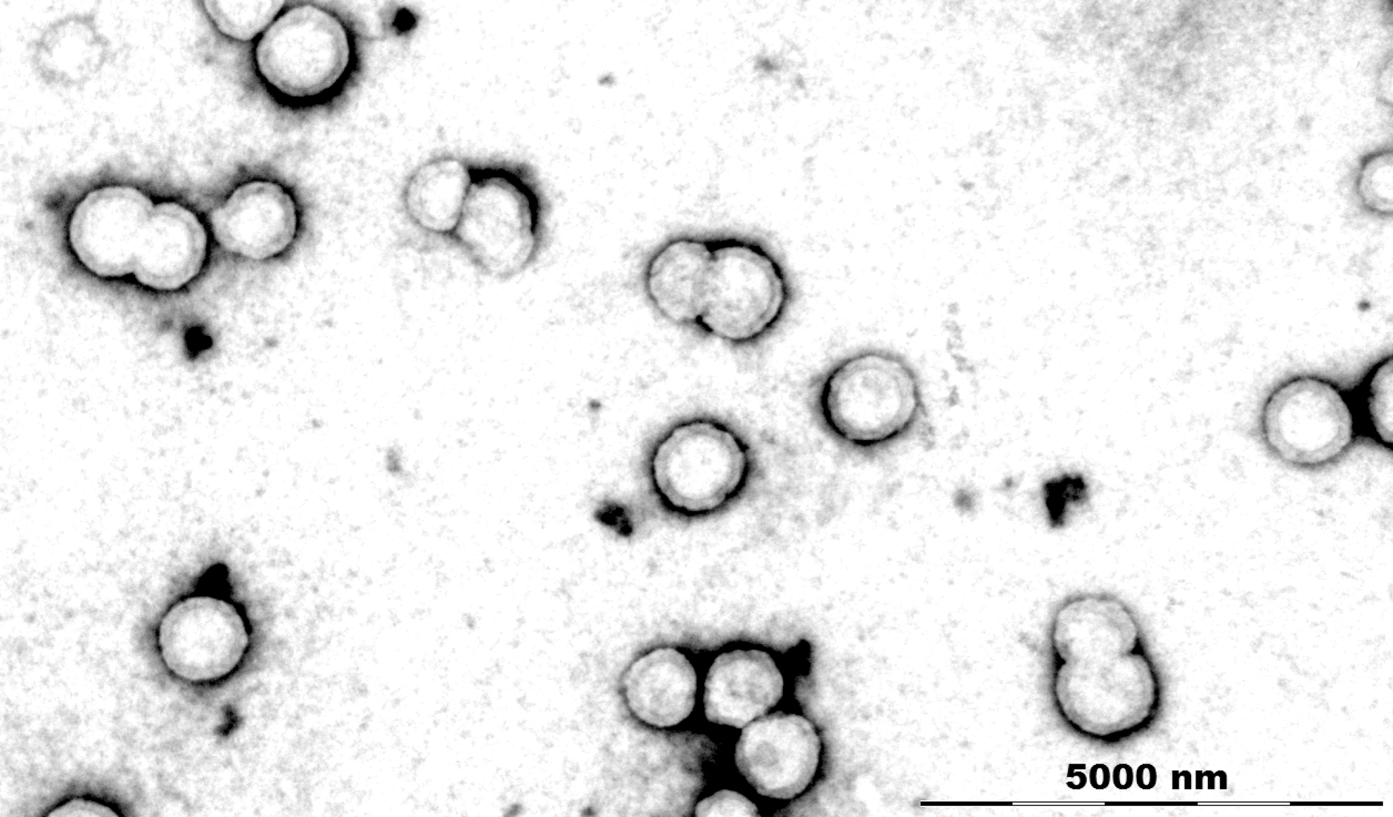
Transmission electron microscope image of the 170Tm-tin-dioxide microparticles for estimation of particle size distribution and examination of particle morphology. Particles had nearly spherical shape and distribution between 1 and 10 μm. 170Tm, Thulium-170.
The size and shape of the particle were found to remain unaltered during the course of entire in vitro particle stability investigation period. DLS confirmed that the mean whole-particle fractions of the microparticles have the hydrodynamic diameters in the range of 1.4–3.2 μm during the 9-month follow-up and mean (average) particle sizes were between 1.6 and 2.6 μm. The analyses clearly demonstrated that the storage at room temperature did not produce a remarkable difference in the mean values of size distributions.
The radiolabeled microparticles were obtained as a clearly visible white sedimentation with slightly opalescent and colorless supernatant. The total labeling volume of the preparation was maintained at 600 μL and 214 MBq of 170Tm activity was incorporated in the preparation. This ensured the preparation of radiolabeled microparticles with high radioactive concentration that is essential for using colloidal agents for RSV in order to deliver sufficient dose to the target with minimal leakage.
In ITLC-SG, 170Tm-labeled microparticles were observed to be retained near the point of spotting (R f=0), while 170TmCl3 has migrated toward the solvent front (R f=1) under identical conditions. The ITLC-SG studies revealed that 170Tm-labeled microparticles could be prepared with>99% radiolabeling efficiency under the optimized preparation procedure mentioned earlier. Presence of Tm-170-labeled degradation products could not be detected between R f=0.1 and R f=1. These results were further corroborated through TLC scanning studies. A typical TLC chromatogram obtained using the TLC scanner is shown in Figure 2.
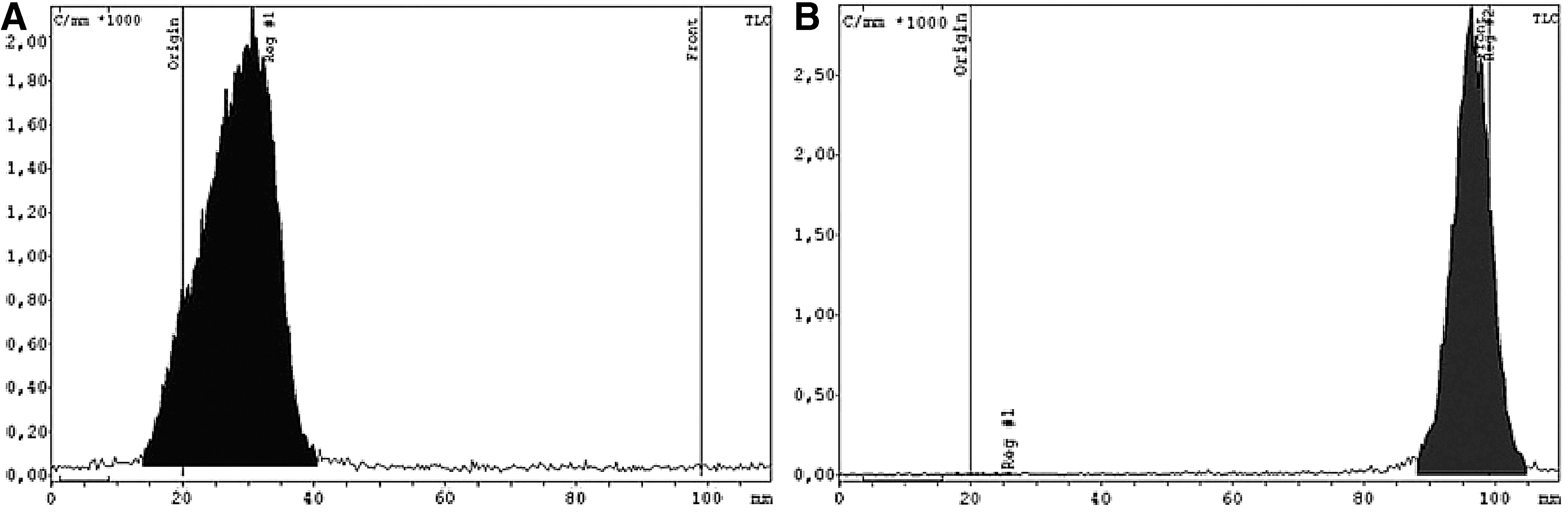
TLC chromatograms of
The radiolabeled microparticles, prepared following the optimized protocol, showed high in vitro radiochemical stability at room temperature. The labeling efficiency of the preparation was found to be >98% after 1 hour, 3 hours, 24 hours, 2 days, and 7 days and >93% after 2 weeks, 4 weeks, 5 weeks, 9 weeks, 14 weeks, and 9 months of storage, respectively. The produced radiocolloid showed high in vitro serum stability also during the 1-month incubation. In the canine serum sample, the labeling efficiency was found to be >98% after 1, 3, and 24 hours; >91% after 2 and 7 days; and >89% after 2 and 4 weeks.
Results of biological studies
Biological evaluation of the radiolabeled microparticles was carried out in 3 healthy, male Beagle dogs. In the experimental animal, any clinical side-effects or signs of histopathological changes of the joints were not recorded during the examinations. The preparation was intraarticularly administered into the right knee joint of the animals and detailed SPECT-CT images of the animals were recorded 4 hours postinjection. SPECT-CT studies provided high-resolution fusion images that clearly and conspicuously showed the localization of the radiolabeled particulates in the knee joints as well as leakages of the particles, if any from the joint with the progress of time. Figure 3 represents representative dorsoventral and lateral SPECT-CT fused images of the animals recorded 4 hours postadministration. In this figure, CT layers appear in gray scale, while SPECT layers have colored tones (brighter tones represent higher activity accumulations). The SPECT-CT images recorded at various postadministration time points indicated the lack of leakage of the injected radiocolloid outside the joint.
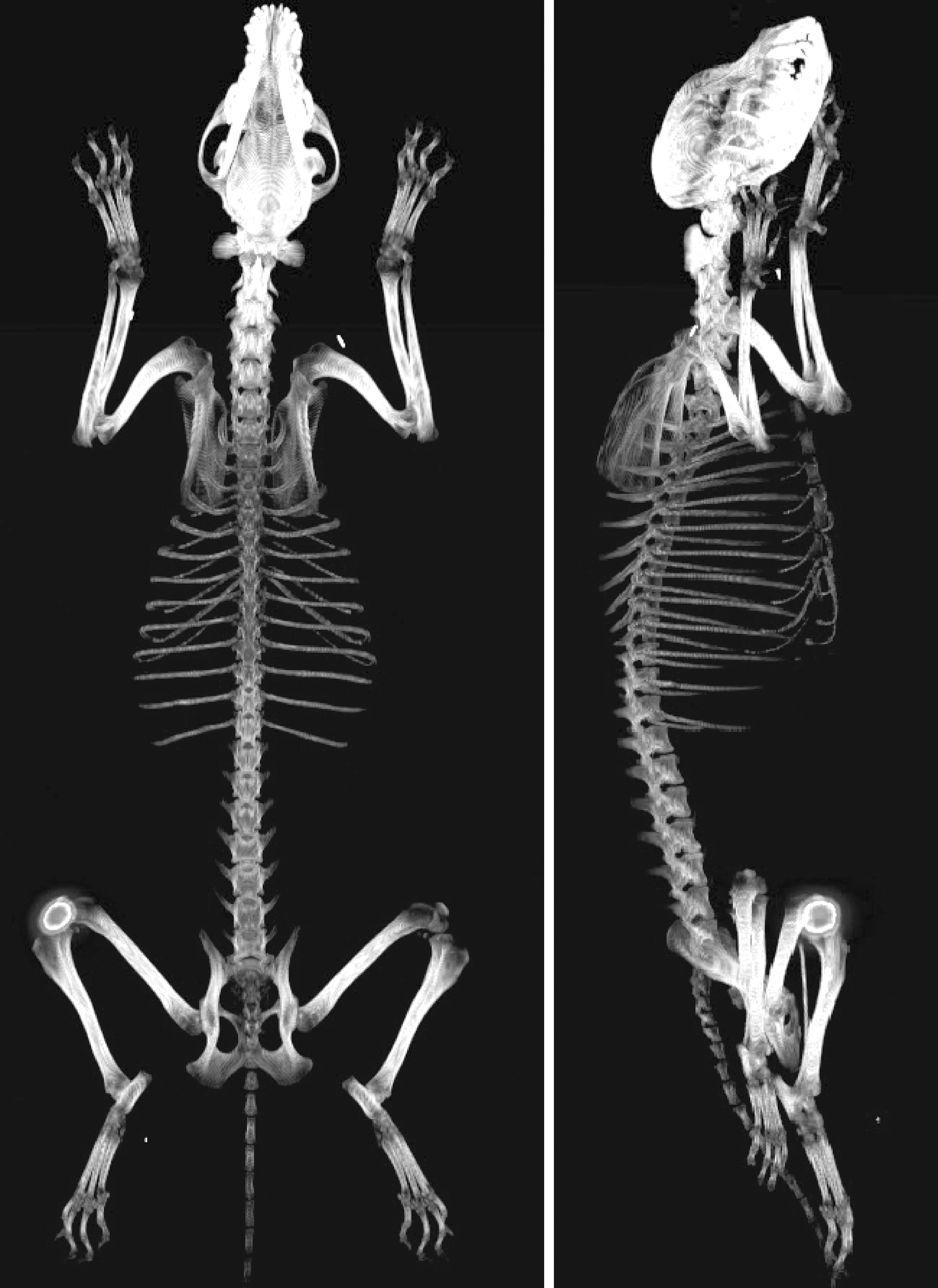
Dorsoventral and lateral fused SPECT-CT whole-body images of Beagle dog recorded after 4 hours of administration of 178 MBq 170Tm-labeled microparticles (volume 500 μL). Radiocolloid accumulation is displayed in the right knee joint.
The aforementioned findings were also corroborated through the serial SPECT studies, which also showed very low leakage of radioactivity from the knee joints after intraarticular injection. Figure 4 represents ventrodorsal whole-body images of the animals recorded at various time points to 9 months postinjection. In these images different activity densities appear in gray scale with darker tones represent higher activity accumulation. Body outlines of the animals, which were drawn using a normal photo from the view of the γ detector, are also added with these images in order to illustrate the actual position of localization of activity in the body of the animals. It is evident from these images that majority of the administered activity remained at the knee joint till 9 months postinjection while a small fraction was leaked out and accumulated in the inguinal lymph nodes. Uptake in the inguinal lymph nodes could be observed in the scans from 24 hours postinjection time points onward and a maximum of 8% injected activity was found in the lymph node during the 9-month follow-up. Apart from this, detectable uptakes were observed in liver but it was always <2% throughout the study. No other organs and tissues exhibited any detectable amount of injected activity during the 9-month follow-up. Examinations of blood, urine, and fecal samples have reinforced the lack of leakage of the injected product; only negligible amounts of radioactivity were found (under the detectable range) in the collected samples.

Serial whole-body scans (with body outlines) of Beagle dog recorded at various postinjection time points (up to 9 months) after the administration of 178 MBq 170Tm-labeled microparticles (volume 500 μL). Radioactive accumulations were displayed in gray scale with darker tones representing higher activity. Apart from knee joint, inguinal lymph node uptake could be observed from 24 hours postadministration.
Moreover, no significant changes were observed in hematological and biochemical parameters as well as in comet assays before and after administration of the radiolabeled microparticles (Table 1). All these biological evaluations indicate that the product had no undesirable side-effects and thus might be safe for further evaluation.
Dosimetric estimations
Dosimetric estimations were calculated to the treated knee joints and the inguinal lymph nodes. Apart from the mentioned organs, only negligible liver uptakes could be identified in the whole-body scans. Moreover, during the 9-month (270 days) observation time, there was not any detectable activity in other organs and tracts, which could be used further for quantitative evaluations. Pharmacokinetic behavior of the injected 170Tm-tin-dioxide microparticles in the treated knee joints of the Beagle dogs is shown in Figure 5. A bi-exponential retention with no discernible uptake phase could be identified from the pharmacokinetic properties of the injected microparticles. The resulted biological time was T 1bio=123.3 days for the slow-washout component and T 2bio=0.8 days for the rapid-washout component.

Accumulation and pharmacokinetic properties of injected 170Tm-labeled microparticles (average values±SD) in the treated knee joints of the Beagle dogs during the 9-month period.
On the other hand, inguinal lymph node activities had bi-exponential retention with a discernible uptake phase and a single washout phase (Fig. 6). For the lymph nodes, the calculated biological time was T 1bio=72 days (biologic uptake component) and T 2bio=0.5 days was the biologic disappearance component. The absorbed dose for target organs per unit of cumulated activity in source organs (S) (which is a physical quantity that depends on the nature of the radiations and their absorption characteristics as well as on the anatomic model) was calculated using the MIRD tables, unit density sphere model. Table 2 provides the estimated values for the unit imparted dose in Gy per MBq unit. Therefore, assuming that the Beagle dog knee had 100 g mass (diameter of sphere model d=5.58 cm), in the case of intraarticularly injected 170Tm-tin-dioxide microparticles, the absorbed dose per injected value activity ratio was found about 3.3 Gy/MBq.

Accumulation and pharmacokinetic properties of injected 170Tm-labeled microparticles (average values±SD) in the inguinal lymph nodes of the Beagle dogs during the 9-month period.
Estimated absorbed dose/injected activity density values using sphere spatial model.
Conclusions
The main objective of the present work was to develop a new 170Tm radionuclide carrier colloid–based therapeutic radiopharmaceutical that may serve as a potential and inexpensive alternative to other products presently used in RSV (mostly colloidal suspensions labeled with 90Y, 153Sm, 165Dy, 166Ho, 169Er177Lu, 186Re, 188Re, and 198Au radionuclides). Toward this, production of 170Tm was achieved with high radionuclidic purity and sufficient specific activity using inexpensive natural thulium oxide target. 170Tm-labeled tin-dioxide microparticles were prepared following a simple method that yielded high radiochemical purity and adequate in vitro stability. In vivo studies carried out in normal healthy Beagle dogs revealed high in vivo stability of the preparation along with significant retention of activity in the knee joints till 9 months postinjection. Effective dose received in the knee joint was also calculated. This preliminary study revealed the potential of the developed agent toward its application as an alternative agent for RSV. These types of 170Tm-labeled particles could be useful for locoregional administration in hepatic cancer also. However, further investigations will be required in biological models for unraveling the true potential of the developed agent.
Footnotes
Disclosure Statement
There are no existing financial conflicts.