
Abstract
The PET radiopharmaceutical [18F]Fluromisonidazole ([18F]FMISO) is presently the agent of choice for the clinical imaging of tumor hypoxia. Considering the logistic advantages of 99mTc and wider availability of SPECT machines, a 99mTc-radiopharmaceutical for this purpose constitutes an attractive choice. In the work presented here, a misonidazole analogue was radiolabeled with 99mTc(CO)3 core and the complex was evaluated in Swiss mice bearing fibrosarcoma tumor. The results obtained are compared with the biodistribution of [18F]FMISO carried out in the same tumor-bearing animal model. Misonidazole-99mTc(CO)3 complex showed significant uptake and retention in tumor. Notably, the rate of clearance of misonidazole complex from the tumor was slower than that of [18F]FMISO. The maximum tumor/muscle ratio obtained with misonidazole-99mTc(CO)3 complex was significantly higher than that of [18F]FMISO. The study constitutes a positive step toward the development of a 99mTc-analogue of [18F]FMISO.
Introduction
The role of hypoxia in tumor propagation, malignant progression, and resistance to therapy is well documented. 1 –9 Presence of hypoxia has been correlated to poor prognosis in several types of cancers such as advanced squamous cell carcinoma of cervix, 2 –4 head and neck cancer, 5 –7 adenocarcinoma of pancreas, 8 soft tissue sarcoma, and so on. 9 Thus, in the clinical management of cancer, detection of hypoxia has assumed significance. Nitroimidazoles are known to undergo selective reduction and accumulation in hypoxic cells. 10 Therefore, majority of radiopharmaceuticals evaluated for the detection of hypoxia are based on nitroimidazole. At present, [18F]Fluromisonidazole ([18F]FMISO), a 2-nitroimidazole analogue, is the radiopharmaceutical of choice for the detection of hypoxia in the clinic. It is a PET-radiopharmaceutical that allows unambiguous detection of clinically significant regional hypoxia in cancerous tissues. 10 Though other 18F-labeled nitroimidazole radiopharmaceuticals like [18F]FETA, 11 [18F]EF1, 12 [18F]EF5, 13 –16 [18F]FAZA, and others 17,18 are reported, [18F]FMISO continues to be the primary choice for detection of hypoxia.
Preparation of [18F]FMISO requires cyclotron facility and specialized modules, which significantly increases the cost of this radiopharmaceutical. Other problems associated with [18F]FMISO include its uptake in brain and slow clearance from nontarget organs, which compromises the quality of the image obtained. 14 Delaying image acquisition to improve target to nontarget ratio is of limited scope because of the short half-life (109.8 minutes) of 18 F. Considering these factors, development of a 99mTc-radiopharmaceutical that can provide diagnostic information equivalent to or better than [18F]FMISO assumes significance. Other advantages with 99mTc include its easy availability (99Mo/99mTc generator), low cost, optimal half-life (6 hours), and the possibility of developing a lyophilized kit formulation for the preparation of the 99mTc-radiopharmaceutical.
At present, the advantage of PET over SPECT imaging is the superior spatial resolution of the images obtained with the former, which is critical for planning hypoxia-directed external beam radiation therapy. 19 However, advances being made in hardware and image reconstruction algorithms have significantly improved the spatial resolution of images obtainable with SPECT. 20 In the near future, it may be possible to utilize the information available from SPECT images for planning hypoxia-directed radiotherapy, which is another compelling reason for developing a 99mTc-radiopharmaceutical for hypoxia imaging applications.
The search for a 99mTc-radiopharmaceutical for hypoxia imaging started couple of decades ago. Several 99mTc-agents, prepared utilizing the diverse chemistry of 99mTc, have been evaluated for hypoxia imaging applications with varying levels of success. 21 –44 BRU59-21 is a prominent 99mTc-complex that has undergone phase-I clinical evaluation, but could not be taken any further. 38 Our group has also worked on different 99mTc-labeled nitroimidazole complexes, employing some of the recent approaches of radiolabeling with 99mTc, but again with limited success. 39 –45 Most of the radiopharmaceuticals evaluated for targeting hypoxia have utilized one of the three nitroimidazoles, 2-, 4-, or 5-nitroimidazole, the 2-nitroimidazole being the most common choice. The primary reason for this preferential choice is the molecular property, single electron reduction potential (SERP), which decides the efficiency of reduction of nitroimidazole in hypoxic cell. The SERP of a number of nitroimidazole derivatives has been reported by Peter Wardman. Typical values of SERP of unsubstituted 2-, 4-, and 5-nitroimidazole are −418 mV, −527 mV, and −450 mV with respect to standard hydrogen electrode (SHE) in aqueous solution, respectively. 46 Between two nitroimidazoles, the one with a more positive SERP is expected to undergo faster reduction in hypoxic cells and in this context the 2-nitroimidazole becomes the obvious choice. Misonidazole is a 2-nitroimidazole derivative with a SERP value of −389 mV 46 (against SHE). This compound was initially developed as a radiosensitizer. [18F]FMISO is its fluorinated analogue, which is clinically used as a radiopharmaceutical for the detection of tissue hypoxia. To the best of our knowledge, there is no report so far on the preparation and evaluation of a 99mTc-labeled misonidazole derivative that, in a way, should be the logical starting point toward the development of a 99mTc-agent equivalent to [18F]FMISO.
Unlike labeling with radiohalogens like 18 F, radiolabeling with 99mTc requires a chelator in the target molecule for radiolabeling with 99mTc. The choice of chelator depends on the radiolabeling strategy. Versatile chemistry of 99mTc offers several options for radiolabeling a biomolecule. 47 Among different approaches, the [99mTc(CO)3(H2O)3]+ precursor, introduced by Alberto et al., 48 is particularly useful for radiolabeling small molecules at low ligand concentration, yielding complexes with high radiochemical purity (RCP) and in vivo stability. Chelators having O, N, or S donor atoms are most appropriate for radiolabeling molecules with 99mTc(CO)3 core. The three substitution labile water molecules in [99mTc(CO)3(H2O)3]+ precursor complex facilitated the formation of a pseudo-octahedral complex with appropriate monodentate, bidentate or tridentate ligands. 49 In the present work, an iminodiacetic acid (IDA) derivative of misonidazole (miso-IDA), a tridentate ligand having two “O” and one “N” donor atoms, was synthesized and radiolabeled using [99mTc(CO)3(H2O)3]+ precursor complex. After characterization using appropriate techniques, this complex was evaluated in Swiss mice bearing fibrosarcoma tumor. Biodistribution of [18F]FMISO was also carried out in the same animal model and the results are discussed.
Materials and Methods
General
2-Nitroimidazole was purchased from Aldrich. 2,3-Epoxypropyl phthalimide, N-ethyl-diisopropylamine (DIEA), and tert-butyl bromoacetate were purchased from Fluka. All other reagents were of analytical grade and used as such without additional purification. Silica gel plates (silica gel 60 F254) used for thin layer chromatography (TLC) and silica gel (60–120 mesh) used for column chromatography were obtained from Merck. [18F]FMISO was obtained from Radiation Medicine Centre, Mumbai, India. Sodium pertechnetate was eluted from 99Mo/99mTc alumina column generator using normal saline. The [99mTc(CO)3(H2O)3]+ precursor was prepared using Isolink® carbonyl kit vial obtained as a gift from Mallinckrodt Medical B. V. High-performance liquid chromatography (HPLC) analyses were performed on a JASCO PU 2080 Plus dual pump HPLC system, with a JASCO 2075 Plus tunable absorption detector and a Gina Star radiometric detector system, using a C18 reversed phase HiQ Sil (5 μm, 4×250 mm) column. IR spectra were recorded on a JASCO-FT/IR-420 spectrophotometer. 1 H-NMR spectra were recorded either using a 300 MHz Varian VXR 300S or 300 MHz Bruker AvanceII NMR spectrometer. Mass spectra were recorded on a Varian 500MS Ion trap mass spectrometer.
Synthesis of 2-(2-hydroxy-3-(2-nitro-1H-imidazol-1-yl)propyl)isoindoline-1,3-dione
(1)
To 2-nitroimidazole (0.5 g, 4.43 mmol) in absolute ethanol (20 mL), 2,3-epoxypropyl phthalimide (1 g, 4.87 mmol) was added and the reaction mixture was heated at reflux. Upon completion of the reaction (cf. TLC), the pale-white precipitate obtained was filtered. The precipitate was washed with cold ethanol (5 mL) followed by water (5 mL) and dried. Compound
Synthesis of 1-amino-3-(2-nitro-1H-imidazol-1-yl)propan-2-ol hydrochloride (2)
To compound
Synthesis of N,N-bis(tert-butoxycarbonylmethyl)-[(2-hydroxy)-3-(2-nitroimidazolyl)]propyl amine (3)
To a solution of compound
Synthesis of 2-{N-carboxymethyl, N-[(2-hydroxy)-3-(2-nitroimidazolyl)propyl]amino}acetic acid (4)
Compound
Radiolabeling
Preparation of [99mTc(CO)3(H2O)3]+ precursor complex
The preparation of [99mTc(CO)3(H2O)3]+ precursor complex involved the addition of 1 mL of freshly eluted Na99mTcO4 from 99Mo/99mTc alumina column generator to the Isolink kit vial and heating the vial at 95°C for 20 minutes. After cooling and re-equilibrating to atmospheric pressure, the pH of the reaction mixture was adjusted to 7 using 1:3 mixture of 0.5 M phosphate buffer (pH 7.4):1 N HCl.
Preparation of 99mTc(CO)3 complex
About 100 μL of freshly prepared [99mTc(CO)3(H2O)3]+ precursor was added to 900 μL of 10−3 M solution of the ligand in phosphate buffer (pH 7.4) and incubated for 30 minutes in a water bath at 70°C. The reaction mixture was cooled to room temperature and characterized by HPLC.
Quality control
High-performance liquid chromatography
The RCP of the [99mTc(CO)3(H2O)3]+ precursor complex and miso-IDA-99mTc(CO)3 complex was assessed by HPLC with a C18 reversed phase column. About 15 μL of the test solution (∼0.037 MBq) was injected into the column and elution was monitored by observing the radioactivity profile. Aqueous 0.05 M triethylammonium phosphate (TEAP) buffer, pH=2.5 (Solvent A) and methanol (Solvent B) were used as the mobile phase. Both the solvents were filtered through 0.22 μm filter. The elution started with 100% A from 0 to 6 minutes. At 6 minutes the eluent was switched to 75% A and 25% B and at 9 minutes to 66% A and 34% B followed by a linear gradient 66% A/34% B to 100% B from 9 to 20 minutes. Up to 30 minutes the eluent remained at 100% B before reverting back to the initial conditions. Flow rate was maintained at 1 mL/min. Percentage RCP (%RCP) of 99mTc(CO)3 complex was calculated from the peak area measurements of the HPLC chromatogram.
Partition coefficient (LogP o/w)
About 100 μL of the labeled compound was mixed with 0.9 mL of water and 1 mL of n-octanol on a vortex mixer for about 3 minutes and then centrifuged at 3500 g for 5 minutes to effect clear separation of the two layers. About 0.8 mL of the n-octanol layer was withdrawn and equal volume of water was added. The mixture was vortexed again and centrifuged as described above. Equal aliquots in triplicate were withdrawn from n-octanol and aqueous layer and radioactivity associated with each layer was determined in an NaI(Tl) counter. The partition coefficient was calculated using the equation: LogP o/w=Log[(counts in n-octanol layer)/(counts in aqueous layer)].
In vitro serum stability
About 50 μL of the labeled compound was added to 0.5 mL of human serum and this mixture was incubated at 37°C. Equal aliquots (100 μL) were drawn at 1, 2, and 3 hours and an equal volume of ethanol was added to precipitate the serum proteins. The mixtures were centrifuged and the supernatants were analyzed by HPLC to assess the stability of the complex in serum.
In vivo studies
All procedures performed herein were in strict compliance with the national laws governing the conduct of animal experiments. Solid tumors were developed in Swiss mice by implantation of HSDM1C1 murine fibrosarcoma. The fibrosarcoma cells were procured from National Centre for Cell Science (NCCS), Pune, India. Cells were cultured at 37°C and 5% CO2 atmosphere in Dulbecco's modified Eagle's medium supplemented with 10% fetal bovine serum. About 106 cultured cells in 100 μL volume were subcutaneously injected on the dorsum of every animal. The tumors were allowed to grow till they reached ∼10 mm in diameter after which the animals were used for in vivo studies. For the biodistribution studies, the radioactive preparation (∼0.37 MBq in 100 μL volume) was intravenously administered through the lateral tail vein of the animal. Individual set of animals (n=3) were utilized for studying the biodistribution at different time points (30, 60, and 180 minutes). At the end of respective time periods, animals were sacrificed and the relevant organs excised for measurement of retained activity. The organs were weighed and the activity associated with each organ was measured in a flat-bed type NaI(Tl) counter with energy window set for 99mTc (140keV±10%). The activity observed with each organ or tissue at different time points were expressed as a percentage injected dose per gram (%ID/g).
Determination of stability of nitroimidazole complexes in vivo
The blood collected from the tumor-bearing animal at 3 hours postinjection (p.i.), after measuring the activity associated with it, was allowed to clot and the serum is separated. Serum proteins are then precipitated using ethanol and separated by centrifugation. Supernatant is then analyzed by HPLC.
Results and Discussions
Primary reason for selecting a tridentate IDA as the chelator ligand for the synthesis of misonidazole derivative is as follows. We had recently evaluated a series of differently substituted nitroimidazole-99mTc(CO)3 complexes having different values of SERP, overall charge, and lipophilicity. 37 We observed that blood clearance of the nitroimidazole complex was strongly depended on the nature of the tridentate ligand present in it. Nitroimidazole-99mTc(CO)3 complexes having diethylene triamine (DETA) and aminoethyl glycine (AEG) as the tridentate chelators were observed to clear very fast from blood, whereas the nitroimidazole-IDA complexes cleared slowly. Consequently, the tumor uptake showed by DETA and AEG complexes was lower than the corresponding nitroimidazole-IDA complex. These observations indicated that tridenate IDA may be a more suitable ligand to avoid faster blood clearance of the radiotracer.
Miso-IDA derivative was synthesized in four steps (Fig. 1). The 2-nitroimidazole and 2, 3-epoxypropyl phthalimide were coupled in the first step to obtain the basic misonidazole framework (
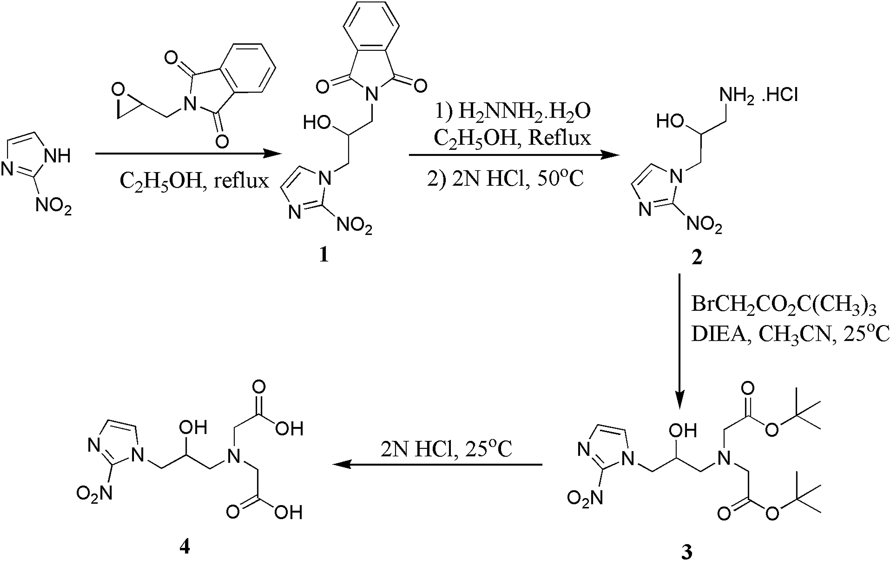
Synthesis of miso-IDA ligand. IDA, iminodiacetic acid.
Miso-IDA was characterized by IR, 1 H-NMR, and mass spectrometer. 1 H-NMR was consistent with the expected structure of miso-IDA and (M-H)− peak at m/z 301.1 served as an additional evidence for the formation of the expected ligand. The [99mTc(CO)3(H2O)3]+ precursor complex was prepared using Isolink™ carbonyl kit vial obtained as a gift from Mallinckrodt Medical B. V. Miso-IDA was subsequently radiolabeled using [99mTc(CO)3(H2O)3]+ precursor to obtain miso-IDA-99mTc(CO)3 complex. The [99mTc(CO)3(H2O)3]+ precursor complex and miso-IDA-99mTc(CO)3 complex were analyzed by HPLC. Typical elution profiles obtained are shown in Figure 2a and 2b. The peak corresponding to [99mTc(CO)3(H2O)3]+ precursor complex appeared at 3.7 minutes while that of miso-IDA-99mTc(CO)3 complex appeared at 13.9 minutes. The only other minor impurity observed in the HPLC elution profile of miso-IDA-99mTc(CO)3 complex was the unreacted [99mTc(CO)3(H2O)3]+ precursor complex. Radiolabeling yield was consistently more than 95%. The RCP of miso-IDA-99mTc(CO)3 complex calculated from the peak area measurements was 96.5%±1% (n=6). Typical specific activity of miso-IDA-99mTc(CO)3 complex used for biodistribution is 107.2±1.2 μCi/μmol of the ligand (n=6). Lipophilicity of miso-IDA-99mTc(CO)3 complex (LogP o/w) was determined following a reported procedure 50 and it was found to be 0.17. In comparison, the LogP o/w value of [18F]FMISO was 0.41. 10 The miso-IDA-99mTc(CO)3 complex showed no signs of decomposition when incubated in human serum for 3 hours at 37°C.
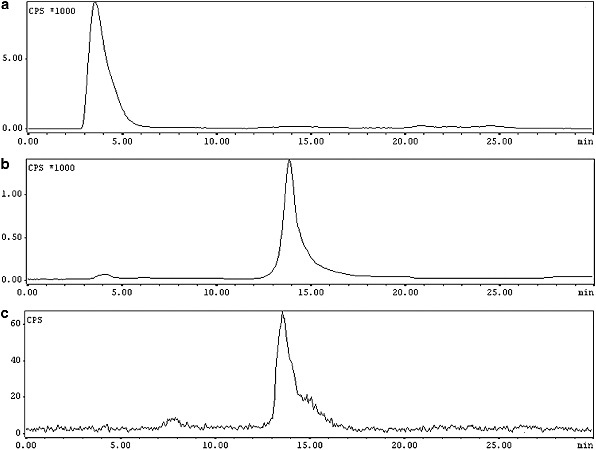
High-performance liquid chromatography elution profile of
The miso-IDA-99mTc(CO)3 complex and [18F]FMISO (obtained from Radiation Medicine Centre, Mumbai, India) was evaluated in Swiss mice bearing fibrosarcoma tumor. The in vivo distribution of these two complexes in Swiss mice is shown in Table 1. It could be observed that major clearance of activity for both the complexes was through hepatobiliary route. Fraction of [18F]FMISO activity cleared through renal route was slightly higher than that of miso-IDA-99mTc(CO)3 complex though the latter was hydrophilic than the former. Uptake of [18F]FMISO activity in organs such as heart, lungs, spleen, and bones was also higher than that of miso-IDA-99mTc(CO)3 complex at every time point studied. This could be explained considering relatively higher lipophilicity of [18F]FMISO (LogP o/w=0.41) 10 compared to miso-IDA-99mTc(CO)3 complex (LogP o/w=0.17). However, activity accumulated in these organs cleared with time.
[18F]FMISO, [18F]Fluromisonidazole; %ID/g, percentage injected dose per gram; IDA, iminodiacetic acid; SD, standard deviation.
Tumor uptake of [18F]FMISO and miso-IDA-99mTc(CO)3 complex at different time points are shown in Figure 3. It could be observed that [18F]FMISO showed significantly higher tumor uptake than miso-IDA-99mTc(CO)3 complex at all the time points (30, 60 and 180 minutes). Apparently both complexes showed bi-phasic clearance from tumor, with a relatively faster initial phase between 30 and 60 minutes p.i., followed by a slow clearance phase after 60 minutes p.i.

Tumor uptake observed with miso-IDA-99mTc(CO)3 complex and [18F]FMISO at different time points. [18F]FMISO, [18F]Fluromisonidazole.
The observed tumor uptake pattern is similar to many other studies reported earlier. 39 –45 The decrease in tumor activity between 30 and 60 minutes p.i. (Fig. 3) could be attributed to the clearance of unreduced radiotracer from the tumor, initiated by the reversal of concentration gradient between blood pool and intra-tumoral environment due to the clearance of the radiotracer from blood. This is evident from Figure 4, which shows the blood clearance of miso-IDA-99mTc(CO)3 complex and [18F]FMISO from blood at different time points. It could be observed that at 30 minutes p.i., blood activity of miso-IDA-99mTc(CO)3 complex was lower than [18F]FMISO. This indicates faster clearance of miso-IDA-99mTc(CO)3 complex from the blood pool in the initial phase (0–30 minutes) of the study. Relatively slower clearance of [18F]FMISO from blood in the initial phase could be attributed to its protein binding owing to higher lipophilicity (LogP o/w=0.41) 10 compared with miso-IDA complex (LogP o/w=0.17). The blood clearance of both radiotracers was through hepatobiliary route, which is evident from the presence of significant levels of activity in liver and intestine (Table 1). Earlier, we had observed that the rate of clearance of radiotracer from blood had a significant effect on its uptake and retention in tumor. 37 Lower tumor uptake of miso-IDA-99mTc(CO)3 complex compared with [18F]FMISO could be attributed to the faster blood clearance of the former in the initial phase. Faster blood clearance leads to inadequate distribution of the radiotracer in tumor at the same time decreases the time the radiotracer actually spends in hypoxic tumor cells to undergo reduction and trapping.

Clearance of miso-IDA-99mTc(CO)3 complex and [18F]FMISO from blood at different time points.
Slow clearance of activity from tumor after 60 minutes p.i. could be attributed radiotracer reduced and trapped in hypoxic tumor cells. Steadily decreasing activity levels in muscles (Table 1) and slow clearance of activity from tumor (Fig. 3) is an additional positive indication of hypoxia-specific reduction occurring in tumor.
The miso-IDA-99mTc(CO)3 complex did not showed any degradation upon incubating in human serum for 3 hours. To identify the activity associated with the circulating blood pool, blood drawn from the animal at 3 hours p.i. was processed to precipitate serum proteins and the supernatant was analyzed by HPLC. The HPLC analysis showed that major activity in blood is due to the intact miso-IDA-99mTc(CO)3 complex (Fig. 2c).
The tumor activity observed with miso-IDA-99mTc(CO)3 complex and [18F]FMISO at 60 minutes p.i. cleared slowly with time. However, miso-IDA-99mTc(CO)3 complex cleared slower than [18F]FMISO. With [18F]FMISO, ∼45% of tumor-associated activity observed at 60 minutes p.i. was cleared at 180 minutes p.i. During the same interval only 28% clearance in tumor activity was observed for miso-IDA-99mTc(CO)3 complex. A possible explanation for the slower clearance of tumor activity in the case of miso-IDA-99mTc(CO)3 complex may be that their reduction products formed in hypoxic cells are less permeable across the cell membrane than that of [18F]FMISO. In this work, however, no attempt was made to detect or identify the reduction products of either radiotracer.
The variation in tumor to blood ratio (TBR) and tumor to muscle ratio (TMR) observed with miso-IDA-99mTc(CO)3 complex and [18F]FMISO is shown in Table 2. While the TBR of [18F]FMISO gradually increased with time, that of miso-IDA-99mTc(CO)3 complex showed only marginal improvement, attaining a maximum value of 1.15 (0.05) at 180 minutes p.i. Though initial blood activity (30 minutes p.i.) of miso-IDA-99mTc(CO)3 complex was lower than that of [18F]FMISO, its subsequent clearance from blood was slow [0.84 (0.13)%ID/g retained in blood even at 180 minutes p.i.], which adversely affected the TBR.
The TMR obtained with miso-IDA-99mTc(CO)3 complex gradually increased with time reaching a maximum value of 93(0.36) at 3 hours p.i. (Table 2). This could be attributed to fast clearance of the radiotracer from muscles with time. Maximum TMR obtained with [18F]FMISO was only 4.84 (0.90) at 180 minutes p.i.
Conclusions
In terms of absolute tumor uptake, miso-IDA-99mTc(CO)3 complex was not at par with [18F]FMISO. However, very high TMR and relatively slow clearance from tumor are the two advantages observed with miso-IDA-99mTc(CO)3 complex. A possible hint for improving tumor uptake and TBR of miso-IDA-99mTc(CO)3 complex could be obtained from the blood clearance pattern of [18F]FMISO. [18F]FMISO showed initial high blood pool activity, which facilitated better distribution in tumor and provided sufficient tumor residence time for the radiotracer to undergo reduction and trapping in hypoxic cells. Subsequently, gradual clearance of blood activity improved the TBR. It would be an interesting idea to see whether significant improvement in overall biodistribution is possible by structural modification of miso-99mTc(CO)3 complex to mimic the blood clearance pattern of [18F]FMISO.
Footnotes
Acknowledgments
Authors acknowledge all staff of the Radiochemistry Section, Isotope Production and Applications Division, BARC for providing Molybdenum-99 isotope. Authors also acknowledge Mr. Saikat Nandy and other staff of the Radiation Medicine Centre, Mumbai for providing [18F]FMISO for our work.
Disclosure Statement
No competing financial interests exist.