
Abstract
The greatest hurdle in cancer treatment is the metastasis of primary tumors to distant organs. Our knowledge on how different immune cells, in the absence of exogenous stimulation, prevent tumor metastasis in distant organs is poorly understood. Using a highly metastatic murine lung B16 melanoma cell line BL6-10, we employed naive mice that genetically lack CD4+ or CD8+ T cells, or are depleted of dendritic cells (DCs) or natural killer (NK) cells to understand the relative importance of these cells in metastasis prevention. Irrespective of the presence of naïve CD4+ T, CD8+ T, DCs, or NK cells, lungs, which act as primary site of predilection for B16 melanoma, readily developed numerous lung BL6-10 melanoma colonies. However, their absence led to B16 melanoma metastasis in variable proportions to distant organs, particularly livers, kidneys, adrenals, ovaries, and hearts. NK cells mediate prevention of BL6-10 metastasis to various organs, especially to livers. Mechanistically, CD40L signaling, a critical factor required for DC licensing and CD8+ cytotoxic T lymphocyte (CTL) responses, was required for CD4+ T cell-mediated prevention of systemic BL6-10 metastasis. These results suggest that the composition and functions of different immune cells in distant tissue microenvironments (distant organs other than primary sites of predilection) robustly mediate natural resistance against melanoma metastasis. Thus, harnessing these immune cells' responses in immunotherapeutics would considerably limit organ metastasis.
Introduction
Cancer immune surveillance is an important host protection process to inhibit carcinogenesis and primary cancer formation. 1 The central roles of both innate and adoptive immune cells such as natural killer (NK), CD4+ and CD8+ T cells, and dendritic cells (DCs) have been identified in cancer immune surveillance. 1
Melanoma is the most aggressive and metastatic form of skin cancer. 2 Most deaths occur from disseminated, therapy-resistant tumors disrupting major organ function. 3 Melanoma metastasis is a complex process requiring melanoma cell detachment from the primary tumor and migration to secondary sites in the body through the lymphatic or blood circulatory systems. 4,5 Cancer metastasis results from a nonrandom process, in which organ selectivity by the tumor cells is largely determined by factors such as chemokines that are expressed at the remote organs that eventually turn into preferred sites of metastasis formation. 6 In accordance with the “seed and soil” hypothesis, cancer metastatic cells function as “seeds” and a particular organ microenvironment or niche serves as the “soil.” 7 It is then accompanied by secretion of factors including proteases, cytokines, and chemokines from tumors and tissues or immune cells at the secondary sites, performing autocrine and paracrine roles to promote metastatic tumor cell growth. 8 Therefore, immune cells including both innate and adoptive immune cells in distant organ environments, which contribute to immune surveillance, should also be considered as another part of the “soil” components. However, how these different innate and adoptive immune cells such as NK and T cells such as CD4+ and CD8+ T cells and antigen-presenting cells such as DCs contribute to natural resistance against tumor metastasis in distinctive organs has less been studied.
In this study, we selected a highly lung metastatic B16 melanoma cell line BL6-10, 9 as the “seed,” and assessed the potential relationship between innate and adoptive immune cells and distinctive organ metastasis after BL6-10 cells were i.v. injected into wild-type C57BL/6 and CD4+ or CD8+ T cell knockout mice or mice with depletion of NK cells or DCs.
Materials and Methods
Reagents, tumor cells, and mice
Fluorescein isothiocyanate (FITC)-conjugated anti-CD4, anti-CD8, anti-CD11c, anti-Gr1, and anti-NK1.1 antibodies (Abs) were obtained from BD-Biosciences (San Diego, CA) for flow cytometric analysis. Anti-NK1.1, Ab for depletion of NK cells were purified from ascites fluids of PK136 hybridoma cells obtained from American Tissue Cell Collection (ATCC, Rockville, MD). Diptheria toxin (DT) was obtained from Sigma-Aldrich Canada Ltd (Oakville, Ontario, Canada). The highly lung metastatic mouse B16 melanoma cell line BL6-10 was obtained from Dr. Kimura, University of Florida. 9 Wild-type (WT) C57BL/6 (B6) mice and CD4−/−, CD8−/−, and CD40L−/− gene knockout (KO) and transgenic DTR-CD11c mice expressing DT receptor (DTR) under the control of CD11c promoter were purchased from Jackson Laboratory (Bar Harbor, ME). All animal experiments were carried out according to the guidelines of the Canadian Council for Animal Care.
Depletion of NK and DCs
C57BL/6 mice were intraperitoneally (i.p.) injected with anti-NK1.1 Ab (0.2 mg/mouse) at −1, +2, +5, +8, and +11 days relative to tumor inoculation at day 0, 10,11 for depletion of NK cells. Transgenic DTR-CD11c mice were injected with DT (i.p., 4 ng/g mouse body-weight) 1 day before tumor inoculation for depletion of DCs. 12
Adoptive transfer of naïve CD4+ or CD8+ T cells or DCs
The naïve polyclonal CD4+ or CD8+ T cells were isolated from C57BL/6 mice splenocytes by enriching T lymphocytes in nylon wool columns (C&A Scientific, Manassas, VA), and negative selection using anti-CD8 (L3T4) or anti-CD4 (L3T4) paramagnetic beads (DYNAL, Lake Success, NY) as previously described. 13 CD11c+ DCs (∼15×106 cells) purified from splenocytes of WT B6 mice using mouse CD11c positive selection kit as per manufacturer's instructions (Stemcell Tech, Vancouver, British Columbia, Canada) 14 were i.v. injected into DT-treated DTR-CD11c mice. To determine the contribution of immune cells in preventing melanoma organ metastasis, polyclonal CD4+ T cells (∼15×106 cells) purified from WT B6 or CD40L-deficient mice were i.v. transferred to CD4- or CD40L-deficient mice 1 day before and 5 days after tumor challenge. Similarly, polyclonal CD8+ T cells (∼15×106 cells) were i.v. transferred to CD8-deficient mice.
Tumor metastasis study
For experimental organ metastasis study, WT B6, gene KO, and DC-depleted mice or the above mice with adoptive cell transfer (five mice/group) were i.v. challenged with BL6-10 (0.5×10⁶ cells/mouse). 9 Twenty-four days after tumor cell inoculation, mice were sacrificed, and the visible black metastatic melanoma colonies were counted on fresh lungs or various distant organs, and confirmed by histological examination. Metastatic foci too many to count were assigned an arbitrary value of >100.
Results
CD4+ T environment prevents systemic B16 melanometastasis in multiple distant organs
In this study, we selected a highly lung metastatic B16 melanoma cell line BL6-10, 9 as the “seed,” and assessed the potential relationship between innate and adoptive immune cells and distinctive organ metastasis. We first i.v. injected BL6-10 melanoma cells into WT C57BL/6 mice. Four weeks after BL6-10 cell inoculation, we sacrificed the mice and found numerous black BL6-10 melanoma colonies (>100) in lungs in 5/5 mice (Fig. 1A), whereas no melanoma colony was seen in other organs such as liver, heart, kidney, and ovary (Fig. 1B). Compared to normal lungs, the mouse lungs with numerous melanoma colonies became larger in size. The melanoma colonies were confirmed by histological examination (Fig. 1C). To determine whether endogenous CD4+ T cells mediate natural immunosurveillance against organ metastasis, we also i.v. injected BL6-10 cells into CD4−/− mice with CD4+ T cell deficiency. Compared to WT, CD4-deficient mice showed drastic metastasis in distant organs in variable proportions (Fig. 2A, B): liver showed medium to large tumor colonies of varying numbers; both adrenals had medium to large tumor colonies; one or both ovaries showed large-sized tumor masses; and kidneys and heart also showed few small to medium-sized melanoma colonies. In addition to these organs, serosal layers of thorax and abdomen are frequently affected (data not shown). In contrast, lungs, being the primary site of predilection of both WT and CD4-deficient mice, showed severe tumor development although the degree of development was relatively higher in CD4-deficient mice (Fig. 2A, B). To further confirm the participation of CD4+ T environment, CD4-deficient mice were reconstituted with polyclonal naïve CD4+ T cells 1 day before and 5 days after tumor injection. Indeed, the reconstitution of CD4+ T cells prevented metastasis to all the above organs except lungs (Fig. 2A). Our data suggest that CD4+ T environment prevents systemic B16 melanometastasis in multiple distant organs.
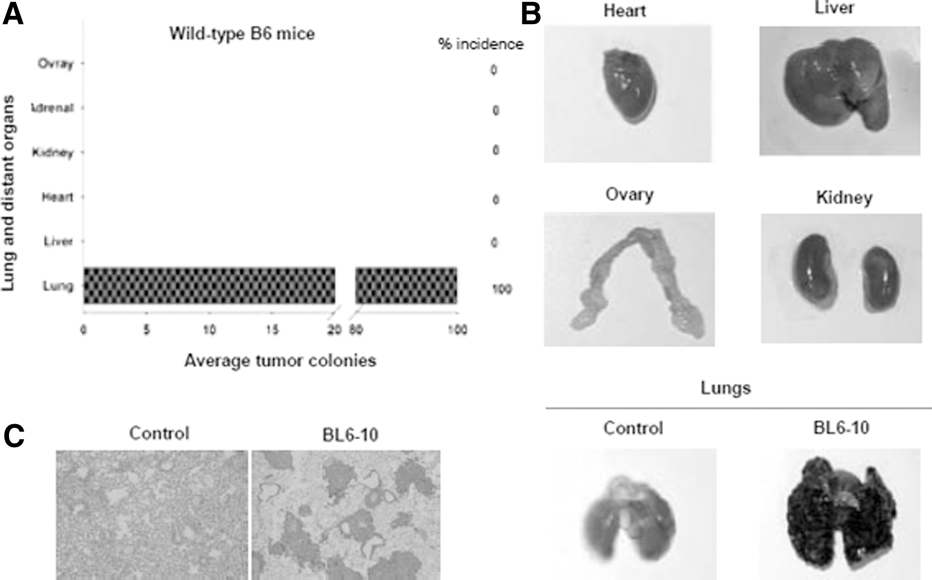
A highly lung metastatic B16 melanoma cell line BL6-10.

CD4+ and CD8+ T cells prevent B16 melanometastasis in distant organs.
Prevention of systemic B16 melanometastasis mediated by CD4+ T cells is via CD40L signaling
To determine the potential involvement of CD40L signaling in CD4+ T cell-mediated systemic B16 melanometastasis prevention, we injected CD40L-deficient mice with BL6-10 cells and then monitored for tumor burden in lung and distant organs. We found that multiple distant organs of CD40L-deficient mice similarly showed systemic metastasis when compared to CD4-deficient mice (Fig. 2B). To further confirm the role of CD40L signaling in organ metastasis, we reconstituted CD40L-deficient mice with polyclonal CD40L-sufficient or CD40L-deficient CD4 T cells 1 day before and 5 days after tumor challenge. Indeed, the provision of CD40L signaling of CD4+ T cells completely abrogated the metastasis in different organs except in lungs (Fig. 2A), indicating that prevention of systemic B16 melanometastasis mediated CD4+ T cells is via CD4+ T cell's CD40L signaling.
Polyclonal CD8+ T environment prevents B16 melanometastasis in distant organs
Next, we determined whether endogenous CD8+ T cells also mediate natural immunosurveillance against organ metastasis. To do this, WT or CD8-deficient mice were i.v. injected with BL6-10 tumor cells and observed for the extent of metastasis of melanoma cells to different organs. In contrast to WT, which showed metastasis only in lungs, CD8-deficient mice showed varying degrees of metastasis in various organs, including lungs (Fig. 2C). Although metastasis in distant organs was observed in CD8-deficient mice, it appears that these organs, particularly liver, kidney, and heart, develop metastasis in a relatively lesser frequency and degree when compared to those of CD4-deficient mice (Fig. 2A). Again, serosal layers of thorax and abdomen were also frequently affected (data not shown). To further confirm the participation of CD8+ T cells, CD8-deficient mice were reconstituted with polyclonal CD8+ T cells 1 day before and 5 days after tumor injection. As expected, the reconstitution of CD8+ T cells completely prevented metastasis to all the above organs except lungs (Fig. 2C). Together, these results suggest the requirement of CD8+ T cell responses in the prevention of organ metastasis.
DCs are required to prevent B16 melanometastasis in distant organs
The participation of adaptive cells, CD4+ and CD8+ T cells, in the prevention of metastasis of B16 melanoma in distant organs prompted us to further assess the potential involvement of DCs initiating CD4+ and CD8+ T cell responses in metastasis prevention. To do this, we employed transgenic (Tg) DTR-CD11c mice and selectively depleted DCs using diptheria toxin (DT) 1 day before tumor injection (Fig. 3A). We found that a single treatment of DT was sufficient to allow B16 melanoma cells to establish in distant organs (Fig. 3B), compared to WT mice with intact DCs (Fig. 1A). Again the lungs of challenged Tg mice showed severe tumor burden. Interestingly, all melanoma metastasis in distant organs disappeared except for lungs (Fig. 3B) when DTR-CD11c DC-depleted mice were reconstituted with WT B6 mouse DCs (Fig. 3A), suggesting that DCs also play a critical role in mediating natural immunosurveillance against organ metastasis.
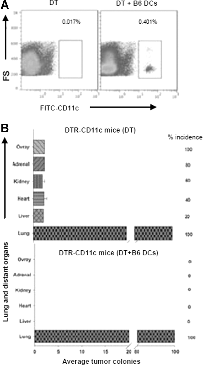
DCs mediate prevention of B16 melanometastasis in distant organs.
NK cells prevent B16 melanometastasis to liver
To assess the involvement of innate immune cells such as NK cells in organ metastasis prevention, following i.p. injection of NK1.1 antibody for NK cell depletion (Fig. 4A) before and after BL6-10 tumor challenge, we found that melanoma metastasis were observed in numerous organs with varying proportions (Fig. 4B). Next to lungs, livers of NK-depleted mice were the most severely metastasized, and they contained more than 100 BL6-10 melanoma colonies of varying sizes in each liver (Fig. 4C), which were confirmed by histologic examination (Fig. 4D), suggesting the importance of NK cells in liver metastasis prevention. In contrast, WT mice treated with the irrelevant isotype-matched control Ab showed tumor metastasis only in lungs (Fig. 4B).

NK cells are critical to prevent B16 melanometastasis in distant organs, especially in livers.
Discussion
The process of tumor metastasis can be divided into four steps. The first step is represented by tumor cell epithelial-mesenchymal transition, in which tumor cells acquire fibroblastoid characteristics that allow them to invade epithelial linings and basal membranes, thus reaching blood vessels. 15 The second step is represented by intravasation of tumor cells into blood vessels. Inflammation may promote this step by production of mediators that increase vascular permeability. This is followed by the third step, in which metastatic tumor cells travel throughout the circulation. It has been estimated that only 0.01% of tumor cells that enter the circulation eventually survive and result in micrometastasis in distant organs. 16 The fourth step involves the interaction of integrin-mediated metastatic progenitors with immune, inflammatory, and stromal cells, leading to tumor cell proliferation in the distant organs. 17
To date, most studies have mainly focused on studying the role of immune cells such as active CD4+ and CD8+ effector T cells, 13,18 DCs 19 and NK cells 20 –22 in anti-tumor immunity. However, the role for endogenous naïve immune cells in natural resistance against tumor metastasis in distant organs is less clear. Hence, to determine whether endogenous naïve CD4+ and CD8+ T cells mediate natural immunosurveillance against tumor organ metastasis, we chose a mouse B16 melanoma model, in which WT B6 mice i.v. injected with a highly lung metastatic B16 melanoma cell line BL6-10 readily established BL6-10 melanoma metastasis in lungs, but not in other organs. 9 The predominant localization of BL6-10 melanoma cells in lungs has been shown to be associated to Met72 glycoprotein highly expressed on BL6-10 cells since anti-Met72 Ab treatment for blocking Met72 adhesion significantly reduced BL6-10 melanoma lung metastasis. 23 This model thus allowed us to assess if melanoma cells initially established in lungs could also establish melanoma metastasis in distant organs in the absence of specific components in cellular arms of the immune system.
Based upon this model, we found that both naïve CD4+ and CD8+ T cells, DCs, and NK cells prevent systemic B16 melanometastasis in multiple distant organs. Our data thus indicate that the adoptive cellular arm of the immune system plays a critical role in mediating natural immunosurveillance against systemic organ metastasis, which is consistent with some previous reports showing a similar widespread metastasis of melanoma into distant organs in immune deficient mouse models. 24,25 The enhanced malignancy rate in immunosuppressed patients, together with the increased occurrence of tumors in mice deficient in various types of immune cells, 26,27 also suggest the role for immune cells in natural surveillance against tumor initiation.
The regulation of CD4+ T cell-mediated humoral and cellular immunity requires CD40L signaling. 13,28 Hence, we then determined whether CD40L signaling plays any role in CD4+ T cell-mediated B16 melanometastasis prevention. To do this, we first injected CD40L-deficient mice or CD4-deficient mice with BL6-10 melanoma cells, which were also reconstituted with naïve CD40L-sufficient or deficient CD4+ T cells, and then monitored for melanoma burden in lungs and distant organs. Our study revealed that reconstitution of CD40L-deficient mice with polyclonal CD40L-sufficient, but not CD40L-deficient CD4 T cells completely abrogated the melanoma metastasis in different organs except in lungs, indicating that CD40L signaling acts as an important costimulatory molecule in CD4+ T cell-mediated prevention of systemic organ metastasis.
Innate immune cells such as NK cells regulate the responses of cell-mediated immunity, either by promoting or suppressing immune responses. 26 Hence, we sought to determine the impact of NK cell depletion on B16 melanoma organ metastasis under natural conditions. Following i.p. injection of NK1.1 antibody, NK cell depletion was ensured before and after BL6-10 tumor challenge. Strikingly, upon sustained NK cell depletion, the melanoma metastasis was observed in numerous organs with varying proportions. It is well known that certain types of immune cells predominate in certain organs. For example, NK cells predominate in livers, compared to other organs, constituting more than 30% of total liver immune cell population. 29 Accordingly, livers of WT B6 mice depleted of NK cells showed extensive metastasis when compared with livers of mice lacking CD4+ or CD8+ T cells or DCs, suggesting that impairment of certain immune components such as NK cells could influence melanoma metastasis in specific organs such as livers, which is consistent with the previous study indicating that modulating NK cell activity by IL-12/IL-18 therapy enhanced antitumor immunity in livers. 30 It has been reported that IFN-γ production by NK cells is critical for natural resistance of B16 melanoma metastasis in mice via direct inhibition of tumor cell proliferation and activation of antitumor host mechanisms. 21,31
According to seed and soil hypothesis, certain organs provide fertile environment for tumor cells to establish metastasis (i.e., melanoma to lungs and colon cancer to liver). This theory is further supported by recent findings, which showed that such propensity is due to interactions of adhesion molecules and chemokines with specific receptors present in tumors and certain organs. 6,32,33 Furthermore, the discovery of cancer stem cells, which could act as seeds for organ metastasis, provided insights into our understanding of metastasis. 34 Our study further advanced these observations by demonstrating that, under natural conditions without exogenous stimuli, these immune cells including both innate and adoptive immune cells do prevent melanoma metastasis to distant organs. As with most cancers, it is the metastasis to distant organs, rather than primary melanoma, 3 which is causing major deaths at a shocking rate worldwide. At present, treatments, such as surgery, chemotherapy, or radiation therapy, are failing due to resistance of cancers to these therapies. Given their key role for modulating cellular arms of the immunity, our efforts should be aimed to develop novel cancer immunotherapeutics to boost organ-specific responses of immune cells such as NK cells to prevent the recurrence of metastasis.
Conclusion
Our results suggest that the composition and functions of different immune cells in foreign tissue microenvironment (distant organs other than primary site of predilection) robustly mediate natural resistance against metastasis, and thus, harnessing these immune cells' responses in immunotherapeutics would considerably limit organ metastasis.
Footnotes
Acknowledgments
This study was supported by research grants of the Canadian Institutes of Health Research and Jiangsu Provincial Special Program of Medical Science (BL2012023).
Disclosure Statement
No competing financial interests exist.