
Abstract
Imidazolium salts have antitumor potential and toxicological effects on various microorganisms. The authors' aim is to synthesize a new imidazolium salt and to assess its pharmacokinetic and antitumor potentials by in vitro and in vivo studies. In this study, bis(trifluoromethanesulfonyl) imide (ITFSI) was synthesized and labeled with 131I using the iodogen method. The efficiency of radiolabeling was determined with high yield (95.5% ± 3.7%). Pharmacokinetic properties of the compound were investigated in albino Wistar rats using radiolabeled compound. The radiolabeled compound (131I-ITFSI) has been stable during a period of 3 hours in human serum. The uptake of 131I-ITFSI reached maximum in the spleen, liver, and blood at 60 minutes, large intestine and heart at 30 minutes, and ovary at 120 minutes. It is observed that intracellular uptake of the radiolabeled compound is higher in the CaCo-2 (colon adenocarcinoma tumor) cell line than HEK-293 (human epithelial kidney) cell line. In further study, antitumor potential of ITFSI on a colon adenocarcinoma tumor-bearing animal model may be investigated.
Introduction
Imidazolium salts can be found in natural products and isolated from the roots of plants such as Lepidium meyenii. Imidazolium salts are known as a type of ionic liquid and green solvents due to their low vapor pressure and high chemical stability. 1–2 In literature, it was shown that imidazolium salts have antitumor potential and toxicological effects on various microorganisms. Imidazolium salts could be diversified by attaching different groups to imidazolium salt stem. They have a wide range of applications as ligands for metal complexes or organocatalysts in synthetic chemistry owing to their easily tunable characteristics such as amphiphilicity, lipophilicity, and solubility. Toxicity caused by chemotherapeutic agents is the main challenge in cancer treatment, so to address this problem, great effort has been focused on the development of new anticancer drugs. Imidazolium salts may be utilized as an anticancer drug due to their amphiphilic structures in which the hydrophilic cationic segments could have strong interactions with many biological components. Cytotoxic effects of two imidazolium alkaloids, which were isolated from the roots of Lepidium meyenii, were assayed in human cancer cell lines by Cui et al. One of the naturally occurring imidazolium salts displayed promising results against the human bladder carcinoma (UMUC3), human breast carcinoma (MDA231), and human pancreatic adenocarcinoma (PACA2) cell lines. 3 In another study, the antitumor activity of many alkyl imidazolium salts was evaluated on human tumor cell lines by Malhotra and Kumar. 4 They found that the chain length of the alkyl substitution at the 3-position of the imidazole (IMS) ring plays a pivotal role in increasing the antitumor activity. It was observed that IMS with alkyl-substituted chain lengths shorter than C-7 was not active against tumors. However, IMSs with an alkyl chain of C-12 were determined to be effective on all 60 tumor cell lines and showed low cytotoxicity.
Alternatively, Gopalan's group reported that antitumor properties of some imidazolium salts were investigated first in a xenograft mouse model. Their IC50 values for the hepatocellular carcinoma cells were determined to be 100–120 μM. When the imidazolium salts were used for treatment of Huh7 HCC xenograft mouse model, they led to shrinkage in the tumor volume. 5
For tracking the metabolic pathway of the compounds through the body, radiolabeled compound is used owing to the simplicity of its labeling procedure. Radioiodine complexes are used in pharmacokinetic studies. 6–7 In the present study, the authors aimed to synthesize a new imidazolium salt and to evaluate its pharmacokinetic by in vivo and antitumor potentials by in vitro studies.
Materials and Methods
Materials
All chemicals used at cell studies were purchased from Biological Industries. Iodogen was supplied from Sigma-Aldrich. Thin-layer chromatography cellulose gel (ITLC-F plastic sheets 20 × 20) and other chemicals were purchased from Merck. Cell culture studies were performed in Thermo MSC Advantage 1.2 Laminar Air Flow. An Olympus Japan inverted and light microscope was used to count cells. Thermo Multimode microplate reader was used for determining IC50 values of cell cultures.
Preparation of the imidazolium-TFSI salt, [octyl-bis(3-methylimidazolium) di (bis(trifluoromethane) sulfonimide)]
A mixture of 5.52 g (15.1 mmol) of 1,8-diiodooctane and 2.42 mL (30.2 mmol) of 1-methylimidazole in 10 mL of toluene was refluxed (110°C) for 12 hours (Fig. 1). After cooling to room temperature, the yellow-colored lower phase was separated from the upper phase and washed a few more times with 5 mL toluene. The raw product was dissolved in CH2Cl2 and filtered, and then the solvent evaporated in a rotary evaporator. Obtained imidazolium iodide salt and equimolar amount of bis(trifluoromethane)sulfonimide lithium salt were mixed in 40 mL water and heated to 70°C for 30 minutes. A cloudy white precipitate formed immediately, and stirring was continued for an additional 24 hours at room temperature. CH2Cl2 was added to the mixture and the product was extracted from water. Organic solvent was removed in a rotary evaporator and a brown viscous liquid was obtained, 10.9 g, 86.5% yield.
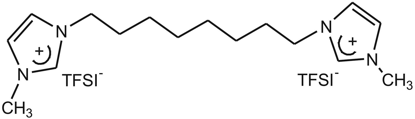
Chemical structure of imidazolium-(trifluoromethanesulfonyl) imide (TFSI) salt.
Radiolabeling
Fifty microliters of the imidazolium-TFSI salt (ITFSI) (0.13 μM) was diluted with 400 μL phosphate buffer (pH = 7) and the solution was put into an iodogen-coated tube. The compound was labeled with 131I (5.5 Bq) through the iodogen method (1 mg) and the mixture was incubated for 30 minutes. 8 –12
Quality control studies
Thin-layer radiochromatography
The labeling efficiency of 131I-labeled ITFSI (131I-ITFSI) was tested by thin-layer radiochromatography (TLRC). After 30 minutes of incubation, 131I-labeled compound was spotted on cellulose-coated plastic (ITLC-cellulose) sheets (1 × 10 cm; Merck) and the sheets were developed in two different mobile phases. The developed ITLC-cellulose sheets were removed from mobile phases and allowed to dry. Then, the sheets were scanned on a TLC scanner (BioScan AR-2000).
Lipophilicity
Analysis of the lipophilicity of 131I-labeled compound was carried out by mixing 50 μL of 131I-labeled imidazolium-TFSI in 3 mL of n-octanol and water. The mixture was mixed in magnetic mixer for 1 hour. For separation of the phases, the mixture was centrifuged (2500 rpm, 5 minutes). Then, 500 μL of both organic and water phases was taken and counted using a Cd (Te) RAD 501 single-channel analyzer.
Human serum stability study
The radiolabeled compound was added on fresh human serum and incubated at 37°C. The quality control was checked by taking samples at various times (30 minutes, 1–3, and 24 hours) using the TLRC method.
Biodistribution
The animal experiment was approved by the Animal Ethics Committee of Dokuz Eylül University. In vivo study of 131I-ITFSI was carried out using 9 female albino Wistar rats. The imidazolium-TFSI salt (500 μg) was labeled with Na131I through the iodogen method. The radiolabeled imidazolium-TFSI salt (specific activity: 268.12 MBq/μmol) (300 μL) was intravenously injected through the tail vein after weighing the rats. Three animals were sacrificed at each time point (30, 60, and 120 minutes postinjection) under anesthesia following injection. Organs of interest were excised, weighed, and counted by the Cd (Te)-RAD-501 single-channel analyzer.
Cell culture
Cell culture studies were carried out using the CaCo-2 cell line [ATCC HTB-37, tissue: human colon, colorectal adenocarcinoma] and HEK-293 cell line [ATCC HEK293T/17 human epithelial kidney]. These cells were cultured in MEM nonessential amino acid solution containing 100 IU/mL penicillin G, 100 mg/mL streptomycin, and 10% heat-inactivated fetal bovine serum (FBS) and maintained at 37°C in an incubator containing 5% CO2.
Cytotoxicity assay of imidazolium-TFSI salt
XTT (2,3-bis-(2-methoxy-4-nitro-5-sulfophenyl)-2H-tetrazolium-5-carboxanilide salt) assay was used to determine the IC50 value of imidazolium-TFSI salt in CaCo-2 and HEK-293 cell lines. The number of viable cells was determined by means of the Cell Proliferation Kit XTT. The study was performed in a 96-well culture plate by seeding each well with 1 × 10⁵ cells. Following 2 days of incubation, 50–300 μM compounds prepared with MEM solution (without FBS) were added to each well of the 96-well plate. After 24 hours, the solvent media on the cells were removed and replaced with MEM solution containing FBS. After 1 hour of incubation, 50 μL solution from the XTT kit was put in the wells. Three hours later, absorbance of the samples in wells was measured in a microplate reader (Varioskan Flash Multimode Reader-Thermo) at 450/630 nm. Then, the percentage of cytotoxicity was calculated.
Intracellular uptakes of 131I-imidazolium-TFSI salt
The in vitro cellular uptake of 131I-labeled imidazolium-TFSI salt was performed using the CaCo-2 cell line and HEK-293 cell line as control group. The cells were placed in 24-well culture plates (1 × 10⁵ cells in each well) at 37°C in an incubator for 2 days. The medium on the cells was removed and the wells were washed twice with 0.9% NaCl solution. After that radiolabeled imidazolium-TFSI salt (25 μM, activity: 0.9 Bq), which was diluted with MEM (without FBS), was added on the cells. In the same conditions, the authors assayed intracellular uptake of Na131I as the control group. Indeed, in this step, it was checked whether the uptake was caused by free iodine or radioiodinated compound. After 1 hour of incubation, the wells were counted by the Cd (Te) RAD 501 signal-channel analyzer. Then, the radioactive medium on cells was removed. The cells were washed once after that same amount of 0.9% NaCl solution was added in each well. After the wells were counted, the data were analyzed and the percentage of uptake was calculated.
Statistical analyses
Differences in the mean values of measured activities were evaluated statistically using the SPSS 16.0 program (Univariate Variance Analyses and Pearson Correlation). Probability values of p < 0.05 were considered to be significant.
Results
1H NMR results of the imidazolium-TFSI salt, [octyl-bis(3-methylimidazolium) di (bis(trifluoromethane) sulfonimide)]
1H NMR (CH3OD) δ ppm: 8.83 (s, 2H, NCHN), 7.61 (t, J = 1.6 Hz, 2H, NCHCHN), 7.55 (t, J = 2 Hz, 2H, NCHCHN), 4.22 (t, J = 7.6 Hz, 4H, NCH2CH2), 3.94 (s, 6H,–NCH3), 1.97–1.84 (m, 4H, NCH2CH2), 1.41 (brs, 8H, -CH2CH2CH2CH2-). 13C NMR (CD3OD) δ ppm: 136.42 (NCHN), 124.73 (TFSI), 123.67 (NCHCHN), 122.91 (NCHCHN), 121.57 (TFSI), 118.36 (TFSI), 115.18 (TFSI), 49.32 (NCH2CH2), 35.27 (–NCH3), 29.52 (–CH2 CH2–), 28.33 (NCH2 CH2–), 25.63 (NCH2CH2 CH2).
Quality controls of 131I-labeled ITFSI
The labeling efficiency of ITFSI was determined using the TLRC method. Rf values of 131I-labeled ITFSI and Na131I were determined as 0.08 and 0.80, respectively, when mobile phase 1: 2-propanol/n-butanol/ammonium hydroxide (2:1:1) was used. While mobile phase 2: chloroform/acetic acid 9:1 was being used, the Rf values of 131I-labeled ITFSI and Na131I were obtained as 0.07 and 0.20, respectively. Labeling of the imidazolium-TFSI salt was performed by using 1 mg iodogen oxidation agent, pH = 7, room temperature, and reaction time of 30 minutes. The efficiency of the radiolabeling was determined as 95.5% ± 3.7%.
Serum stability study
Serum stability study was performed to determine the stability in the biological system of radiolabeled imidazolium-TFSI. While radiolabeling efficiency at first 30 minutes was 98.5% ± 0.7%, at the end of 1440 minutes, it decreased to 70% ± 14.1% (Fig. 2). It was observed that the labeled compound was stable during a period of 180 minutes.

In vitro stability of 131I-ITFSI at different times after labeling.
In vivo study
The percent of radioactivity per gram of organs (%ID/g) was calculated and is shown in Figure 3. The uptake of 131I-ITFSI reached also maximum in the spleen (%ID/g:1.2 ± 0.16 at 60 minutes), blood (%ID/g:0.23 ± 0.06, p < 0.05 at 60 minutes), heart (%ID/g: 0.23 ± 0.07, p < 0.05 at 30 minutes), lung (%ID/g: 0.64 ± 0.12, p < 0.05 at 120 minutes), and ovary (%ID/g: 1.03 ± 0.09, p < 0.05 at 120 minutes).
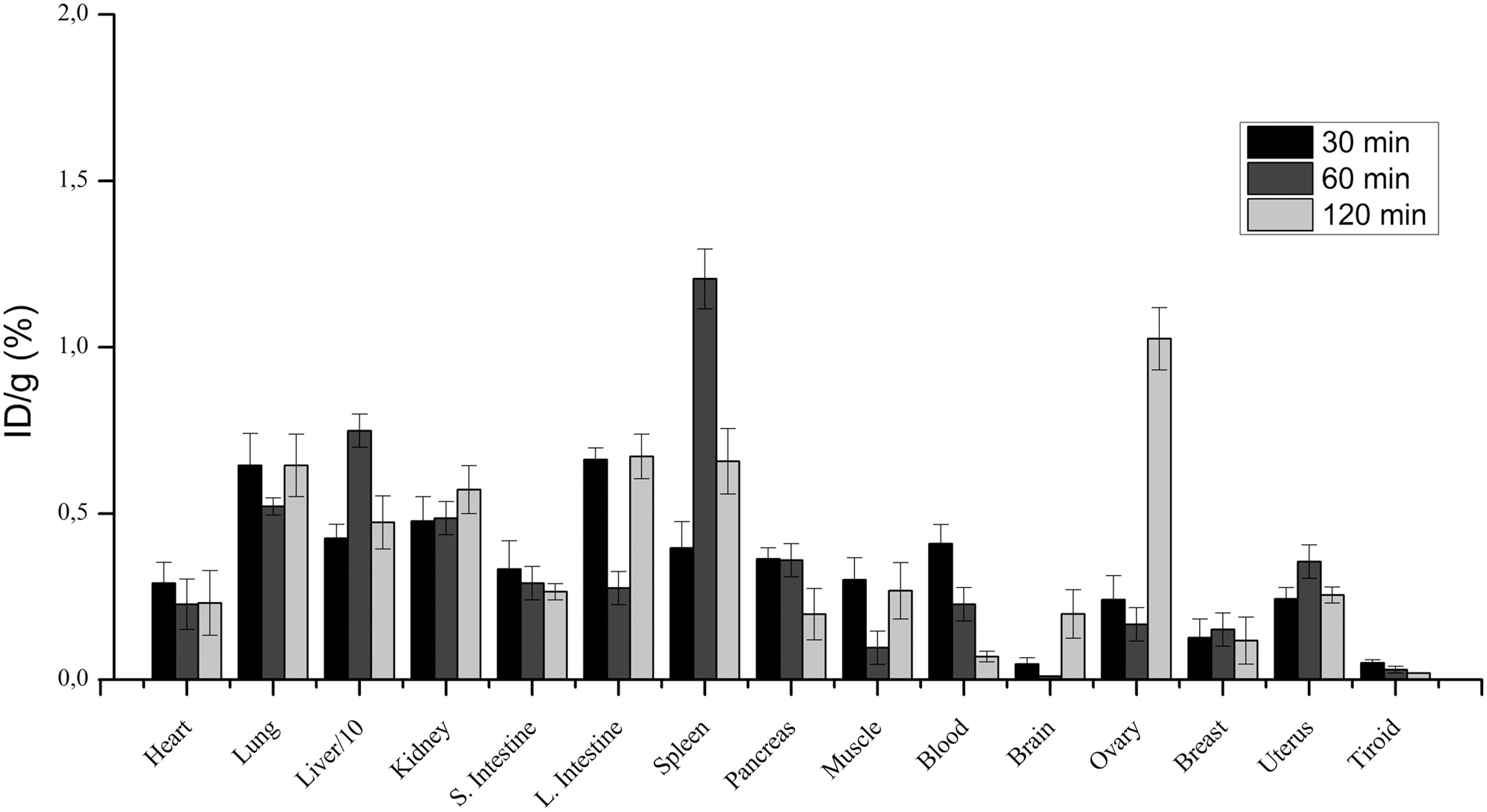
Biodistribution results of 131I-ITFSI.
The accumulation activity of the labeled compound in the liver at 60 minutes postinjection was %ID/g 7.5 ± 0.5. While the activity was increasing in large intestines (l. intestine) (0.30% ± 0.03%), the liver uptake (%ID/g 4.7 ± 0.38) decreased at 2 hours. Mostly, the radiolabeled compound eliminated through the kidney and bladder because of the poor lipophilicity of the radiolabeled compound (log p = 0.99). 13 As can be seen from Figure 3, low activity of the thyroid gland was observed, which is an indication of the stability of 131I compound against biological decomposition. While the uptake in blood was decreasing with time, the accumulation was almost stable in the small intestine (s. intestine). It can be concluded that the accumulation in s. intestine does not depend on the uptake in blood.
In vitro study was performed in the CaCo-2 cell line (colon adenocarcinoma tumor) and HEK-293 cell line as control group. According to XTT cytotoxicity test results, the cell 50% viability was determined to be 200 μM TFSI in the CaCo-2 cell line. After determination of the IC50 value, the intracellular uptakes of 131I-TFSI in CaCo-2 and HEK-293 cell lines were evaluated. Uptake of 131I was examined in CaCo-2 and HEK-293 cell lines simultaneously and compared with each other. As can be seen from Figure 4, the uptake of labeled compound in CaCo-2 cell lines is 9.7% ± 2.6%. On the other hand, the uptake of 131I-ITFSI was determined as 1.8% ± 0.7% in HEK-293 cell lines (Fig. 4). The results of uptake experiments show that the uptake of 131I was low in both cell lines.
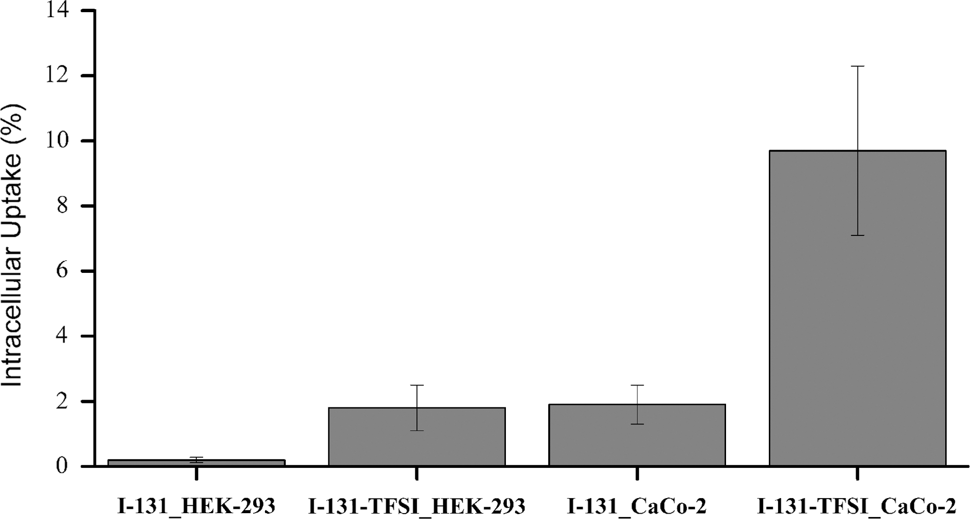
Intracellular uptake of 131I-ITFSI in CaCo-2 and HEK-293 cell lines.
Discussion
According to biodistribution results, the uptake of 131I-ITFSI was observed in lung, liver, intestines, and spleen in normal rats. In the study by Kurtdede et al., the biodistribution study of 131I-labeled 4-benzoyl-1-(4-carboxyphenyl)-5-phenyl-1H-pyrazole-3-carboxylic acid (P3CA) was investigated in albino Wistar rats. It is observed that the maximum uptake of 131I-P3CA had been determined in lungs, stomach, and spleen at 15 minutes. 14 The biodistribution results are similar to Kurtdede's results.
In the current study, IC50 values of ITFSI in the CaCo-2 cell line were determined as 200 μM. Haque et al. studied with ortho/paraxylyl linked bis-imidazolium salts and Ag(I) N-heterocyclic carbene (NHC) complexes. In the study, cytotoxicity tests of all compounds against human colorectal cancer (HCT 116) and breast cancer cell lines (MCF-7) were assayed. The IC50 value ranges were determined as 5.6–20.3 μM for HCT 116 and 1.12–6.38 μM for MCF-7. 15 As seen, IC50 values show changes depending on the structure of imidazolium salts and cell line.
In this study, it was observed that the uptake of 131I-ITFSI in tumor cells was about five times higher than epithelial cells, whereas Cui et al., reported that imidazole alkaloids isolated from Lepidium meyenii have not been active against human colon adenocarcinoma (HT-29), human prostate adenocarcinoma (PC3), and human kidney carcinoma (A4982LM) cell lines. 3 Indeed, imidazolium salts might have different antitumor activity depending on their structures. 2 To figure out the mechanism of the anticancer behavior of imidazolium salts, more in vivo experiments are required.
Pharmacokinetics of the labeled compound was demonstrated in normal rats and antitumor potential in the CaCo-2 cell line in vitro. Further studies may be designated on testing animals with colon adenocarcinoma tumor in vivo.
Conclusion
In this study, tissue distributions of imidazolium-TFSI salt in normal female rats and antitumor potential on the CaCo-2 cell line were evaluated. The radiolabeled compound showed quite high intracellular uptake in the CaCo-2 (colon adenocarcinoma tumor) cell line. The antitumor potential of ITFSI should be investigated through in vivo study in the future.
Footnotes
Disclosure Statement
No competing financial interests exist.