




Abstract
For the growth and spread of a tumor beyond 2 mm, angiogenesis plays a crucial role, and association of various integrins with angiogenesis is evidential. The aim of the study was radiolabeling of DOTA-chelated RGD (arginine-glycine-aspartic acid) peptide with 68Ga for PET imaging in locally advanced breast carcinoma. DOTA-RGD was incubated with 68GaCl3, eluted in 0.05 m HCl. Elution volume, peptide amount, and reaction pH were studied. Radio-ITLC, gas chromatography, endotoxin, and sterility testing were performed. Serial (n=3) and whole-body (n=2) PET/CT imaging was done on patients post i.v. injection of 111–185 MBq of 68Ga-DOTA-RGD. Maximum radiolabeling yield was achieved with 3 mL elution volume of 15–20 μg peptide at pH 3.5–4.0 with 10 minutes of incubation at 95°C. Product samples were sterile having 99.5% radiochemical purity with residual ethanol content and endotoxins in injectable limits. Intense radiotracer uptake was noticed in the tumor with SUVmax 15.3 at 45 minutes in serial images. Physiological radiotracer uptake was seen in the liver, spleen, ventricles, and thyroid with excretion through the kidneys. The authors concluded that 68Ga-DOTA-RGD has the potential for imaging α,vβ3 integrin-expressing tumors.
Introduction
Angiogenesis is an essential condition for tumor growth and metastasis. Tumors cannot grow beyond 2 mm in size due to the limited supply of oxygen and nutrients, as well as the need for new blood vessels for the same. Tumors secrete angiogenic factors, which activate the endothelial cells and induce endothelial procreation and novel vessel generation cascade. G-protein-coupled receptors for angiogenesis-modulating proteins, vascular endothelial growth factor (VEGF) receptors, integrins, and VEGF regulate the angiogenesis processes. 1,2
Integrins are the important components for the adhesion of cells to extracellular matrix proteins. Along with the migration of cells, integrins also regulate the cellular access and withdraw from the cell cycle. 3 Among the various types of integrins characterized, αvβ3 integrin acts as a receptor for distinct extracellular matrix proteins with the uncovered repeated sequences (motifs) for RGD (arginine-glycine-aspartic acid) tripeptide binding. The αvβ3 integrin is usually expressed in relatively low levels on mature endothelial and epithelial cells, but is overexpressed in many tumors, including melanomas, osteosarcomas, glioblastomas, neuroblastomas, prostate, breast, and lung carcinomas. 4 –8 Due to this peculiar characteristic, αvβ3 integrin has been identified as an enticing molecular target for the noninvasive monitoring of fast-growing malignant cells as well as for the assessment of their response to therapeutic interventions. 9,10 Several animal studies using radiolabeled RGD peptides have been done to study αvβ3 integrin-targeting molecules for imaging various types of cancerous lesions by SPECT and PET. 11 –13
Preclinical trials of several radiotracers have been done in tumor-bearing animal models. A 99mTc-labeled linear and cyclic RGD peptide study demonstrated excretion through the hepatobiliary and urinary system along with high liver uptake, which was observed as a major impediment toward its widespread clinical applications, possibly because of the lipophilic nature of the 99mTc-chelate.11 99mTc-NC100692 is a 99mTc-labeled cyclic RGD peptide reported to have high αvβ3 integrin-binding affinity. 12 Of the various 18 F radiotracers evaluated, 18 F-AH111585 and 18 F-Galacto-RGD have entered in to clinical trials for noninvasive imaging of the αvβ3 integrin expression. 14,15 However, due to high preparation cost and lack of dedicated preparative modules for routine radiosynthesis, the clinical utility of these 18 F-labeled molecules is limited. As a result, there is ample scope in developing new agents with superior pharmacokinetic properties, which could be easily formulated at a hospital radiopharmacy.
68Ga, a generator-based radionuclide, provided a significant breakthrough in imaging of malignancies with good quality PET images and is independent of the onsite availability of a cyclotron. A recent study demonstrated the successful use of 68Ga-labeled cyclic RGD peptide derivatives in animal tumor models. 16 It is documented that an increase in peptide multiplicity increases the radiotracer tumor uptake and retention of radiolabeled RGD peptide derivatives. This trend is observed from derivatives having monomer to tetramer RGD derivatives. A steady increase of uptake in other nontarget organs, for example, the liver, intestine, and kidneys, has also been reported depending upon the number of RGD molecules. A comparison of biological behavior of radiolabeled RGD monomers, dimers, and tetramers revealed that dimers exhibit rapid tumor localization and significant tumor retention with excellent target/nontarget ratios and are the most suitable candidates for tumor imaging. 17 –19
Recently, A kit-based 68Ga-NOTA RGD preparation was used to compare the role of 68Ga-NOTA RGD PET/CT and widely used 18 F-FDG PET/CT in breast cancer patients. 20 However, no radiosynthesis has been explained for in-house preparation.
Taking into account the favorable characteristics of the radiotracer, herein the authors report the development of the optimized protocol for the preparation and characterization of the radiotracer at the hospital radiopharmacy setup using a commercial 68Ge-68Ga generator and its preliminary clinical investigations in human patients. The purpose of this study was to explore the potential of 68Ga-DOTA-RGD tracer as a noninvasive tool for assessment of neoangiogenesis in tumors expressing αvβ3 integrin.
Materials and Methods
68Ga was obtained from 999 MBq 68Ge-68Ga generator (ITG) for all the clinical studies. The dimeric RGD peptide conjugate, DOTA-E[c(RGDfK)]2, that is, DOTA-RGD, was custom synthesized by ABX Advanced Biochemical Compounds, Germany. Suprapure HCl and HPLC grade ethanol, water, and acetonitrile were procured from Merck, Germany. Sodium acetate was purchased from Sigma-Aldrich. C-18 Sep-pack cartridges were purchased from Waters, Ireland. Radioactivity assay of 68Ga was done using a properly calibrated dose calibrator (CRC 25 PET; Capintec). ITLC-SG paper (Merk), pH paper (Merk), and TLC scanner (EZ-Scan) with multimode radiation detector (Omni Rad) were used for chromatography. Sterility test was done using tryptic soya broth medium (Himedia). Pyrogenicity was checked by the point-of-use test system (PTS; Charles River). Ethanol content was assessed using a gas chromatograph (Varian 3500) based on flame ionization detection. The patient imaging was done using a dedicated PET/CT scanner.
Radiolabeling of DOTA-RGD with 68Ga
Radiolabeling of DOTA-RGD was done by changing reaction volumes (2–4 mL), amount of peptide (5–30 μg), and at varied pH (3.0–5.0). The reaction was carried out at 95°C for 10 minutes.
The reaction mixtures were cooled to room temperature and a small aliquot was withdrawn for determining the radiolabeling yield. Subsequently, the solution was passed through the C-18 column preconditioned with 5 mL 70% ethanol solution with a flow rate of 1 mL/min. The column was then washed with 10 mL water. The labeled product was finally eluted with 1 mL of ethanol from the C-18 cartridge, followed by 9 mL of normal saline. The final product was collected in a sterile vial after passing through a 0.22-μm filter and subjected to quality control tests. Characterization of radiolabeled purified peptide was done using matrix-assisted laser desorption–ionization time-of-flight (MALDI-TOF) mass spectrometry (Brukers Daltonics).
Quality control procedures
Quality control procedures were done as described earlier. 21 The radiolabeling efficiency and radiochemical purity of 68Ga-DOTA-RGD before and after purification using the C-18 cartridge were determined by using ITLC-SG with 0.5 M sodium citrate solution (pH-5.5) as the mobile phase. Retardation factor (Rf) was determined by using the TLC scanner.
Sterility of the radiolabeled preparation was established by following the procedure described earlier. 21 Briefly, 1.5 mL of each sample was inoculated in tryptic soya broth and incubated at 37°C and turbidity was observed up to 7 days for detecting the presence of any growth. Endotoxin testing was also done using PTS. The test was sensitive to 0.01 endotoxin unit (EU)/mL. The sample (25 μL) was added to each well in the PTS cassette and incubated at 38°C for 15–20 minutes for the reaction to take place, and then the observations were noted with 50%–200% spike recovery.
Chemical purity was ascertained by gas chromatography of decayed samples. In this procedure, the temperature of the injector, oven, and detector was 140°C, 80°C, and 200°C, respectively. The flow rates for hydrogen and air were 30 and 290 mL/min, respectively. The concentration of ethanol was calculated based on the area under the curve of the sample and ethanol standard (4000 ppm).
Patient studies
Ethical clearance was obtained from the Institutional Ethics Committee (IEC) for patient study (Ref No, Histopath/13/NK/2421, dated 12/8/13). Written informed consent was obtained from all the patients enrolled in the study. Five locally advanced breast carcinoma (LABC) patients (all female; mean age of 55 years; range 33–63 years) referred for 18 F-FGD PET/CT study were enrolled in this study (Table 1). Before the start of any definitive treatment (chemotherapy/surgery), patients were subjected to 18 F-FGD PET/CT, followed by 68Ga-DOTA-RGD PET/CT imaging on the second day. Whole-body images (base of skull to mid-thigh) were acquired in 3D mode, 45 minutes after intravenous (i.v.) injection of 111–185 MBq of 68Ga-DOTA-RGD. Data acquisition was performed on a hybrid PET/CT scanner (Discovery STE-16; GE Healthcare), with CT parameters being 120 kV, 350 mA, rotation time of 0.5 seconds, and slice thickness of 3.75 mm for diagnostic CT, 512×512 pixel matrix, and pixel size of about 1 mm. A 128×128 pixel matrix with a slice thickness of 3.25 mm was used for PET acquisition. CT-based attenuation correction of the emission images was employed. Iterative methods, ordered subset expectation maximization and filtered back projection, were used for reconstruction of PET and CT images, respectively.
IDC, infiltrating duct carcinoma; LABC, locally advanced breast carcinoma; LMP, last menstrual period.
To determine the maximum uptake time of 68Ga-DOTA-RGD, serial PET images were acquired at 1 and 4 minutes (30 sec/bed position), 10 minutes (45 sec/bed position), 15 and 30 minutes (60 sec/bed position), and 45 minutes (90 sec/bed position) postinjection of 68Ga-DOTA-RGD in patients (n=3). The patients were asked to void before acquiring the image at 45 minutes. The second and subsequent CT images were acquired using low current (10 mA). For semiquantitative analysis, a region of interest was drawn around the site of abnormal 68Ga-DOTA-RGD uptake. The semiquantitative measurement of lesion activity was represented as the standard uptake value (SUVmax).
Based on the serial image findings, the whole-body PET/CT images of patients (n=2) were acquired only at 45 minutes (90 sec/bed position). Image findings were evaluated independently by two experienced nuclear medicine physicians on a per patient basis. Disagreement, if any, was resolved by consensus. The effectiveness of 68Ga-DOTA-RGD PET/CT in defining bone involvement was assessed in the skull, orbit, axial skeleton, and appendicular skeleton. A lesion was judged positive if nonphysiological tracer uptake was seen.
Results and Discussion
Optimization of radiolabeling parameters
To optimize the parameters to obtain maximum radiolabeling yield, DOTA-RGD conjugate was labeled with 68Ga at varied physiochemical conditions mentioned in the experimental section. The optimal peptide amount, pH, and eluent volume for good radiolabeling yield of ∼90% or higher were 20–25 μg (Fig. 1A), 3.5–4.0 (Fig. 1B), and 3 mL (Fig. 1C), respectively. The radiochemical purities of the labeled product prepared under optimized conditions were ∼90% and >99%, before and after C-18 cartridge purification, respectively, as determined by the ITLC technique (Fig. 2A, B). The maximum specific activity of 68Ga-DOTA-RGD was found to be 0.9 mCi/μg of RGD.

Showing the effect of peptide concentration
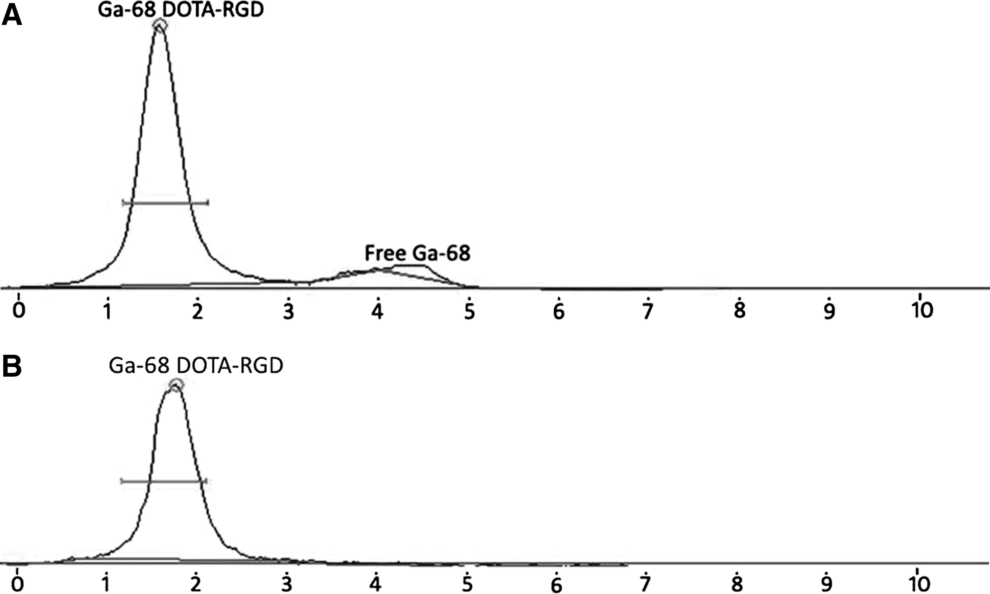
ITLC scan showing two peaks before
Chemical and biological quality control
The residual ethanol content in the radiolabeled product after purification was found to be between 1998 and 2492 ppm at retention time of 3.99 minutes in gas chromatogram (Fig. 3A, B). No turbidity was observed in samples even after 7 days of incubation in tryptic soya broth and that confirmed the sterility of the product. The endotoxins in all the decayed samples tested were found to be between 3.01 and 3.71 EU/mL. MALDI-TOF results also confirmed labeling of RGD peptide with 68Ga (Fig. 4A, B).

Gas chromatogram showing ethanol standard (4000 ppm)
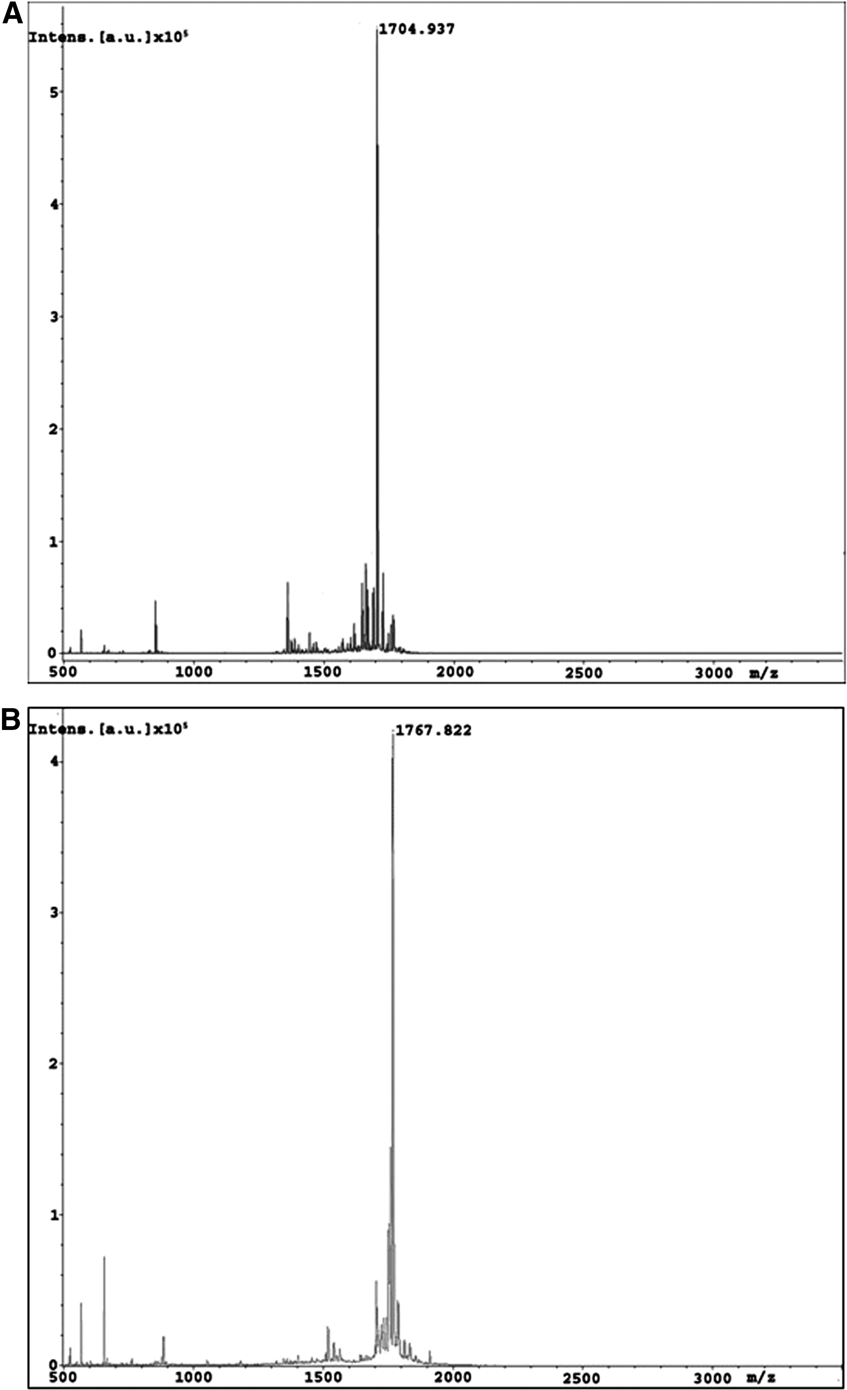
Mass spectra of pure DOTA-RGD tripeptide
Patient studies
PET/CT imaging using 68Ga-DOTA-RGD was performed in patients having LABC. High radiotracer uptake was noticed in the tumor with SUVmax (1 minutes) 8.42, SUVmax (4 minutes) 11.86, SUVmax (10 minutes) 10.43, SUVmax (15 minutes) 10.00, and SUVmax (30 minutes) 13.75, with maximum uptake SUVmax 15.3 at 45 minutes in serial images (Fig. 5). Physiological uptake of the radiotracer was noticed in the liver and spleen with kidneys being the route of excretion of 68Ga-DOTA-RGD in all patient studies. Diffuse uptake was seen in bilateral ventricles and thyroid. Good tumor-to-background ratio was observed in the images of 68Ga-DOTA-RGD, as shown in Figures 5 –7.
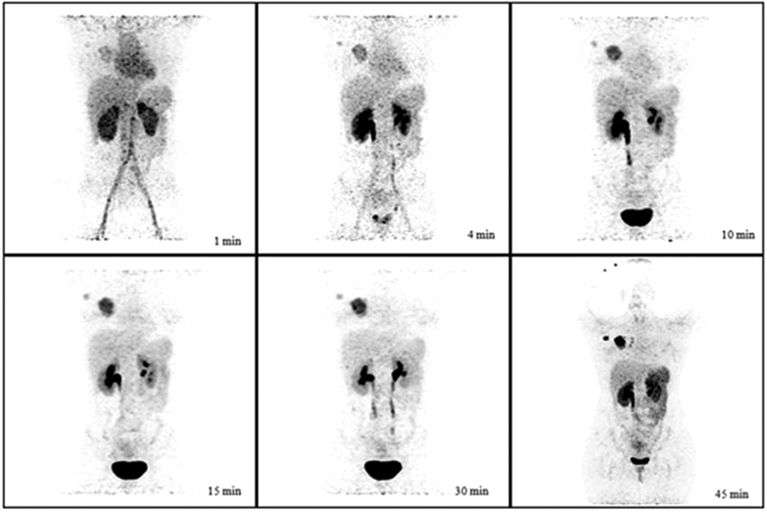
Maximum intensity projection (MIP) positron emission tomography serial images of a 33-year-old female patient with infiltrating duct carcinoma of right breast post i.v. injection of 68Ga-DOTA-RGD (111–185 MBq). Significant tracer uptake is noticed in the breast lesion. SUVmax at 1, 4, 10, 15, 30, and 45 minutes was 8.42, 11.86, 10.43, 10.00, 13.75, and 15.30, respectively. Significant tracer uptake is noted in the right axillary lymph node (SUVmax at 45 minutes, 15.62). The image at 45 minutes was taken postvoid.

Positron emission tomography images of a 63-year-old female with infiltrating duct carcinoma of left breast 45 minutes post i.v. injection of 111–185 MBq 68Ga-DOTA-RGD. Maximum intensity projection (MIP image)

Positron emission tomography images of a 60-year-old female with infiltrating duct carcinoma of left breast 45 minutes post i.v. injection of 111–185 MBq 68Ga-DOTA-RGD. Maximum intensity projection (MIP) image
68Ga-DOTA-RGD was labeled in-house with good radiochemical yields. Moreover prepurification of 68Ga, after elution from the generator, was not required. 68Ge-68Ga generator used in this study has a silica-based column and high affinity for 68Ge. The breakthrough of 68Ge was also studied as reported earlier. 21 For good labeling yield, elution volume, peptide amount, and pH play a crucial role. In the present study, radiolabeling of dimeric RGD was done with 68Ga using DOTA as a bifunctional chelator. The appropriate peptide amount for good radiolabeling yield in these experiments was 15–20 μg. Below this concentration, the radiolabeling yield was very low (18%–30%) (Fig. 1A). The amount of free 68Ga indicated that the peptide concentration was not enough to exhaust all 68Ga present in the reaction solution. The further increase in peptide amount did not affect the labeling yields and coeluted with 68Ga-DOTA-RGD. However, the earlier study suggested 20 μg as a minimum peptide. 15 Another study mentioned that radiolabeling efficiency can be improved even with lower peptide amounts, provided the reaction volume is reduced, 21 but the maximum activity with the generator was observed in the first 3 mL when the elution was done in 1-mL serial fractions. Therefore, the authors have taken 3 mL as optimal elution volume for the preparation of 68Ga-DOTA-RGD (Fig. 1C). Varied pH range has been reported in literature, but the authors found consistent high labeling yield with pH 3.5–4.0 15,21,22 (Fig. 1B). The amount of free 68Ga was more than the bound 68Ga-DOTA-RGD below pH 3.5. At pH above 4.0, although the free 68Ga was not recovered, an overall low yield was observed because free 68Ga was hydrolyzed at higher pH and was not available for radiolabeling of peptide present in the reaction solution. The hydrolyzed 68Ga remained bound to the C-18 cartridge and high activity was observed in the cartridge after purification of 68Ga-DOTA-RGD.
The endotoxin content in all the decayed samples tested was within the permissible limits (<175 EU), ranging from 3.01 to 3.71 EU/mL. Absence of microbial growth in tryptic soya broth after incubation for 7 days indicated sterility of the samples tested. The quality control results indicated that this formulation was good for intravenous administration.
In this study, substantial tumor uptake was noticed at 4 minutes with SUVmax 11.86 and maximum tumor uptake was noticed at 45 minutes (SUVmax 15.3) postinjection of radiotracer (Fig. 5). The same trend was seen in all 3 patients. Physiological radiotracer uptake was observed in the liver and spleen along with diffuse uptake in bilateral ventricles and thyroid (Figs. 6 and 7). The radiotracer was observed to be predominantly excreted through the renal route in all patients and the target-to-background ratio increased with time. The role of αvβ3 integrins in tumor-induced angiogenesis and metastasis was explored to study the tumor receptor status and therapy planning in an animal model. 10 In another study, enhanced in vitro tumor affinity with multimeric RGD was noticed. 16 Tumor uptake is more in case of tetrameric RGD compared with dimeric RGD, but due to higher uptake in kidneys and intestine, it cannot be used for imaging of abdomen tumors. Roed et al., in a rat study of 99mTc-NC100692 (RGD-containing peptide) found phenylalanine excreted in urine and intact compound in blood. 23 However, in the human study, only intact compound was found in the urine. 24 An 18 F-AH111585 study in breast cancer patients suggested rapid blood clearance from the blood pool with good tumor uptake and demonstrated good in vivo stability of radiotracer with no adverse effects on the patient. 13 However, the cost of production is very high as an on-site cyclotron and synthesis module is required for preparation of fluorinated radiopharmaceuticals. 25 This study will contribute to a cost-effective and easy in-house radiosynthesis of a neovasculature imaging agent. No ready-to-use costly kits were used in this study. 68Ga-DOTA-RGD showed good in vivo stability and demonstrated good tumor-to-background ratio for easy interpretation.
Conclusion
68Ga-DOTA-RGD has the potential to be a promising agent for noninvasive molecular imaging of neoangiogenesis in tumors expressing αvβ3 integrins.
Footnotes
Disclosure Statement
No conflicting financial interests exist.