
Abstract
The present article describes the preparation of β-emitter lutetium-177-labeled zirconia colloid and its preliminary physicochemical and biological evaluation of suitability for local radionuclide therapy. The new 177Lu-labeled therapeutic radiopharmaceutical candidate was based on the synthesis mode of a previously described zirconia nanoparticle system. The size and shape of the developed radiopharmaceutical compound were observed through a scanning electron microscope and dynamic light scattering methods. The radiocolloid had a 1.7 μm mean diameter and showed high in vitro radiochemical and colloid size stability at room temperature and during the blood sera stability test. After the in vitro characterizations, the product was investigated in the course of the treatment of a spontaneously diseased dog veterinary patient's hock joint completed with single-photon emission computed tomography (SPECT) imaging follow-up measurements and a dual-isotope SPECT imaging tests with conventional 99mTc-methanediphosphonic acid bone scintigraphy. In the treated dog, no clinical side-effects or signs of histopathological changes of the joints were recorded during the treatment. SPECT follow-up studies clearly and conspicuously showed the localization of the 177Lu-labeled colloid in the hock joint as well as detectable but negligible leakages of the radiocolloid in the nearest lymph node. On the basis of biological follow-up tests, the orthopedic team assumed that the 177Lu-labeled zirconia colloid-based local radionuclide therapy resulted in a significant and long-term improvement in clinical signs of the patient without any remarkable side-effects.
Introduction
Localized irradiation of the synovial lining of a joint has emerged as one of the most successful modality for the treatment of rheumatoid arthritis. 1 Local radiation therapy can be effected by intra-articular administration of suitable β-emitting radionuclides, which are bound to colloidal size range particles. This treatment is known as radiosynovectomy or radiosynoviorthesis (RSV). Numerous β-emitting (usually lanthanide) radioisotopes were used for RSV purpose such as 90Y, 153Sm, 188Re, 165Dy, 166Ho, 169Er, and 170Tm. 2 –6 The authors report here the first trials of 177Lu isotope-labeled (T 1/2 = 6.73 days, E β(max) = 0.49 MeV, E γ = 208 keV [11%]) zirconia-based colloid particles: their preliminary physicochemical and biological evaluation for suitability as a new potential therapeutic agent candidate for radiation synovectomy.
Zirconia, a ceramic material, is used for the synthesis of nanoparticle systems 7,8 and for other biomedical applications, for example, in dental implants. 9 It is known to be biocompatible, 10,11 and these preliminary experiments have shown that zirconia is also biodegradable under certain circumstances. 12 Zirconia crystal lattice is known to host yttrium or other lanthanide ions by replacement of Zr4+ ions to Y3+ or Ln3+ ions. This phenomenon is used widely to stabilize the monoclinic crystal structure of zirconia during sintering procedures, and therefore, this characteristic makes the zirconia-based colloid system suitable for viable binding and carrying of the radiolanthanide 177Lu for the intra-articular injection of therapeutic dose to the joints as well.
Besides biocompatibility and biodegradability, the most important attributes of radionuclide carrier colloids, microparticulates focusing as the carrier of radionuclides for intra-arterial therapy, are appropriate particle size distribution and high radiochemical stability to overcome the possible in vivo leaching of the radioisotope. 13 In the present study, the authors have explored the possibility of a zirconia particulate system above 1 μm effective mean particle size.
In this present study, the new product was investigated in the course of the radionuclide therapy of a spontaneously diseased dog veterinary patient's hock joint. As previously, the objective of treatment of a veterinary patient was to provide a preliminary preclinical proof of concept of a potential prospective human medical application and pharmaceutical before further detailed evaluations. 12,14,15
Materials and Methods
Chemicals
177Lu isotope (radiochemical purity >99.9%) was obtained from the Radioisotope Centre POLATOM in the form of 0.04 M HCl solution of lutetium chloride (LuCl3). For the control examination, the lyophilized MDP kit was obtained from Medi-Radiopharma Co. Ltd. Absolute ethanol (a.r., >99.7%, <0.2% water; Reanal), cesium chloride (CsCl; Specpure; trace metal basis), and zirconium (IV) butoxide (TBOZ, 80 wt% in butanol; Sigma-Aldrich) were used for ZrO2 synthesis. TBOZ was kept and manipulated under argon gas. Ultrapure water was collected from a Milli-Q System. HEPES buffer solution (1 M; 59205C) was purchased from Sigma-Aldrich.
Preparation of 177Lu-labeled zirconia particles
The 177Lu-labeled colloid was based on the previously prepared and described zirconia nanoparticle system. 16,17 Briefly, 100 mL of absolute ethanol was stirred at 300 rpm and heated to 60°C in a closed glove box flushed with argon gas. Freshly prepared 0.1 M CsCl solution (0.4 mL) was pipetted into the ethanol. After homogenization, 3.25 mL of TBOZ was quickly added to it and the glass vial was closed. The solution became white within 1 minute. The sol was centrifuged thrice at 4000 rpm (10 minutes). The supernatant was discarded and the sediment resuspended in ethanol by means of an ultrasonic homogenizer (150VT; BioLogics). The obtained ZrO2 sol was dried at 60°C and stored in powder form. Before the radiolabeling, 0.1 mg of it was resuspended in 300 μL 0.01 M HEPES buffer (pH 7.0) and ultrasonicated for 30 minutes. Then, 5 μL of 177LuCl3 in 0.04M HCl (activity: 316 MBq) and 50 μL 0.1 M HEPES buffer were added to the resuspended colloid, and the 177Lu containing colloid suspension was incubated at room temperature for 15 minutes.
In vitro characterization of labeled zirconia particles
To find out the in vitro stability of the 177Lu-labeled zirconia colloid, the product was stored at room temperature up to 1 week postpreparation and its radiolabeling efficiency and particle size distribution were evaluated at different time intervals (1 hour, 8 hours, 24 hours, 168 hours). Labeling efficiency was examined by thin layer chromatography (ITLC-SG, silica gel impregnated glass fiber plates; Gelman Sciences, Inc.). The serum stability of the produced radiocolloid was evaluated in vitro in canine blood serum. A 20-μL aliquot of labeled compound (13 MBq) was added to 500 μL of serum. The sample was incubated at 37°C and labeling efficiency was measured at different times (1 hour, 8 hours, 24 hours, 168 hours) using the same ITLC-SG method.
Morphological investigation of the zirconia particles was carried out on a ZEISS EVO 40XVP scanning electron microscope (SEM, tungsten cathode, 20 kV accelerating voltage). A diluted sample of the original zirconia sol was dropped and dried on carbon-coated copper grids. Particle size distribution measurements were carried out in an Avid Nano w310i dynamic light scattering (DLS) instrument equipped with a fiber coupled laser diode (λ = 660 nm) and a silicon avalanche photodiode detector at fixed angle of 90°. Input parameters were as follows: solvent refractive index: 1.330, viscosity: 0.891 Pas, temperature: 25°C, laser power: 25%, and optical attenuation: 3%. Ten runs of 10 seconds were recorded. The number weighted size distribution function is presented. The suspension was ultrasonicated for 5 minutes before the measurement.
Biological evaluation of prepared compound
An 11-year-old spayed Cavalier King Charles Spaniel was referred to the Institute with chronic, destructive, therapy-resistant osteoarthritis in the left hock joint. The dog had been walking three-legged for 6 months, and the handling orthopedic veterinary team offered amputation to the owners. The patient's blood tests showed increased white cell number (19.6 g/L) with lymphocytosis (5.0 g/L) and elevated C-reactive protein (28 mg/L) and alkaline phosphatase (ALP): 613 IU/L levels. These latter findings and the physical examinations revealed painful, swelling hock joint and atrophic muscles in the left hind leg.
The radionuclide therapy was performed by injecting 200 μL of radiolabeled compound intra-articularly directly to the left hock joint of the dog. The 178 MBq injected activity was selected by the extrapolation from the human-related EANM Procedure Guidelines. 18 After intra-articular application of compound, the hock joint was immobilized with a bandage, the animal was taken into a cage for 3 days, and only short walks were allowed for her. Bandage and the overall physical conditions (and joint flexibility) were checked twice daily.
Serial whole-body single-photon emission computed tomography (SPECT) images were taken with a human SPECT camera (AnyScan, Mediso, Inc.) 1, 8, 24 hours and 1 week, 1 month, and 3 months postadministration to detect the potential activity leakage from the hock joint and thus the potential degradation of radiocolloid before the proper time. Twenty-four hours, 1 week, and 1 month postinjection control, SPECT examination was performed by dual-isotope SPECT imaging; before the SPECT investigations, 99mTc-methanediphosphonic acid (MDP) (Medronic acid) injection (Medi-Radiopharma Co. Ltd.) was applied intravenously to the patient, and the localizations and biodistributions of the two radiopharmaceuticals were recorded simultaneously. Resulted scans were evaluated visually and by region of interest (ROV)/volume of interest (VOI) analysis using Interview Software (Mediso Ltd.).
Besides the SPECT imaging follow-up, serial blood and urine samplings were carried out for radioactivity measurements to find out the possible excretion of the injected activity through these pathways. One, three days, 1, 2, 4 weeks, and 3 months posttreatment blood samples were also collected for carrying out hematological, biochemical, and comet assay studies to check the possible radiotoxicological effects. (Hem. parameters studied: WBC, RBC, hemoglobin, hematocrit [PCV], MCV, MCH, MCHC, platelets, neutrophils, lymphocytes, eosinophils, monocytes, and basophils; Biochem. parameters studied: AST, ALT, ALP, total bilirubin, total prot, alb, glob, alfa amilase, glu, chol, urea, creatinine, P, Na, K, Cl, Ca).
The treated animal was kept in compliance with all applicable sections of the Hungarian Laws No. XXVIII/1998 and LXVII/2002 on the protection and welfare of animals and animal welfare directions and regulations of the European Union. The study was also approved by the Governmental Ethics Committee (permission No. 22.1/609/001/2010). The informed owners of the only referred dog patient declared their consent to the study.
Results and Discussion
In vitro characterization results
The size and shape of the radiocolloid were observed through SEM and DLS and it was found that the zirconia particles, which are originally nearly 380 nm in diameter, 17 were partially dispersed in the HEPES buffer, and by this means the authors could obtain loosely packed inorganic aggregates (Fig. 1). More than the micron size aggregates remain in the slurry with a mean diameter of 1.7 μm according to the DLS size distribution function (Fig. 2).The resulted aggregates possessed a higher surface area (16 m2/g) 17 and a lower solid content than single zirconia particles of the same diameter (specific surface area ≈11 m2/g). 16 The advantage of such a construction against compact particles of micron-sized diameter is as follows: lower quantity of extraneous material applied regarding to a larger colloid surface area for the grafting of 177Lu radionuclides (higher drug/carrier dose). The lower solid content may be of interest for the long-term degradation and elimination of the carrier colloid as well.

Scanning electron microscope images of 177Lu-labeled zirconia colloid: partially packed nanoparticular aggregates are observable.
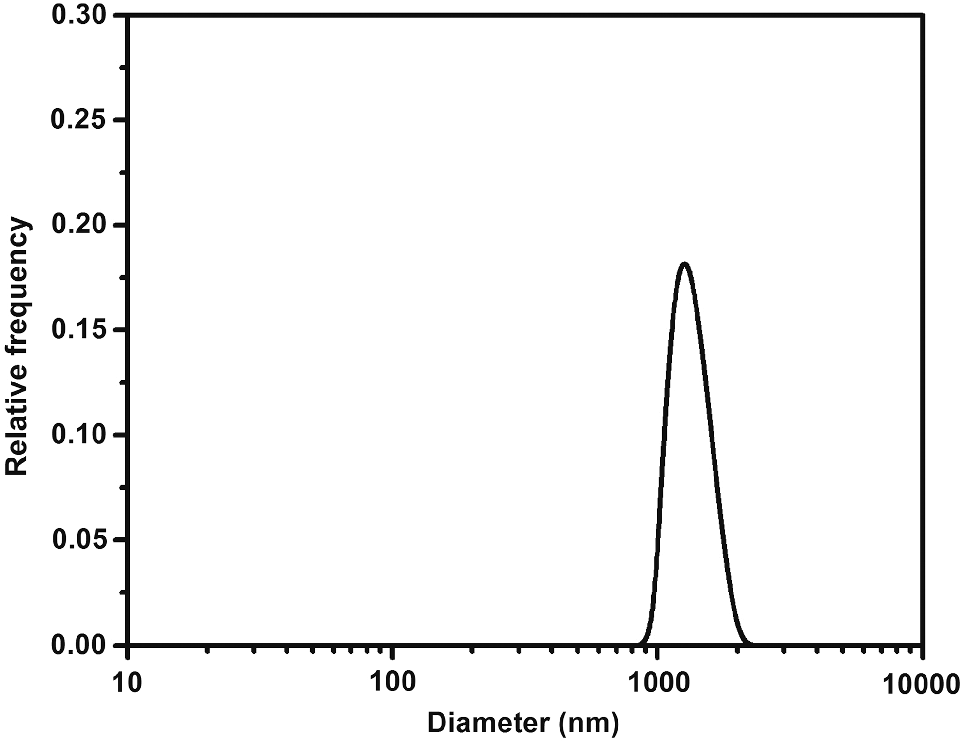
The resulted effective particle size distribution of 177Lu-labeled zirconia colloid: mean diameter (MD) = 1.7 μm.
The radiolabeled colloid showed high in vitro radiochemical and colloid size stability. The labeling efficiency of the sample incubated at room temperature was found to be above 99% after 1, 8, and 24 hours and 97% after 1 week of storage. The produced radiocolloid showed high in vitro serum stability also during the 1-week incubation. In the canine serum sample, the labeling efficiency was found to be >99% after 1, 8, and 24 hours and 98% after 1 week. DLS analyses demonstrated that the storage at room temperature did not produce a remarkable difference in the particle size distributions. The mean hydrodynamic diameter values of the colloidal product were found in the range of 1.4–2.5 μm during the 1-week follow-up. Results suggested that 177Lu-labeled zirconia particles do not disintegrate under the effect of self-β-radiation or by the storing in different physiological conditions at different temperatures.
Results of biological study
In the treated dog, no clinical side-effects were recorded during the treatment. The SPECT images were recorded at 1, 8, 24 hours and 1 week, 1 month, and 3 months postadministration time points. SPECT follow-up studies clearly and conspicuously showed the localization of the 177Lu-labeled colloid in the hock joint as well as negligible leakages (below 1% of total injected activity was measured by ROI/VOI analyses) of the radiocolloid in the nearest lymph node. Figures 3 and 4 represent dorsoventral dual-isotope SPECT images of the veterinary patient recorded simultaneously 24 hours and 1 month postadministration using 99mTc-MDP as control radiopharmaceutical. Figures 3A and 4A show the only detectable source of the 177Lu in the whole-body SPECT scans, while Figures 3B and 4B show parallel recorded whole-body 99mTc-MDP distributions. The exact localization of the hock joint is signed by retrospectively added reference lines in the paired scans. Examinations of blood and urine samples have reinforced the lack of leakage of the injected product; also, only negligible amounts of radioactivity (under 0.01% values of total ID) were found in the collected samples. Moreover, no significant changes were observed in hematological and biochemical parameters, as well as in Comet assays before and after administration of the labeled compound. All these biological evaluations indicate that the 177Lu-labeled zirconia colloid had no undesirable side-effects and thus might be safe for further evaluation.
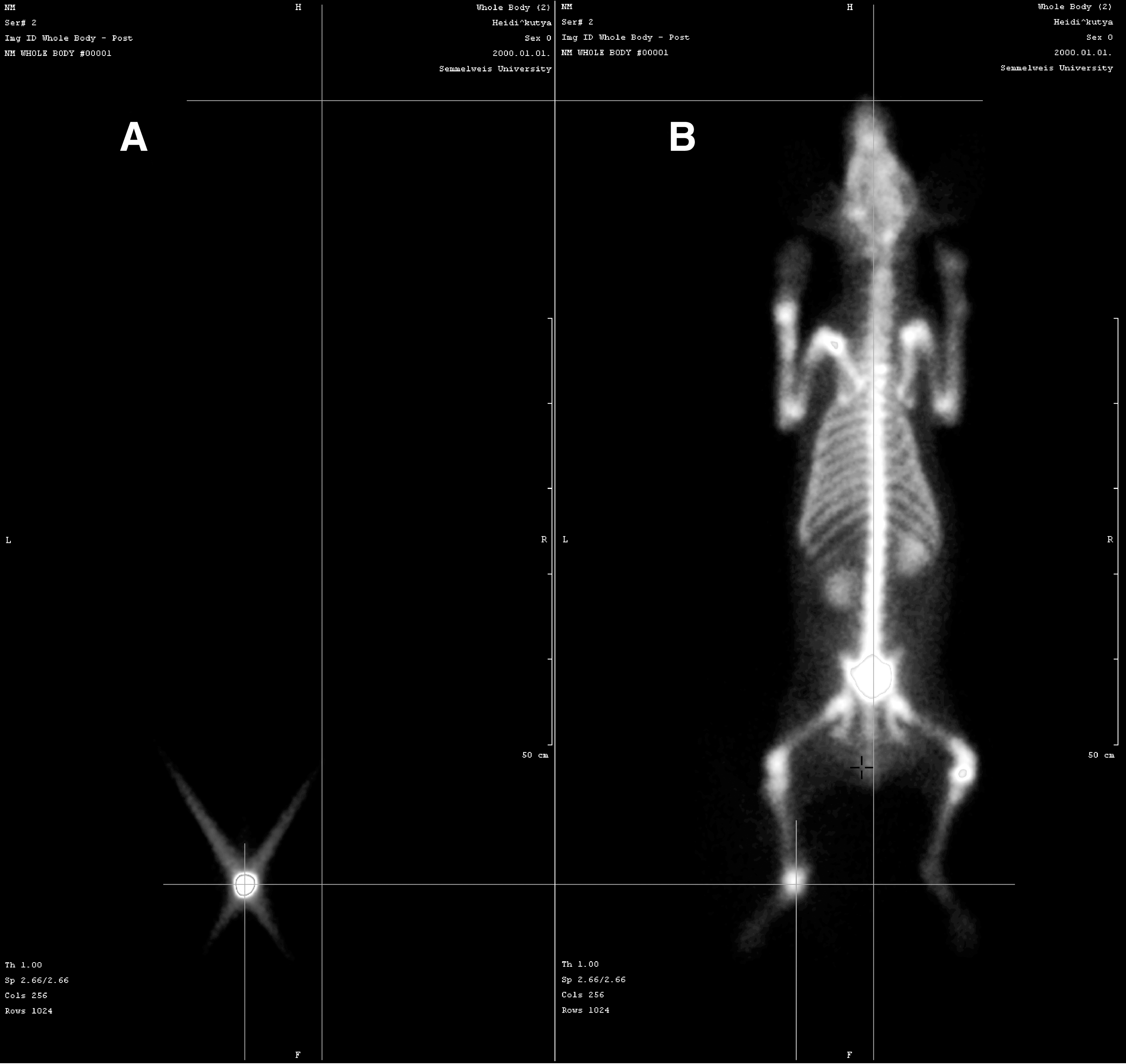
Dual isotope SPECT images of dog veterinary patient.

Dual isotope SPECT images of dog veterinary patient.
Three days after treatment, the bandage was removed and the patient was agitated progressively to move. One week to 3 months after completion of treatment, there was a significant improvement in clinical symptoms, for example, the dog could walk for hours with only a slight limp, no swelling, and joint flexibility as in contralateral hock joint. Follow-up bone scintigrams 1 month and 3 months posttreatment revealed normal 99mTc-MDP radiopharmaceutical uptakes (see Figs. 4B and 5). On the basis of follow-up tests and owner's semiquantitative evaluations, the orthopedic team assumed that local 177Lu-labeled zirconia colloid-based radionuclide therapy resulted in a significant and long-term improvement in clinical signs without any remarkable side-effects.
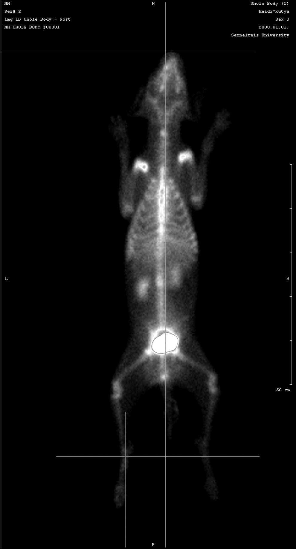
99mTc-MDP whole body SPECT scan of dog veterinary patient 3 months posttreatment.
Conclusions
The objective of the present work was to develop a new 177Lu-bearing therapeutic radiopharmaceutical candidate using biocompatible, inorganic carrier colloid, which may serve as a potential alternative to other radiocolloidal products presently used in radiation synovectomy. The 177Lu-labeled colloid was prepared following a simple radiolabeling method of a previously originated zirconia nanoparticle system. The radiocolloid had high radiochemical purity and adequate in vitro stability. The new product was investigated in the course of treatment of a spontaneously diseased dog veterinary patient. In vivo studies revealed high in vivo stability of the preparation along with significant retention of activity in the treated hock joint till 1 week postinjection. This preliminary study revealed the potential of the developed agent toward its application as an alternative agent for radiation synovectomy.
Footnotes
Disclosure Statement
No competing financial interests exist.