




Abstract
Introduction:
Natural killer (NK) cells are a subset of lymphocytes in humans that release cytokines such as tumor necrosis factor alpha and interferon gamma-γ during infection. NKG2D is one of the most important stimulating NK receptors binding MIC-A, MIC-B, and ULBPs, which leads to activation of NK cells against tumor cells. In this study, the authors evaluated the effect of G2 adjuvant on gene expression and delivery of NKG2D receptor on NK cells in peripheral blood.
Materials and Methods:
Peripheral blood mononuclear cells were isolated from venous blood obtained from healthy volunteers after adding G2 adjuvant within 12, 24, and 48 hours of incubation. Then, total RNA was extracted from the cells, cDNA synthesis was performed, and gene expression was evaluated by real-time PCR. In addition, NK cells were stained with the appropriate monoclonal antibodies, and the receptors expressed on cell surface were quantified.
Results:
G2 adjuvant leads to upregulation of gene expression and increases the expression of NKG2D receptor on the surface of NK cells after incubation.
Conclusion:
The findings of this study demonstrated that G2 adjuvant can increase NK cell cytotoxicity. It may play an important role in killing tumor cells, preventing tumor growth and metastasis.
Introduction
Natural killer (NK) cells are a subset of lymphocytes in humans and mice with an important role in innate and adaptive immunity. 1 In humans, NK cells comprise 10%–15% of peripheral blood lymphocytes, 2,3 and are found in a number of tissues such as the liver, lungs, gastrointestinal tract, and uterine decidua. 4 Human NK cells are divided into two subgroups based on function and phenotypes. Over 90% of NK cells in peripheral blood are CD56,dim CD16,+ and perforin,+ and the remaining are CD56,bright CD16, and perforin. 5 CD56bright cells express CXCR3 chemokine receptor, which migrates toward the inflammatory tissues and lymph nodes. 4 CD56dim cells circulate in the peripheral blood and have cytotoxic activity through perforin and granzyme production. 1 These cells release cytokines such as tumor necrosis factor alpha (TNF-α) and interferon gamma (IFN-γ) during infection. These cytokines stimulate and increase the activity of innate and adaptive immune responses. Moreover, these cytokines have exhibited an important role in cytotoxic activity against cancer and viral infections. 6
The function of NK cells is dependent upon stimulatory and inhibitory receptors. In humans, NK receptors are divided into three groups based on their structure, including killer cell immunoglobulin-like receptors (KIRs), natural cytotoxic receptors, and C-type lectin-like receptors (CTLRs). 7 Most inhibitory immunoglobulin-like and CTLRs bind to classical and nonclassical MHC class I molecules on normal cells and transmit the inhibitory signal through tyrosine motifs in cytoplasmic tail of the receptor within the cell. 7 NKG2D is one of the most important stimulatory NK receptors binding the MHC class I proteins such as MIC-A, MIC-B, and ULBPs. MHC class I-like ligands are present on cancer cells as well as virus-infected cells, which leads to stimulation of NKG2D receptor on NK cells. 8 Activation of the cells subsequently increases the expression of NKG2D receptor on NK cells. 9 NKG2D receptor binds to a 10 kDa messenger protein in the membrane known as DAP10. This subunit contains an immunoreceptor tyrosine-based activation motif (ITAM), a messenger motif that leads to activation of NK cells and cytotoxicity against cancer. 10,11
In cancer patients, NK cells are activated and converted to lymphokine-activated killer (LAK) cells with a higher cytotoxic activity, 12 increasing lytic proteins such as perforin and granzyme against target cells. Moreover, LAK cells express Fasl and TRAIL receptors that may be involved in lysis of cancer cells. These components of cytokine TNF receptors play an important role in apoptosis. 13
Thus, NK cells are an important component of innate immunity against infection and disease at an early stage and play an important role in the activation of cellular immune function as well as destruction and elimination of cancer cells. The majority of cancer patients receive chemotherapy, immunotherapy, or radiation therapy after surgery to prevent recurrence or metastasis. Cancer immunotherapy has been developed during the last few decades. Recent evidences showed that immunotherapy by tumor infiltrating lymphocytes increases the inflammatory response and decelerates the progression of cancer. 14 Several methods are used for immunotherapy, including treatment with NK cells, in which activated NK cells play an important role in controlling metastasis in cancer patients. Metastasis of cancer cells occurs in patients who have a defect in activation of these cells. 15 G2 adjuvant contains buffalo spleen extract stimulating TH cells, which has been developed by Saleh Mohaghegh Hazrati in Iran. G2 adjuvant causes improvement of breast cancer in animal models. This adjuvant was also used in patients with asthma and allergy and showed acceptable results. 16
NK cells play a crucial role in primary defense against infection, activation of innate and acquired immunity, as well as prevention of cancer metastasis. NKG2D receptor is one of the most important stimulatory receptors of NK cells, and G2 adjuvant has a stimulatory effect upon T cells. Therefore, in this study, the authors evaluated the effect of G2 adjuvant on gene expression and delivery of NKG2D receptor on NK cells in peripheral blood.
Materials and Methods
Preparation of the adjuvant
G2 adjuvant has been prepared from buffalo spleen lipid and registered as a patent in the Iranian Patent Office as Immune System Activator vaccine (Invention Register No.: 36679 and Date of Invention October 28, 2006). The preparation method of adjuvant is as follows: spleen is divided into small pieces and centrifuged at 800 g for 30 minutes after several days of exposure to dilute alcohol. The supernatant containing various types of lipids, glucose, cholesterol, and triglycerides is known as G2 adjuvant. 16
Isolation of peripheral blood mononuclear cells
Peripheral blood mononuclear cell (PBMC) were isolated from heparinized venous blood obtained from four healthy volunteers. Peripheral blood cells were isolated using 1.077 g/mL Ficoll–Paque density gradient (Biochrome) and were centrifuged for 30 minutes at 900 g at a temperature of 4°C. The mononuclear cell layer was transferred to a centrifuge tube. The cells were washed by a sterile RPMI 1640 medium (three times the volume of the mononuclear cell layer) and centrifuged at 18°C–20°C for 10 minutes at 400 g. The supernatant was removed and the cells were resuspended in the RPMI 1640 medium. Viability of PBMC was determined by trypan blue staining. Cells with more than 90% viability were used for examination. 17
Cell treatment
To investigate the effect of tested agents, optimal effective concentrations were established to be used in experiments. An optimally effective concentration of G2 adjuvant has been established at 400 mg/mL with serial dilutions. PBMC were cultured at a final concentration of 500,000 cells/mL in 12-well plates containing the RPMI 1640 medium, including 10% fetal calf serum (FCS) and 100 mg/mL of antibiotics (penicillin/streptomycin) at a temperature of 37°C and 5% CO2 in humidified cell culture plates. The cells were incubated for 12, 24, and 48 hours after addition of G2 adjuvant (concentration 400 mg/mL). Gene expression and delivery of receptors were subsequently measured. 18
RNA extraction
Total RNA was extracted using the RNA purification kit (Jena Bioscience) according to the manufacturer's instructions. The concentration and quality of extracted mRNA were spectrophotometrically determined at 260 nm and 260/280 nm wavelength ratio, respectively. Furthermore, the sample was electrophoresed in 1% agarose gel to confirm the purity and check the absence of DNA and quality of RNA.
Reverse transcription was performed using a transcriptor. First, cDNA synthesis was performed using a synthesis kit (Thermo Scientific) according to the manufacturer's instructions. Oligo (dT) primer was mixed with template RNA and the mixture was incubated at 65°C for 5 minutes. Transcriptase reaction buffer, transcriptase, deoxynucleotide mix, and RNase inhibitor (all from Thermo Scientific) were added to the tube containing template RNA-primer. Total mixture was incubated at 42°C for 60 minutes and at 70°C for 5 minutes. The rest of extracted RNA and cDNA was stocked at −70°C.
Real-Time PCR
Real-Time PCR was performed by ABI (Applied Bio systems 7500) kit SYBR green amplification (TaKaRa) at denaturation temperature of 95°C for 30 seconds and annealing/elongation at 57°C for 15 seconds at 40 cycles. Two specific primers were used for GAPDH (Forward: 5′-CACCATCTTCCAGGAGCGAG-3′), (Reverse 5′-GCAGGAATTGCTGAT-3′) and NKG2D (Forward: 5′-ACTGTGGCCCATGTCCTAAA-3′), (Reverse 5′-GGTTGGGTGAGAGAATGGAG-3′). Real-Time PCR data were analyzed using REST-RG software.
Flow cytometry
Flow cytometry is a common method to detect the presence of receptors and molecules on cell surface. After incubation, the cells were washed in phosphate-buffered saline containing 1% bovine serum albumin. To evaluate delivery of receptors on the surface of NK cells, the cells were examined by three-color flow cytometry using the appropriate monoclonal antibodies, including anti-CD3-PE-CY5 (eBioscience), anti-CD56-FITC (eBioscience), and anti-NKG2D-PE (eBioscience) (FACScom; BD Bioscience). 19 Afterward, the results of flow cytometry were analyzed using WinMDI v2.8 software.
Results
The cell samples were studied to determine the expression of NKG2D on NK cells. It was observed that G2 adjuvant affects NK cell activity and increases the expression of NKG2D gene, which plays a role in cytotoxic activity of NK cells against the target tissue. Increase expression of NKG2D gene at 12, 24, and 48 hours is shown in Figure 1. The expression of gene responsible for NKG2D in cells treated with adjuvant G2 compared to the control group (untreated) after incubation is shown in Figure 1.
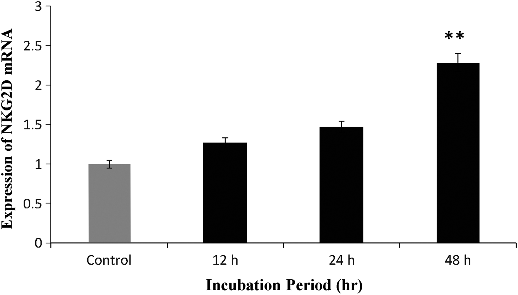
Fold increase of mRNA NKG2D gene expression relative to unexposed controls in human NK cells after 12, 24, and 48 hours of incubation in the presence of concentration (400 mg/mL) of G2 adjuvant. The results are the mean ± SEM of three independent experiments. **p = 0.001 significant difference between the treated and independent experiments. **p = 0.001 significant difference between the treated and untreated (control) groups after 48 hours of incubation in the presence of G2 adjuvant. SEM, standard error of the mean.
Flow cytometry
The delivery of receptors on the surface of NK cells was evaluated relative to unexposed controls (untreated) after 12, 24, and 48 hours of incubation in the presence of 400 mg/mL G2 adjuvant. The flow cytometry data were examined by WinMDI v2.8 software and the obtained gate mean (GM) was analyzed by IBM SPSS 22 software and T-test. The mean and standard deviation of NKG2D + NK cells and the intensity of expressed GM for NKG2D receptor are shown in Table 1. It was observed that the delivery of NKG2D receptor on the surface of NK cells treated with G2 adjuvant was significantly increased in comparison to the control group (Untreated) (p = 0.004). In these Figures, delivery rate of receptors in the samples treated with G2 adjuvant was compared to control, indicating a significantly increased delivery rate of receptors in the samples treated with G2 adjuvant compared to control at the level of NK cells.
p < 0.001 between the treated and untreated groups.
Histograms investigating NKG2D on the surface of NK cells
The range of expression of the NKG2D is shown in M1 index shown in Figure 2. The results showed that gene expression was increased with increasing period of incubation, and maximum NKG2D gene expression was observed after 48 hours of incubation.
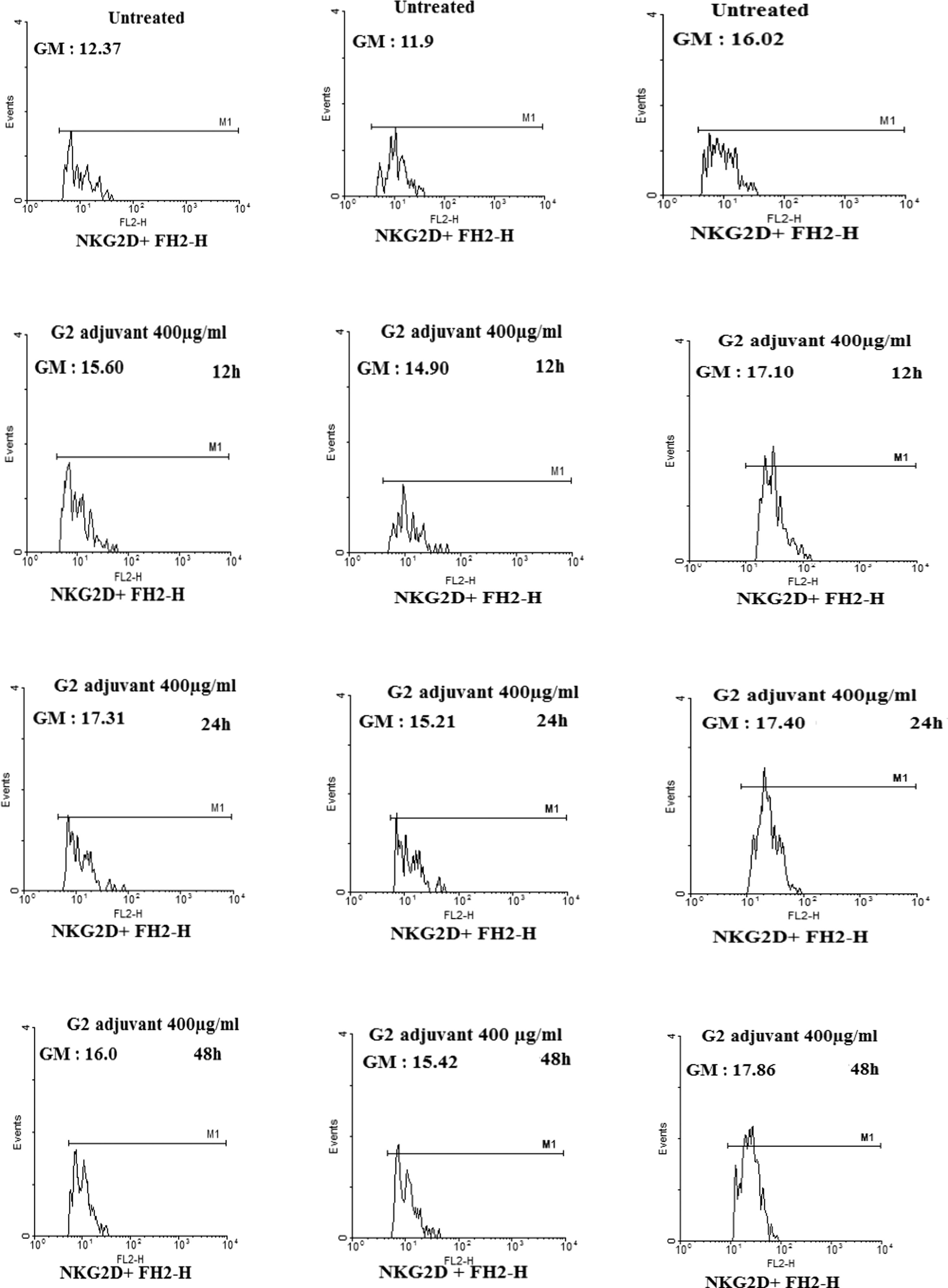
Increased delivery of NKG2D receptor on the surface of peripheral blood NK cells. Freshly isolated cells were cultured for 12, 24, and 48 hours in medium in the absence and presence of G2 adjuvant (400 mg/mL concentration). Significant difference was observed between the treated and untreated groups (p = 0.004).
Discussion
Previous studies have confirmed the role of G2 adjuvant in the stimulation of immune cells such as T lymphocytes. This adjuvant contains buffalo spleen extract and improves breast cancer in animal models. 20 As a natural adjuvant, G2 adjuvant can be used in treatment and recovery from several diseases. After treatment of asthma patients with G2 adjuvant, about 70% of the patients were recovered, which may indicate the important role of this adjuvant in control of asthma through affecting TH2 cells. 21 Besides, G2 adjuvant controls the allergic response through preventing the increase in eosinophils and basophils, inhibiting TH2-related responses. 22 Neamati et al. reported that this adjuvant regulates the balance between TH1 and TH2 and can induce the stimulation of TH1 cells. In addition, G2 adjuvant plays a crucial role in the production of IL-2 and IFN-γ as well as activation of macrophages to regulate proinflammatory responses through stimulation of the TH1 cells. 16
Hazrati et al. reported that G2 adjuvant increases the production of IFN-γ by stimulating TH1 cells, and that increasing level of this cytokine leads to increased activity of cell-mediated immunity. 20,23 Subsequently, these cytokines can help patients recover from breast, brain, and prostate cancers by inhibiting or reducing the expression of matrix metalloproteinases. 24,25 G2 adjuvant can also stimulate cell-mediated immunity and increase the production of IFN-γ to recover patients from viral infections. 26 In another study on cancer patient, Hazrati et al. found that the tumor mass was gradually shrunk and even disappeared completely after immunotherapy using G2 adjuvant, and suggested that this adjuvant can contribute to cancer elimination and patient recovery through stimulating and activating immune cells such as NK cells against cancer. 27
NK cells serve as the first line of defense against a wide range of pathogens, especially viruses, playing an important role in the immune response against tumor cells in both humans and mice. 28 NK cells play an important role in both innate and acquired immunity, performing four major functions, including cytotoxic activity through secretion as well as release of perforin and granzyme, chemokine and cytokine secretion, TRAIL and Fas ligand-induced apoptosis, and antibody-dependent cell-mediated cytotoxicity. 3 NK cells trigger cytotoxicity through stimulatory receptors such as NKG2D, which identifies its ligands on the tumor cells as well as virus-infected cells, activating NK cells to lyse target cells. 29
In the present study, the expression of NKG2D receptor on NK cells was evaluated in the presence of stimulating TH1 cells called adjuvant G2, and the results showed that the gene expression and delivery of NKG2D receptor were increased. Moreover, it causes increased activation of NK cells, which consequently increases the lethality of these cells in viral infections and cancers. Future perspectives may propose the activation of NK cells in patients with cancer to stimulate the production of cytokines by triggering innate and acquired immunity as well as production and secretion of cytotoxic mediators such as perforin and granzyme, which play an important role in clinical development by way of apoptosis of tumor cells and virus-infected cells and inhibition of tumor cell growth as well as metastasis.
Mincheva-Nilsson et al. showed that the expression of genes encoding NKG2D receptor is regulated under the influence of cytokines and vaccines. Interleukin (IL)-12 increases the expression and availability of NKG2D receptor and DAP10 in NK cells. NKG2D receptor is one of the important receptors for cancer diagnosis, which binds to target cells through its ligands and causes stimulation and activation of NK cells. 30 Jamieson's study showed that LPs, IFN-γ, and IFN-α/β contributed to stimulating the expression of NKG2D receptor. Increasing NKG2D receptor expression is considered as a factor affecting the innate immunity, which may ultimately lead to increased cytotoxic activity of NK cells against viral infections and tumors. 31
Osada showed that the use of dendritic cell (DC) vaccine can activate NK cells against tumor cells and virus-infected cells, and observed that culturing NK cells in the presence of DCs significantly increased the expression of genes encoding NKG2D, resulting in increasing NK cells cytotoxicity against tumor cells and virus-infected cells. 32,33 The results of the present study are consistent with these studies.
The results of another study showed that IL-15 increased the expression of genes encoding NKG2D in addition to increasing effective cytotoxic molecules such as TRAIL and FasL. Furthermore, increased expression of genes encoding NKG2D, which sends activation signal into the cell, leads to activation of NF-κB transcription factor, 34 increasing the production of proinflammatory cytokines such as IL-1 and IL-6 as well as decreasing anti-inflammatory cytokines. Furthermore, recent data have shown that NF-κB inhibits the expression of inhibitory cytokines such as IL-10, but increases the expression of INF-α, IFN-γ, and TNF, 34 which can direct the differentiation of T cells into Th1, stimulating NK and T cytotoxic (CTL) cells against cancer through production of IL-2. 35 Therefore, NF-κB causes increased cytotoxicity of NK cells through several mechanisms, including stimulating the production of perforin and granzyme as well as members of cell death receptors, which play a role against tumor and infectious pathogens. 36
Zhou et al. showed that DNA vaccines can increase the expression of NKG2D receptor on NK cells and cause maturation of DCs, which help increase the activity of NK and CTL cells and change NK cells to LAK cells through cytokine secretion and expression of costimulatory molecules. 37,38 Özdami et al. found that IL-15 also increases perforin and TNF-α, which change NK cells to LAK cells. 38 NK cells play a role in the suppression of early tumor cancer stages, and several studies have indicated that NK cells play an important role in preventing tumorigenesis and may even eliminate the tumor. 39,40 In addition, production of IFN-γ by NK cells leads to differentiation of macrophages into M1 macrophages, which increases reactive oxygen species and nitric oxide activity in macrophages. M1 macrophages increase the apoptosis of cancer cells, inhibit angiogenesis, and prevent tumor growth and progression of cancer. 41 –43 These results are consistent with the present study. In addition to direct effect of NK cells in killing target cells through production of these cytokines, they indirectly play a role in elimination of tumors and infections by affecting other innate immune cells such as DCs and macrophages as well as activating acquired immune cells such as TCD4 and TCD8. Studies have shown that tumor growth is considerably increased in NKG2D-deficient mice during the early stages of malignancy. Moreover, NKG2D has a modulatory role in immune responses as well as limiting the cancer spread and invasion to other tissues, which shows that this receptor plays an important role in early stages of tumorigenesis rather than metastasis. 15,44,45
In contrast to the present study, the study of Sha Hao showed that long-term exposure to estrogen (E2) reduces NK cell activity. E2 also suppresses NK cell killing by reducing the expression of NKG2D stimulatory receptor and reduces the expression of other stimulatory receptors on NK cells such as NKp46 and 2B4 receptors. These functional features have a negative impact on NK cell killing. Overall, this factor leads to decreased secretion of cytotoxic factors such as granzyme B and FasL and is ultimately involved in cancer growth. 46
In conclusion, the use of G2 adjuvant could increase gene expression and delivery of NKG2D receptor on NK cells, which can increase NK cell cytotoxicity against cancers and viral infections. Thus, G2 adjuvant can be used as an activator of NK cells in cancer patients. Besides, G2 adjuvant may play a key role in killing tumor cells by NK cells, preventing tumor growth and metastasis by NK cells as well as killing the virus-infected cells. It stimulates the production of cytokines, activates innate and acquired immunity, as well as production and secretion of cytotoxic mediators such as perforin and granzyme.
Footnotes
Acknowledgments
This study is part of MSc thesis for Nader Dashti Gerdabi. Specific thanks to Vice Chancellor of Research, Ahvaz Jundishapur University of Medical Sciences, for the financial support.
Disclosure Statement
No competing financial interests exist.