
Abstract
The aim of current study is to examine hydroxyurea (HU), which is an antineoplastic drug used for the treatment of leukemia, sickle-cell disease, HIV, psoriasis, thrombocythemia, and various neoplastic diseases in two aspects. The active ingredient hydroxyurea was obtained by purification of the capsule form drug, commercially named as HYDREA. Then, [99mTc(CO)3]+core radiolabeling with HU was performed as first aspect. Quality control studies of 99mTc(CO)3-HU complex were performed by thin-layer radiochromatography and high-performance liquid radiochromatography methods. The results demonstrated that the radiolabeling yield was quite high (98.43% ± 2.29%). Also, 99mTc(CO)3-HU complex has good stability during the 24-hour period. Biological behavior of 99mTc(CO)3-HU complex is evaluated by biodistribution studies on Wistar Albino rats. Fluorescein isothiocyanate (FITC) labeling of HU was performed as second aspect. Fluorometric evaluation of binding efficacy and fluorescence imaging studies on MCF7 and Hela cell lines were carried out. It was thought that the knowledge achieved in this study would contribute to using 99mTc(CO)3-HU complex as an imaging agent, which inhibits the DNA synthesis selectively, by inhibiting ribonucleotide reductase enzyme. It was observed that FITC-HU has noteworthy incorporation on both cell lines.
Introduction
Hydroxyurea (HU) is an antineoplastic compound that was synthesized by Dresler and Stein in 1869. It is used in the treatment of many neoplastic diseases such as chronic, resistant, myelocytic leukemia, ovary carcinoma (recurrent, inoperable, or metastatic), cervical carcinoma, melanoma, meningioma, and in combination with irradiation in treatment of primary squamous cell carcinoma of the head and neck. Nowadays, it can be used to treat leukemia and other malignances, sickle-cell anemia, HIV infection, thrombocythemia, psoriasis, and polycythemia vera. Hydroxyurea is an S-phase cell cycle-specific agent that selectively inhibits DNA synthesis through inhibition of the enzyme ribonucleotide reductase. HU is structurally related to hydroxamic acids. For the purpose of creating more effective antitumor drugs, nowadays, the main focus has shifted to structurally similar hydroxamic acid derivatives. 1 –4
There are various studies about the pharmacology of hydroxyurea and hydroxamic acid derivatives. In 1928, Rosenthal et al. showed that HU inhibits the leukocyte cell growth. Studies in the 1960s have established that hydroxyurea and several related hydroxamates cause inhibition of the synthesis of DNA. 4,5 According to the report by Liebelt et al. in various pharmacokinetic studies performed in human, researchers concluded that hydroxyurea distributes in a volume similar to that of total body water and tends to concentrate in erythrocytes and leukocytes in 2007. In one of these studies, distribution of 14C-hydroxyurea in male CDBA mice and Fischer rats was examined and it is reported that high concentrations of radioactivity were detected in the kidney and bladder. 2 Dogruel and his colleagues performed another study with 13C-HU. They reported that brain uptake of 13C-HU is related to concentration and the main important evidence provided is the blood–brain and blood–cerebrospinal fluid barrier crossing ability of HU. 6
99mTc is important in diagnostic nuclear medicine because of its low cost and easy availability from a 99Mo/99mTc-generator. Its 140 keV γ energy is also ideal for imaging. It is well known that technetium has rich coordination chemistry. For radiopharmaceutical applications, six coordinated technetium (I) and rhenium (I) tricarbonyl complexes [fac-M(CO)3]+ were improved by Alberto et al. 7,8 Efficient labeling of bidentate and tridentate biologically relevant ligands with [99mTc(CO)3(H2O)3]+ was demonstrated. By the displacement of water molecules in the complex with amine, aromatic N-heterocycles and/or carboxylic donors it can be attached to the bifunctional ligand complex biomolecules. The spherical shape of [M(CO)3]+ (M = 99mTc, 188Re) has a relatively strong structure. 9,10
Fluorescein isothiocyanate (FITC) is a fluorescent derivative that has a wide usage for flow cytometry applications. It is reactive toward proteins such as antibodies and lectins due to a suitable isothiocyanate group for labeling. In the current study, FITC labeling of HU was performed. Binding efficacy of FITC-HU was evaluated fluorometrically and fluorescence imaging studies on MCF7 and Hela cell lines were carried out.
The studies with hydroxyurea and derivatives were examined and it is observed that there is no effective radiolabeling and radiopharmaceutical study except 14C-hydroxyurea; the studies mostly focused on the kinetics of the compound. In the present study, the biological behavior of 99mTc(CO)3-HU complex was investigated in vivo and FITC-HU was investigated in vitro. It was thought that this knowledge will pave the way for comprehensive studies about using 99mTc(CO)3-HU complex and similar complexes in diagnosis. Hereby, HU was investigated from two different perspectives by labeling with 99mTc(CO)3 + and FITC.
Materials and Methods
The drug called HYDREA (500 mg) was supplied by Deva Holding A.Ş. (İstanbul, Turkey). Chemicals were generally reagent grade and were obtained from commercial sources. Water was purified by filtration using a Milli-Q Gradient A-10 ultrapure water system (Millipore S.A., Molsheim, France) with conductivity of 18 MΩ cm. Freshly eluted Na99mTcO4 was obtained from the 99Mo/99mTc generator (Monrol, Gebze, Turkey) using saline. Radioactivity was detected using a Cd(Te) detector equipped with a RAD 501 (Turkey) single-channel analyzer. Silica-coated alumina thin-layer chromatography (TLC) plates were purchased from Merck (Merck 5554; Darmstadt, Germany). TLC plates were scanned with a Bioscan AR-2000 radioanalyzer. The high-performance liquid radiochromatography (HPLRC) system for the analyses of labeled compounds consisted of a Shimadzu quaternary gradient pump (Model LC-10Atvp Shimadzu), an automated syringe injector (20 μL loop), and a 5 μL reverse-phase column (RP-C-18 column).
Purification of HYDREA (500 mg)
The active ingredient used in this study was hydroxyurea and it was obtained by purification of the drug, commercially named as HYDREA (500 mg). Two capsules of HYDREA in powder form were added in a solution containing 50 mL acetone and 50 mL methanol and this solution was stirred with the aid of magnetic stirrer. Then, the solution was centrifuged at 2500 rpm for 5 minutes to remove excipients in the drug. By evaporating acetone and methanol in the resulting hydroxyurea solution, pure hydroxyurea in powder form was obtained. Hydroxyurea was kept in the refrigerator at +4°C. In each study, 1 mg/1 mL hydroxyurea sample was prepared.
Radiolabeling HU with [99mTc(CO)3(H2O)3]+
[99mTc(CO)3(H2O)3]+ core was prepared using the method in the literature. 7 4.5 mg Na2CO3, 5.5 mg NaBH4, and 20 mg Na/K tartrate were individually weighed and added to a 10-mL glass vial. The vial was sealed to be airtight. The volume of Na99mTcO4 (with activity of 15 mCi), obtained from 99Mo/99mTc generator, was completed to 1 mL with saline and argon gas was passed through for 5 minutes. The same procedure was applied to the glass vial, which is previously prepared. Then, CO gas under pressure of 1 bar allowed to flow through the vial for 15 minutes. Na99mTcO4 was transferred to the glass vial with the aid of an injector, and the glass vial, including all of the reducing agents and Na99mTcO4 (in the presence of CO gas), was incubated at 90°C for 30 minutes. After incubation, the solution was cooled down to room temperature by waiting in an ice bath. By adding about 220 μL of 1 M HCL:phosphate buffered saline (PBS) (3:1) solution to the cooled solution, pH was adjusted to 7 and the 99mTc(I) tricarbonyl core was realized. After the reaction, radiochemical purity of 99mTc(I) tricarbonyl core was evaluated by thin-layer radiochromatography (TLRC) and HPLRC methods.
To prepare radiolabeled 99mTc(CO)3-HU complex, the labeling conditions of 99mTc(CO)3-HU complex were optimized in the aspect of pH, reaction temperature, reaction time, and the amount of ligand. With this aim, 100 μL of hydroxyurea stock solution was measured and transferred into an eppendorf tube. The activity of 99mTc(I) tricarbonyl core, prepared previously, was measured and 1 mCi of 99mTc(I) tricarbonyl core was added to the eppendorf tube. Then, the eppendorf tube was placed into a glass tube, which was filled with water and then incubated at 90°C for 30 minutes. After incubation, the solution was cooled down to room temperature by waiting in an ice bath. Radiochemical purity of 99mTc(CO)3-HU complex was evaluated by TLRC and HPLRC methods.
Radiochemical purity analysis
The radiochemical purity of [99mTc(CO)3(H2O)3]+ core and final complex 99mTc(CO)3-HU was evaluated by TLRC and HPLRC chromatography methods. TLRC analyses were performed on silica-coated alumina TLC plates with acetonitrile and serum physiological (SF) as the developing media. TLC plates were scanned with a Bioscan AR-2000 radioanalyzer.
High-performance liquid chromatography (HPLC) experiments were accomplished by the use of a Shimadzu system with quaternary gradient pump (Model LC-10Atvp Shimadzu), an automated syringe injector (20 μL loop), and a 5 μL reverse-phase column (RP-C-18 column). The gradient system consisting of 0.1% trifluoroacetic acid (TFA) with H2O (solvent A) and methanol (solvent B) was used as the eluting solvent. Mobile-phase flow system was applied as linear gradient in the first 3 minutes %100 A; between minutes 3.1–9 %75 A, %25 B; and between minutes 9.1–20 from %66 A (%34 B) until it reaches %0 A (%100 B). The flow rate was maintained at 1 mL/min.
Lipophilicity analysis
To measure lipophilicity (logP) of 99mTc(CO)3-HU complex, n-octanol and pH 7 buffer (PBS) were used. Initially, 300 μL of n-octanol and 300 μL of pH 7 buffer (PBS) were put into a centrifuge tube; to this mixture, 100 μL of 99mTc(CO)3-HU complex was added and the resulting mixture was stirred for 1 minute with the aid of a vortex. Then, the mixture was centrifuged at 2500 rpm for 30 minutes, so that the formation of two phases in the mixture is provided. One hundred microliters of phases was taken both from the upper and lower phase and the activities were counted with the semiconductor solid-state Cd(Te) detector. The partition coefficient was calculated as the ratio of count per second (CPS)/g of octanol to CPS/g of water. Experiments were conducted thrice and averaged.
Stability studies
In vitro stability of the 99mTc(CO)3-HU-complex was determined by incubating 100 μg/37 MBq of the labeled compound at room temperature. The sample was then analyzed at time intervals of 0, 30, 60, 120, 240, and 1440 minutes by TLRC after labeling (n = 3).
Biodistribution studies
All the animal experiments were carried out in compliance with the relevant national laws as approved by the local committee on the conduct and ethics of animal experimentation (the Department of Experimental Research and Surgery and the Animal Research Ethic Committees of the Ege University numbered 2012–080).
For determination of the biological efficacy of 99mTc(CO)3-HU complex in vivo, a biodistribution study was performed on male Wistar Albino rats with a weight range of 130–250 g. 0.2 mL of the 99mTc(CO)3-HU containing 10 μg HU (18.5 MBq) of activity was injected through the tail vein. The studies were carried out at 5, 30, and 60 minutes postinjection for three rats at each time interval. The rats were sacrificed at desired times with a cocktail of ketamine/xylazine (75–l00 mg/kg and 5–10 g/kg) by intraperitoneal injection. All major organs were excised, rinsed, weighed, and counted in the Cd(Te) detector to estimate the percent of injected dose/g of the tissue.
Differences in the mean values of measured activities were evaluated statistically by the SPSS15 program (Univariate Variance Analyses and Pearson Correlation; Chicago, IL). Probability (p) values <0.05 were considered to be significant. A Pearson correlation was carried out between organs for 99mTc(CO)3-HU.
FITC labeling of hydroxyurea
A solution of hydroxyurea was prepared dissolving 3 mg of drug in 1 mL of 0.1 M sodium carbonate. FITC (1 mg) was dissolved in 1 mL of dimethyl sulfoxide and 50 μL of this solution was added into the stock solution of hydroxyurea while stirring gently and continuously. This solution was incubated at 4°C for 8 hours in the dark. After incubation, 50 μM of NH4Cl solution was added and again incubated at 4°C for 2 hours. At the end of the incubation time, 1 μg Xylene cyanol and 5 μL glycerol were added and the solution was passed through the Sephadex G-25 column to separate the unbound FITC.
Fluorometric evaluation of binding efficacy of hydroxyurea on cells
Breast (MCF7) and cervical (Hela) carcinoma cell lines were used for cell culture studies. MCF7 was obtained from the American Type Culture Collection. Both cell lines were grown in the RPMI-1640 medium containing 2 mM
Cells were washed thrice in PBS and 0.5 mL of culture medium containing FITC-labeled hydroxyurea was added to each well. Cells were incubated for 30, 60, 120, and 240 minutes and then washed with PBS thrice.
FITC standard in several concentrations (1, 0.5, 0.25, 0.125, 0.0625, 0.00 mg/mL) were used for calibration. Thermo Varioscan Multimode was used for measurements (Ex: 495 nm and Em: 525 nm). Binding efficiency of FITC-labeled hydroxyurea was calculated.
Fluorescence imaging studies
MCF7 and Hela cell lines were cultured on chamber slide for fluorescence imaging studies. After 24 hours, culture media on the cells were washed with PBS, and FITC-labeled hydroxyurea solution was added on the cells. All these experiments were carried out under dark conditions. Image of the cells was taken at the 4-hour time point using the Olympus BX 53 Fluorescence microscope with green filter (520 nm).
Results and Discussion
Purified hydroxyurea stock solutions were prepared as mentioned in ‘Purification of HYDREA’ section and for this solution, HPLC analysis was performed. Figure 1 shows HPLC of obtained hydroxyurea with a high yield (>98%) by eliminating the other ingredients of the drug.

High-performance liquid chromatography of purified hydroxyurea.
Radiolabeling and characterization
[99mTc(CO)3(H2O)3]+ was prepared according to the procedure reported by Alberto et al. 7 and used for radiolabeling of HU. Radiolabeling HU with [99mTc(CO)3(H2O)3]+ was performed directly with a high yield of 98.70 ± 1.8 in serum physiological developing media verified by TLRC. The Rf value of 99mTc(CO)3-HU was found to be 0.84, while the Rf value of [99mTc(CO)3(H2O)3]+ was 0.03. Having different Rf values of [99mTc(CO)3(H2O)3]+ and 99mTc(CO)3-HU exhibits that hydroxyurea was radiolabeled with [99mTc(CO)3(H2O)3]+. [99mTc(CO)3(H2O)3]+ has three readily exchangeable water molecules for substitution with appropriate donor groups such as aromatic amines, aliphatic amines, and carboxyl groups. 11,12 In the light of this knowledge, NH2 and NH groups of the ligand as proposed conjugation site are shown in Figure 2. The radiochemical purity of [99mTc(CO)3(H2O)3]+ and the 99mTc(CO)3-HU complex was analyzed by reverse phase HPLRC. As seen in Figure 3, the retention times observed were 7.48 and 4.32 minutes, respectively. The HPLRC supports the results obtained from TLRC.
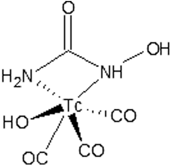
Proposed structure of 99mTc(CO)3-HU.

High-performance liquid radiochromatography of 99mTc(CO)3-HU and [99mTc(CO)3(H2O)3]+.
The value of participation coefficient determined for the 99mTc(CO)3-HU was found as −2.77 ± 0.07 (n = 3). Theoretical logP value of HU was computed as −1.80 ± 0.19 using ACD/LogP Algorithm program. According to these results, it is understood that the experimental lipophilicity value for the 99mTc(CO)3-HU complex is lower than the theoretical lipophilicity of HU.
Figure 4 displays the stability of 99mTc(CO)3-HU for 24 hours with 96% radiochemical purity.
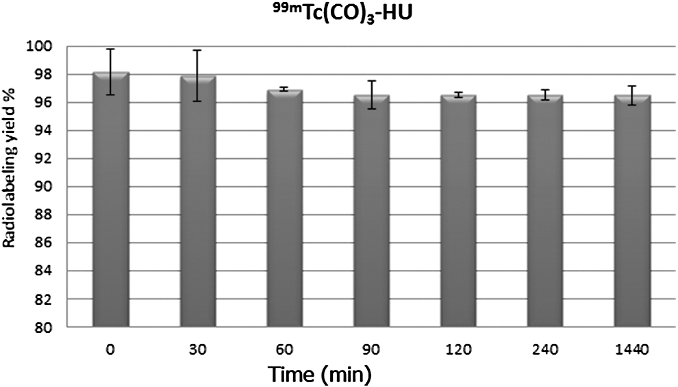
The stability of 99mTc(CO)3-HU complex.
Cell culture
Hydroxyurea is used in the treatment of many neoplastic diseases such as ovarian and cervical carcinoma and melanoma. For this reason, the authors chose MCF7 and Hela cell lines to determine cell uptake ratios of HU with respect to definite time intervals (30, 60, 120, 240 minutes). FITC conjugation to HU was performed, to be able to monitor in vitro behavior of HU during cell uptake and fluorescence imaging studies. Sephadex G-25 column was used to separate the unbound FITC from the solution of FITC-HU. Thus, they can eliminate the interference of unwanted fluorescence signal coming from unbound FITC. According to the results, the cell uptake of FITC-HU was found as 5.42% ± 0.33% on MCF7 and 5.18% ± 0.37% on Hela cells at 30 minutes. The authors could not observe any significant difference up to 240 minutes for both cell lines, as seen in Figure 5. Fluorescence images were taken to support the fluorometric results after FITC-HU treatment of MCF7 and Hela cell lines for a 4-hour incubation. It was observed that FITC-HU was incorporated into both cell lines as seen in Figure 6.
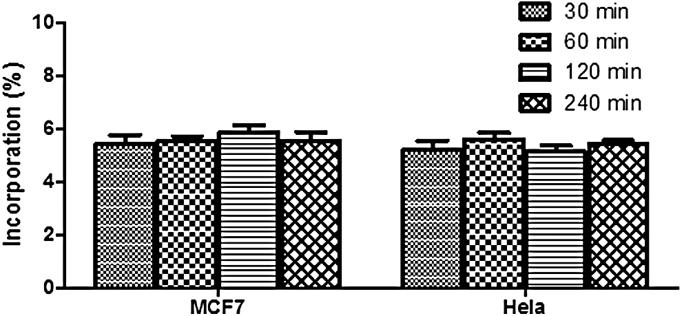
Cell uptake of fluorescein isothiocyanate (FITC)-HU on MCF7 and Hela at distinct time intervals.
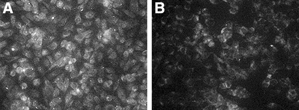
Fluorescence imaging of FITC-HU on
Biodistribution studies
Biodistribution characteristics of 99mTc(CO)3-HU was evaluated using male Wistar Albino rats. Biodistribution results of 99mTc(CO)3-HU on male Wistar Albino rats for different time intervals are shown in Table 1. Obtained injected dose/g tissue percentages are represented in Figure 7. According to the data it is observed that at 5 minutes, 99mTc(CO)3-HU exhibited the highest accumulation in the bladder compared with other organs and a high value of uptake was observed in the lung, liver, and kidney.

Biodistribution of 99mTc(CO)3-HU at distinct time intervals.
HU, hydroxyurea; ID, injected dose.
The results showed that the liver and kidney reached their maximum value of uptake at 30 minutes and decreased at 60 minutes. Also, the bladder had high uptake at 30 minutes and decreased at 60 minutes. In contrast, it is observed that the uptake in the small intestine, large intestine, and stomach increased with time. According to Liebelt's report, Adamson and coworkers examined the biodistribution of 14C-HU on mice and rats. They found that there was high uptake in the bladder compared to the kidneys and intestines and lower uptake in the stomach, heart, spleen, and lungs. 2 The high uptake in the bladder at 5 minutes, in this study, agrees with the results of the biodistribution study of radiolabeled hydroxyurea with C-14. 3
There are limited studies related to the technetium radiolabeled hydroxyurea in the literature. There are several studies, including pharmacokinetic studies of inactive hydroxyurea, on human and mice. One of those reported that the uptake of hydroxyurea by the stomach and intestinal system was very rapid in both healthy and sick individuals. Hydroxyurea tends to accumulate in blood cells and it can easily pass through the blood–brain barrier. 3 In this study, it is seen that the radiolabeled complex, 99mTc(CO)3-HU, has a similar behavior with HU metabolism.
It can be concluded that the presented 99mTc(CO)3-HU spreads fast within tissues and the complex has rapid pharmacokinetics. The high uptake observed in the kidney and bladder at 30 minutes shows that the 99mTc(CO)3-HU complex follows renal clearance.
Variance analysis for each organ showed that there is a significant difference in uptakes between the liver and muscle; kidney, muscle, and head; small intestine, large intestine, and muscle; large intestine, small intestine, stomach, bladder, and testis; prostate and bladder; head, heart, and kidney (p < 0.05).
Conclusion
Hydroxyurea has been successfully labeled by [99mTc(CO)3(H2O)3]+ in this study. The complex was delivered to all tissues and organs quickly, and through renal excretion. The knowledge achieved in this study could contribute to use 99mTc(CO)3-HU complex as an imaging agent.
Limitations
Further studies are required to carry out pharmacokinetic and metabolic studies of 99mTc(CO)3-HU as it is a structurally different entity from unlabeled HU. Because 99mTc(CO)3-HU is now a different chemical compound from HU, it would have a different behavior.
Footnotes
Acknowledgment
This study was financially supported by the Ege University Scientific Research Project No. 2013NBE003 (Bomova Izmir, Turkey).
Disclosure Statement
No competing financial interests exist.