




Abstract
In recent years the authors have reported on 99mTc(CO)3-labeled peptides that serve as carriers for biomolecules or radiopharmaceuticals to the tumors. In continuation of that work they report the synthesis of a pentapeptide (Met-Phe-Phe-Gly-His; pep-1), a hexapeptide (Met-Phe-Phe-Asp-Gly-His; pep-2), and a tetrapeptide (Asp-Gly-Arg-His; pep-3) and the attachment of 3-amino-1,2,4-triazole to the β carboxylic function of the aspartic acid unit of pep-2 and pep-3. The pharmacophores were radiolabeled in high yields with [99mTc(CO)3(H2O)3]+ metal aqua ion, characterized for their stability in serum and saline, as well as in His solution, and found to be substantially stable. B16F10 cell line binding studies showed favorable uptake and internalization. In vivo behavior of the radiolabeled triazolyl peptides was assessed in mice bearing induced tumor. The 99mTc(CO)3-triazolyl pep-3 demonstrated rapid urinary clearance and comparatively better tumor uptake. Imaging studies showed visualization of the tumor using 99mTc(CO)3-triazolyl pep-3, but due to high abdominal background, low delineation occurred. Based on the results further experiments will be carried out for targeting tumor with triazolyl peptides.
Introduction
Technetium-99m (99mTc) is the most widely used radioisotope in diagnostic nuclear medicine. Significant advancement in technetium radiolabeling is associated with the availability of a wide variety of ligand systems as well as bifunctional chelating agents. 1 Further advances have led to the development of organometallic labeling strategy based on the fac-[99mTc(CO)3(H2O)3]+ core, the synthon developed by Alberto et al., for labeling receptor-specific bioactive molecules. 2,3 This core due to its low spin, d 6 electronic configuration of Tc(I), exhibits remarkable stability over a wide range of pH values. Excellent labeling efficiencies with a number of donor groups, including amines, thioesters, phosphines, carboxylates, and thiols have been achieved due to the lability of the three water molecules coordinated to the fac-M(CO)3 moiety. 4
Their laboratory in recent years has made notable attempts toward the development of 99mTc(CO)3-labeled peptides coupled to bioactive pharmacophores to be used as diagnostic radiopharmaceuticals for tumor targeting. 5,6 During the last 10 years several receptor-specific peptides have been radiolabeled with various radionuclides for scintigraphic applications and therapeutic purposes. 7 Small peptides are less likely to be immunogenic, possess rapid blood clearance, and are easy to synthesize and modify. Thus, for the development of target-specific radiopharmaceuticals they are considered to be excellent candidates. 8 L-type amino acid transporter 1 (LAT 1) is responsible for the transport of various natural amino acids, such as methionine, phenylalanine, tyrosine etc. In cancer cells too LAT 1 is found to be highly expressed. Therefore, many natural amino acids and their synthetic analogues are being explored as tumor imaging agents, which have been radiolabeled with different radioactive isotopes. 9,10
Recently the pharmacological activities of 1,2,4-triazoles have been extensively explored. In many cases 1,2,4-triazoles have been found to exhibit significant antitumor activity. 11,12 In an effort to develop radiopharmaceuticals that can localize and image tumors, 99mTc(CO)3-labeled triazole-coupled peptides have been synthesized (Fig. 1), characterized, and evaluated for targeted application. They report, in this study, the synthesis of three peptides, viz. Met-Phe-Phe-Gly-His (pep-1), Met-Phe-Phe-Asp-Gly-His (pep-2), and Asp-Gly-Arg-His (pep-3). The 3-amino-1,2,4-triazole was attached to two of the peptide molecules (Pep-2 and Pep-3) through β carboxylic function of the aspartic acid. The fac-[99mTc(CO)3(H2O)3]+ precursor was used for radiolabeling the above three pharmacophores through His residue. Cellular internalization and binding studies were performed in and αvβ3-receptor-positive B16F10 mouse melanoma and Ehrlich ascites carcinoma (EAC) cell lines. The potentiality of the 99mTc(CO)3-labeled triazolyl peptide pharmacophores as tumor-targeting agent was evaluated by scintigraphic and biodistribution experiments in tumor-bearing mice.
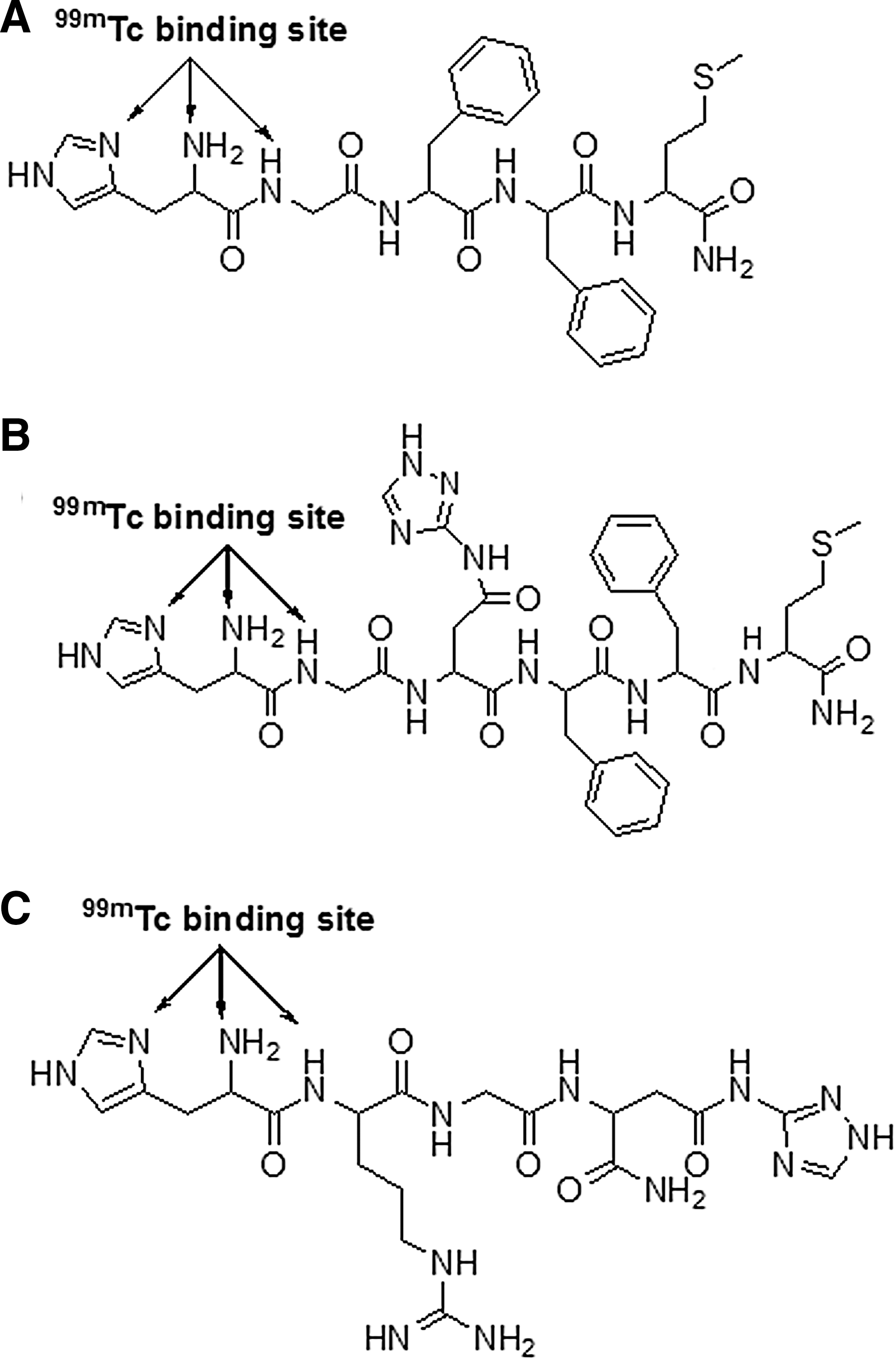
Chemical structures of
Materials and Methods
All Fmoc-amino acids, TBTU [2-(1H-benzotriazol-1-yl)-1,1,3,3-tetramethyluronium tetrafluoroborate] and Rink amide MBHA resin (100–200 mesh) were purchased from Novabiochem.3-amino-1,2,4-triazole, picrylsulfonic acid, trifluoroacetic acid, DIPEA (diisopropylethylamine), thioanisole, and HOBT (1-hydroxybenzotriazole) were purchased from Sigma. 99MoO4 − was procured from Bhabha Atomic Research Center (Mumbai) and 99mTcO4 − was extracted from a 5(M) NaOH solution of it by 2-butanone. Electrospray mass spectrometry (ESI-MS) was recorded on a Waters Micromass Q-Tof microinstrument. ECIL gamma counter (Model LV4755) procured from ECIL was used for the measurement of radioactivities in various samples. High performance liquid chromatography (HPLC) studies on both analytical and preparative were carried out on a Waters Associates HPLC system. Berthold LB500 HERM radio HPLC monitor purchased from Berthold Technologies GmbH was used to analyze radioactivity in the HPLC eluates. All cancer cell line studies were performed using B16F10 mouse melanoma and EAC cell lines.
Synthesis of peptides
Fmoc-protected amino acids with appropriate side-chain protection and Rink amide MBHA resin was used for the synthesis of peptides. 13 0.1 mmol of the Rink amide MBHA resin was treated with 20% piperidine in N,N-dimethylformamide (DMF) under constant nitrogen purging (10 minutes) followed by repeated washing with DMF to remove the Fmoc-protecting group. Amino acid couplings were carried out in DMF medium. A mixture of fivefold excess of Fmoc-protected amino acid [0.5 mmol; Fmoc-Met-OH in case of pep-1 and pep-2 and Fmoc-Asp(OtBu)-OH in case of pep-3], TBTU (0.5 mmol), DIPEA (1 mmol), and DMF (3 mL) was added to the deprotected resin placed in a small glass column under nitrogen purging. Synthesis was carried out as per method 8 described earlier from this laboratory to obtain Met-Phe-Phe-Gly-His-Fmoc (pep-1), Met-Phe-Phe-Asp-Gly-His-Fmoc (pep-2), and Asp-Gly-Arg-His-Fmoc (pep-3). Different protective groups were removed following methods described earlier, crude peptides were finally purified on a Waters HPLC system by semipreparative RPHPLC using Prep C-18 XTerra column (7.8 × 300 mm, 10 μm particle size) at a flow rate of 3 mL/minutes by applying linear gradient solvent system from 100% solvent A (0.1% TFA in H2O) to 15% solvent B (methanol) in 25 minutes. Peptide purity was assessed by analytical HPLC as reported earlier and characterized by ESI-MS.
Synthesis of 1,2,4 triazolyl-peptides
The Fmoc-protected peptides (pep-2 or pep-3; 1 mmol), HOBT (1 mmol), EDCI.HCl (1 mmol), and diisopropylamine (2 mmol) were dissolved in DMF, placed on ice water bath, stirred for half an hour, and treated with 3-amino-1,2,4-triazole (3 mmol). The mixture was stirred at room temperature for 24 hours. The solvent was evaporated in a Rotavapor and treatment with 20% piperidine in DMF was done to remove Fmoc protection. The crude triazolyl peptide was then precipitated out using cold diethyl ether. It was subsequently purified by HPLC as per method described above and characterized by ESI-MS.
99mTc Radiolabeling
The 99mTc-labeled peptide and triazolyl peptide were prepared directly from freshly prepared 99mTc(CO)3(H2O)3 precursor. The organometallic precursor 99mTc(CO)3(H2O)3 was synthesized as per method reported earlier from this laboratory 14 and purity was determined by analytical HPLC following the same method as described under synthesis. The peptide solution (0.1 mL) from a stock (2 mg/mL in nitrogen purged water) was added to 0.3 mL of the freshly prepared 99mTc(CO)3(H2O)3 solution (74–185 MBq). The mixture was heated at 45°C for 10–15 minutes maintaining the pH between 6.5 and 7.0, cooled to room temperature, and analyzed by TLC and HPLC.
Physicochemical Evaluation
In vitro stability studies
Radiochemical stability of 99mTc-labeled ligands in freshly collected rat serum and physiological saline was observed for 24 hours. An aliquot (0.1 mL) of 99mTc-labeled ligand solutions was incubated with 0.9 mL each of serum and saline. Samples were withdrawn at 0, 1, 2, 3, 4, and 24 hours and analyzed by TLC using acetone as the developing solvent and silica gel strips (Merck) as the stationary phase to quantitate the free pertechnetate generated during incubation period. In a separate experiment, the stability of the radiolabeled peptides was also studied by His challenge as per method reported earlier from this laboratory. 14
Lipophilicity
The partition coefficients of the 99mTc(CO)3-labeled peptides between 1-octanol and phosphate buffer (0.025 M, pH 7.4) were determined to measure the lipophilicity of the radiolabeled complexes. An aliquot (0.1 mL) of each of the 99mTc(CO)3-labeled peptides was added to centrifuge tubes containing 1-octanol (1 mL) and phosphate buffer (1 mL). The partition coefficient (P) was determined following the standard protocol and expressed as log P to ascertain the lipophilicity.
Blood clearance studies
In anesthetized and well-hydrated cannulated rats, the radiolabeled peptides were injected through one femoral vein. Blood samples (0.5 mL) were collected at preset time intervals (between 2 minutes and 4 hours) from the other femoral vein, weighed accurately, and counted in a gamma counter. The percentage of injected dose/g (% ID/g) of blood samples at each time interval was determined and plotted against time to generate the blood disappearance curve.
In vitro protein binding study
An aliquot (0.1 mL) of 99mTc(CO)3-labeled peptides were added to 1 mL of freshly collected rat serum and incubated (30 minutes) at room temperature. An aliquot (0.3 mL) of serum was withdrawn, placed in an Amicon Centrifree Ultrafiltration Device in triplicate, and centrifuged for 20 minutes at room temperature. Radioactivity in the 50 μL samples of the serum and the ultrafiltrate was measured in a gamma counter. Bound count was obtained by subtracting the ultrafiltrate count from serum count and serum protein binding was expressed as percent of bound count.
Biological Evaluation
All animal experiments were done in accordance with the national laws approved by the Social Justice and Empowerment Committee for the Purpose of Control and Supervision of Experiments on Animals (CPCSEA), Government of India, New Delhi.
Tissue distribution and imaging studies
Healthy Balb/c mice (25 ± 1.5 g) were injected intramuscularly with freshly collected B16F10 cells (∼1 × 10⁶ per animal) suspended in saline in the thigh of the right hind leg, whereas Swiss albino mice (25 ± 1 g) were injected intramuscularly with EAC cells (∼1 × 10⁶ per animal) in the thigh of the right hind leg. Studies were conducted 14–21 days after implantation, when a palpable tumor in the volume range of 1 ± 0.1 cm3 was developed.
In tissue distribution studies, animals were well hydrated for 1 hour by intraperitoneal administration of saline (0.9%, 2 mL). After another hour, the solution of 99mTc(CO)3-labeled peptide (0.03 mL, 8–12 MBq/kg) was injected in each animal through the tail vein. At 1 and 4 hours following the administration of the radioactive solution, the animals were sacrificed and rest of the methods was the same as reported earlier. The organ uptake was calculated as a percentage of the injected dose per gram of tissue mass (%ID/g) or per organ.
Static images of Balb/c mice bearing B16F10 tumor injected with 99mTc(CO)3-triazolyl pep-3 were acquired at 1 and 4 hours postinjection using a GE Infinia gamma camera equipped with Xeleris Workstation.
Cellular uptake and internalization
The ability of 99mTc(CO)3-labeled peptides to bind and internalize in B16F10 mouse melanoma and EAC cell lines was studied as per the reported method. 15 Briefly, EAC cells (1 × 10⁶) in the ascites form were collected from the peritoneum of Swiss albino mice and washed thrice with the binding medium [20 mM HEPES buffered Hank's balanced salt solution (pH 7.2) containing 0.5% Bovine serum albumin], followed by incubation (37°C) in the same medium containing 99mTc(CO)3-labeled peptides (0.02 mL, 12 MBq/mL) for varying time periods ranging from 30 to 240 minutes. Incubation was stopped after the desired time interval by removing the medium and all other steps were done following exactly the same method reported earlier from this laboratory to get %cellular uptake and %internalized activities. 8,9
The B16F10 mouse melanoma cell lines were cultured in Dulbecco's modified Eagle's medium high glucose supplemented with Fetal bovine serum, penicillin/streptomycin and GlutaMAX at 37°C in a humidified incubator under a CO2 atmosphere and passaged weekly using a trypsin-EDTA solution as reported earlier. Internalization experiments were done as per the method already published. 5
The above experiments were also carried out in the presence of 10 μM c[RGDfV], which is highly selective for αvβ3. The blocking peptide was added to cells (B16f10) before addition of 99mTc(CO)3-triazolyl pep-3 (final concentration 2 nM), and rest of the procedure was the same as reported earlier. 5
All experiments were repeated thrice and the results were expressed as the mean ± standard deviation (SD).
Statistical Analysis
All experiments were repeated for at least three times, each time on a different day. All mean values of animal experiments are expressed as %ID per g of tissues or organs ± SD. All p-values ≤0.05 were considered statistically significant.
Results and Discussion
The 1,2,4-Triazole-coupled peptides radiolabeled with fac-[99mTc(CO)3(H2O)3]+ core were studied for use as tumor-targeted radiopharmaceuticals. Synthesis of three bioactive peptides (pep-1, pep-2, pep-3) bearing His as chelator for the tricarbonyl core was performed by standard solid phase methods in the manual mode using side-chain-protected Fmoc-conjugated amino acids. After HPLC purification the yield of the peptides were around 70%, approximately. The N-terminus of the peptides was chelated with fac-[99mTc(CO)3(H2O)3]+ precursor through histidine. Analytical HPLC determination showed that the purity of the compounds was higher than 95% (Fig. 2a). A single HPLC peak was observed [tR (pep-1) = 12.5 minutes; tR (pep-2) = 11.9 minutes, and tR (pep-3) = 4.8 minutes]. The identity of each peptide was confirmed by ESI-MS (Supplementary Figs. S1–S5). The [M + H]+ peak was obtained at m/z 637.05 for pep-1 (calculated for C31H40N8O5S 637.74), 752.31 for pep-2 (calculated for C35H45N908S 752.85), and 483.39 for pep-3 (calculated for C18H30N10O6 483.30). The 3-amino-1,2,4-triazole was coupled to the free-COOH group of Asp of Fmoc-protected peptides (pep-2 and pep-3) in the presence of HOBT, EDCI.HCI, and diisopropylamine in DMF to form stable amide linkages. The triazolyl peptide was subsequently purified by reversed-phase semipreparative HPLC. For each peptide, the purity was confirmed by analytical HPLC [tR (triazolyl pep-2) = 14.5 minutes; tR (triazolyl pep-3) = 7.1 minutes] and characterization done by ESI-MS. The [M + H]+ peak was found at m/z 819.53 for triazolyl pep-2 (calculated for C37H47N13O7S 819.20) and 550.47 for triazolyl pep-3 (calculated for C20H32N14O5 550.27) as verified by HPLC (Fig. 2b).

Radiolabeling
Pep-1, triazolyl pep-2, and triazolyl pep-3 were radiolabeled using fac-[99mTc(CO)3(H2O)3]+ precursor. The tricarbonyl precursor was successfully prepared in high yield by reducing 99mTcO4 − with sodium borohydride in the presence of CO (g) at atmospheric pressure. The purity (>97%) was confirmed by HPLC showing a retention time of ∼6.7 minutes (Fig. 2b). Radiolabeling was done by the addition of freshly prepared synthon precursor to the aqueous solution of the ligand under nitrogen atmosphere. The pH and temperature of the reaction mixture was maintained between 6 to 6.5 and 40–45°C, respectively. To obtain maximum complexation yield, standardization and optimization studies were performed. Silica gel plates were used for TLC to evaluate the radiochemical purities of the 99mTc(CO)3-labeled peptides using acetone as the mobile phase, where 99mTc(CO)3-labeled compounds remained at the origin (Rf = 0.1) corresponding to >95% activity, and free 99mTcO4 − moved to the solvent front (Rf = 0.9–1.0). All the compounds could be efficiently radiolabeled even at low concentration (10−4 M). RPHPLC further ascertained the radiochemical purity of the compounds, where a single radioactive species was obtained in all the cases, and this remained stable for at least 24 hours after labeling. The radio chromatograms of 99mTc(CO)3-pep-1, 99mTc(CO)3-triazolyl pep-2, and 99mTc(CO)3-triazolyl pep-3 showed single peaks at 18.5, 16.5, and 12.1 minutes, respectively. The 99mTc(CO)3-triazolyl pep-3 containing ARG-GLY-ASP (RGD) peptide consisting of polar amino acids exhibited comparatively lower retention time (12.1 minutes) on HPLC, whereas 99mTc(CO)3-pep-1 and 99mTc(CO)3-triazolyl pep-2 eluted much later from the HPLC column because of the presence of phenylalanine in the peptide chain, which is comparatively a less polar amino acid.
In vitro studies
The in vitro stability of 99mTc(CO)3-labeled compounds incubated at 37°C with either freshly collected rat serum or normal saline was monitored at various time intervals for up to 24 hours. Instant radiolabeling yields of 99mTc(CO)3 complexes of pep-1, triazolyl pep-2, and triazolyl pep-3 were 94.34% ± 0.84%, 93.92% ± 0.20%, and 96.74% ± 1.10%, respectively. The reduction in radiolabeling yield was insignificant (1%–2%) after incubation for 24 hours with saline. The level of free radioactive species generated after 24 hours incubation in rat serum was around 10% in the case of radiolabeled pep-1 and triazolyl pep-2, and about 8% in the case of radiolabeled triazolyl pep-3. The in vitro stability study against ligand exchange as determined through His challenge showed that the presence of His (strong competitor for 99mTc) did not produce any remarkable instability. High transchelation stability (>90% at 24 hours) is well known for most 99mTc-tricarbonyl complexes and this is confirmed in this study for His used for radiolabeling of bioactive pharmacophores.
Lipophilicity of the radiolabeled compounds was determined by distribution in phosphate buffer (pH 7.4) and octanol as described in the experimental section. The log P values of 99mTc(CO)3-pep-1 and 99mTc(CO)3-triazolyl pep-2 were found to be 0.492 and 0.560, respectively, reflecting their lipophilic nature, attributed to the presence of methionine and phenylalanine amino acids in the peptide chain. In contrast, the log P value of 99mTc(CO)3-triazolyl pep-3 was found to be −1.346 indicating the hydrophilic nature of the complex.
Before biodistribution studies, the ability of 99mTc(CO)3-labeled compounds to bind to plasma protein of rat serum was determined. The 99mTc(CO)3-pep-1 and 99mTc(CO)3-triazolyl pep-2 showed moderately high protein binding, the values being 82% and 74%, respectively, whereas 99mTc(CO)3-triazolyl pep-3 showed substantially lower binding (36%), the low protein binding value indicating enough availability of the free drug for binding to the target. It was observed that, in all the cases, the amount of nonspecific binding (<5%) to the ultrafiltration device was negligible. All the radiolabeled compounds cleared readily from blood and exhibited biphasic blood disappearance profile: an initial fast phase followed by moderately slow second phase (Fig. 3).

Blood clearance curves in rats following intravenous injection of 99mTc(CO)3-labeled compounds.
In vivo study
B16F10 and Ehrlich ascites tumor-bearing mice were evaluated to assess the novelty of this approach involving the design of 1,2,4-triazole-based radiopharmaceuticals for tumor targeting. The tumor uptake and organ distribution values of the radiolabeled compounds at 1 and 4 hours are summarized in Table 1. The tumor uptake in B16F10 was substantially high with 99mTc(CO)3-triazolyl pep-3. Accumulation was high initially (1.67 ± 0.02 ID/g at 1 hours) and got gradually reduced with time (1.07 ± 0.03 at 4 hours). Tumor/muscle uptake ratios were 3.44 (1 hours) and 3.87 (4 hours), whereas tumor/blood ratios were 0.69 (1 hours) and 1.05 (4 hours). On the contrary, accumulation in tumor was moderate in the case of 99mTc(CO)3-triazolyl pep-2, the tumor-to-muscle ratio remaining almost unaltered throughout the period of study. Although 99mTc(CO)3-pep-1 exhibited some tumor accumulation initially (0.92 ± 0.10 at 1 hours), it was significantly reduced with time (0.23 ± 0.02 at 4 hours), whereas change in tumor-to-muscle ratio was not pronounced due to clearance of muscle activity with time. The tumor/blood ratio was also not very significant. The 99mTc(CO)3-triazolyl pep-3 being hydrophilic was excreted mainly through the renal route; about 71% of the injected dose was eliminated at 4 hours postinjection. In contrast, 99mTc(CO)3-pep-1 and 99mTc(CO)3-triazolyl pep-2 were eliminated mainly through the hepatobiliary pathway with some renal clearance. The two compounds with comparable log P values were less hydrophilic than 99mTc(CO)3-triazolyl pep-3, thus exhibiting lower urinary excretion.
Results are expressed in percentage of injected dose per organ/tissue (each value is the mean ± SEM of five animals per group).
Percentage of injected dose per gram of tissue.
EAC, Ehrlich ascites carcinoma.
The in vivo behavior of 99mTc(CO)3-labeled compounds was studied in mice bearing Ehrlich ascites tumor. The 99mTc(CO)3-triazolyl pep-3 exhibited some uptake in tumor, but this was less compared to the uptake in mice bearing B16F10 melanoma. However, there was no significant change in tumor/blood and tumor/muscle ratios of 99mTc(CO)3-triazolyl pep-3. The uptake in EAT in mice was also not very significant in case of 99mTc(CO)3-pep-1 and 99mTc(CO)3-triazolyl pep-2. The 99mTc(CO)3-triazolyl pep-3 being hydrophilic was excreted mainly through the renal pathway, whereas the comparatively less hydrophilic 99mTc(CO)3-pep-1 and 99mTc(CO)3-triazolyl pep-2 adopted the hepatobiliary route for their elimination. In the above two tumor models, neither of the radiolabeled compounds exhibited any significant accumulation in stomach indicating no significant decomposition of the 99mTc-labeled complexes in animal systems to produce 99mTcO4 −. Scintigraphic images (Fig. 4) obtained 1 and 4 hours postinjection of 99mTc(CO)3-triazolyl pep-3 into Balb/c mice bearing B16F10 tumor visually confirmed the accumulation of the radiopharmaceutical in tumor. The images also showed high-level background activity in liver, kidney, and urinary bladder as expected from the biodistribution data. However 99mTc(CO)3-pep-1 and 99mTc(CO)3-triazolyl pep-2 did not produce any clear image; comparatively less uptake in the tumor region and remarkably high background activity could be the reason behind this.
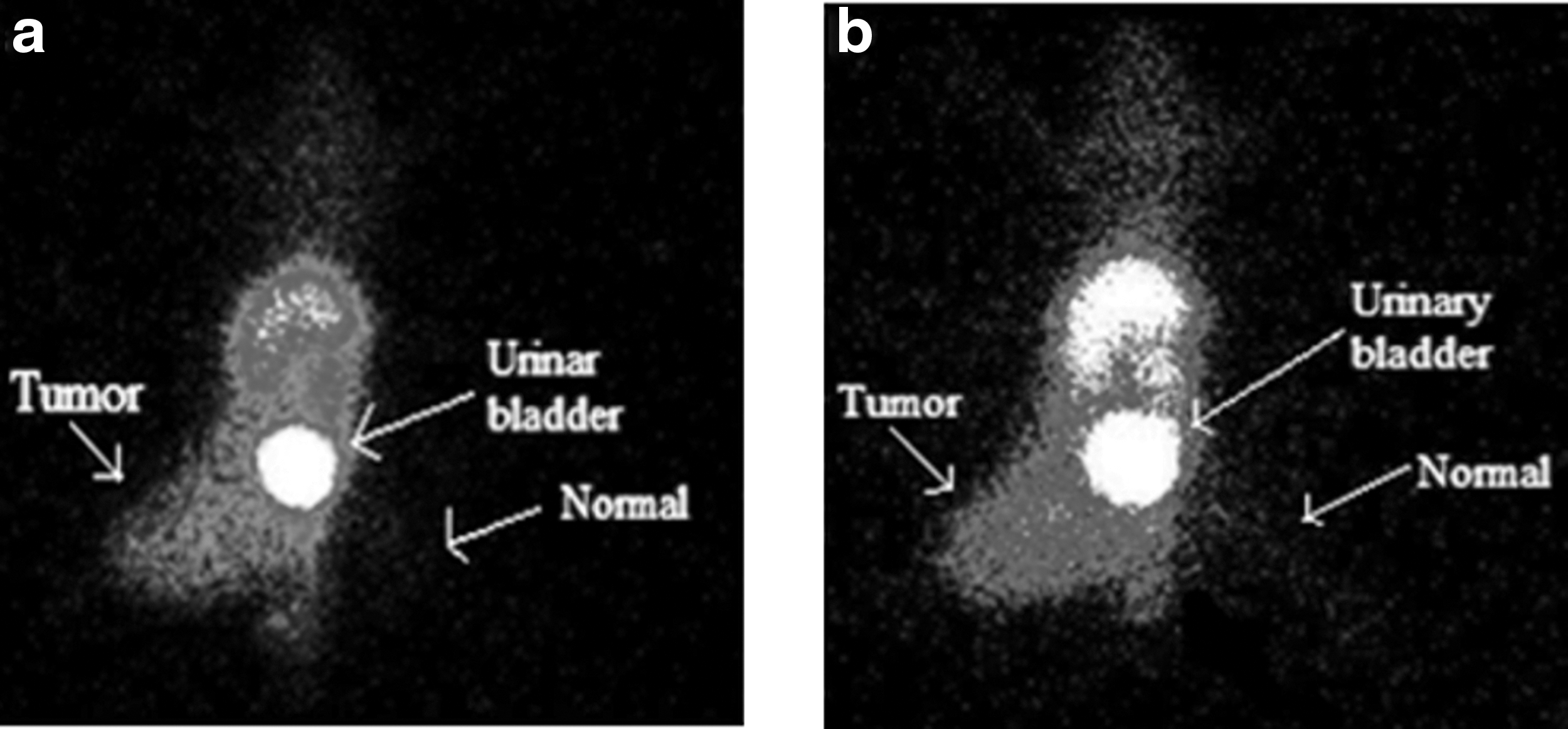
Scintigraphic images of B16F10 tumor-bearing mice at 1 hour
In vitro cell binding and internalization studies
In vitro cell binding and internalization studies were performed by incubating radiolabeled peptide and peptide–triazole conjugates with B16F10 mouse melanoma cell line and EAC cell line for various time periods at 37°C (Fig. 5). Binding of the radiolabeled compounds to the cells increased gradually, attaining a maximum value at 4 hours. No significant increase in binding was observed after that time. The total 99mTc activity associated with the cells after incubation for preselected time interval (30–240 minutes) was measured after washing the cells with phosphate-buffered saline (pH 7.4). The cell-bound activities of 99mTc(CO)3-pep-1, 99mTc(CO)3-triazolyl pep-2, and 99mTc(CO)3- triazolyl pep-3 were about 3.15%–5.6%, 4.12%–9.25%, and 8.22%–15.56%, respectively, following 30–240 minutes incubation with B16F10 mouse melanoma cell line. On the other hand, a relatively lower cellular uptake was observed when the binding studies were performed in EAC cell line. The cell binding capacities of 99mTc(CO)3-pep-1, 99mTc(CO)3-triazolyl pep-2, and 99mTc(CO)3-triazolyl pep-3 with EAC cell line were now 2.5%–3.5%, 2.15%–5.25%, and 4.16%–7.17%, respectively, for the abovementioned time periods. The binding studies were repeated in the presence of ∼100-fold molar excess of unlabeled compounds, which demonstrated 30%–35% reduction in cellular uptake at 4 hours postincubation.

Cell uptake and internalization curves of radiolabeled peptide
In vitro internalization studies were performed to determine the rate and degree of internalization of 99mTc(CO)3-labeled compounds in B16F10 mouse melanoma and EAC cell lines. The results of this study are depicted in Figure 5. It can be observed that in B16F10 cell line in relation to cell-associated activity after 30 minutes incubation, 15%–20% of 99mTc(CO)3-labeled compounds were internalized and after 2–4 hours incubation attaining maximum values of ∼45%, 35%, and 27% for 99mTc(CO)3-triazolyl pep-3, 99mTc(CO)3-triazolyl pep-2, and 99mTc(CO)3-pep-1, respectively. The degree of internalization was not pronounced in EAC cell line. The value was around 12%–18% after 30 minutes of incubation and this became 15%–35% after 2–4 hours incubation.
The residence time of a radiopharmaceutical inside the cell is important for facilitating its targetability and scintigraphic imaging. There exist some differences in the internalization behavior of the radiolabeled compounds. The internalization observed for 99mTc(CO)3-triazolyl pep-3 in B16F10 cell line was fast and high in comparison to the other two. Triazolyl pep-3 contains RGD tripeptide sequence, which explains the high affinity and selectivity of this conjugate for αvβ3-integrin receptors overexpressed on B16F10 mouse melanoma cell line. Competitive binding assays in the presence of 10 μM c[RGDfV] also showed reduction in the binding of 99mTc(CO)3-triazolyl pep-3 to B16F10 cells by 33%. The radiopharmaceutical that enters the cell and resides for a sufficient period of time may produce satisfactory image during scintigraphic studies.
The antitumor activity of 1,2,4-triazole nucleus encouraged the authors to conjugate this moiety to bioactive peptides to develop new potential 99mTc radiopharmaceuticals for targeting tumor. They have synthesized one pentapeptide (pep-1), one hexapeptide (pep-2), and one tetrapeptide (pep-3) with the expectation that methionine/phenylalanine bearing penta- and hexapeptides may undergo transport mediated by LAT 1 that expresses during growth and proliferation of different tumor cells. The tetrapeptide pep-3 bears RGD-tripeptide sequence that may facilitate its transport through the integrin receptor in tumor cell line. The 1,2,4-Triazole was coupled to the side-chain carboxyl function of the aspartic acid of peptides 2 and 3 through stable amide linkage. Aromatic amines have been shown to be the most stable chelation source for tricarbonyl species. Radiolabeling with fac-[99mTc(CO)3(H2O)3]+ core was, therefore, achieved with an N-terminal histidine, an amino acid which was attached to the peptide during solid phase peptide synthesis. Complexation was carried out at atmospheric pressure and reasonable temperatures. The radiolabeled compounds were obtained in high yield and shown to retain high in vitro stability. Biodistribution results indicated a comparatively high accumulation and retention of 99mTc(CO)3-triazolyl pep-3 in B16F10 tumors, demonstrating the possibility of targeting tumor through integrin-positive receptor sites. During scintigraphic studies the tumor region was clearly visible, attributed to the greater hydrophilicity and rapid renal excretion of the radiolabeled species. However, the complex did not show very high uptake in EAT. The tumor uptake and retention in B16F10 and EAC cells for 99mTc(CO)3-pep-1 and 99mTc(CO)3-triazolyl pep-2 were not very significant. The above two compounds do not contain RGD sequence and hence act differently with the receptor on the cell surface. The 99mTc(CO)3-triazolyl pep-3 also exhibited higher cell uptake than the other two. Internalization of 99mTc(CO)3-triazolyl pep-3 was significant (∼45% after 2–4 hours incubation), explaining the extended residence within the cell facilitating its imaging and therapeutic goal.
Conclusions
The diversity of biological response profile of 1,2,4-triazole encouraged the authors to couple this moiety with biologically active peptides for targeted molecular imaging and peptide receptor scintigraphy. Triazolyl peptides were successfully synthesized and radiolabeled with fac-[99mTc(CO)3(H2O)3]+ precursor in high yields under mild conditions. All the radiolabeled compounds showed significant stability, binding, and in vitro and in vivo pharmacokinetic properties. However, during scintigraphic studies only 99mTc(CO)3-triazolyl pep-3 showed a better delineation of the tumor. Attachment of 1,2,4-triazole to other important bioactive peptides as well as in vitro studies with integrin-positive U-87 MG and C6 glioma cell lines are in progress. This strategy for tumor targeting may also create the opportunity to develop peptide receptor-targeted radiotherapy after radiolabeling the pharmacophore with rhenium-188, the β-emitting radionuclide.
Footnotes
Acknowledgments
The work was funded by the University Grants Commission (UGC), the Department of Science and Technology (SB/SO/HS/009/2014), and the Council of Scientific and Industrial Research (India). The authors thank all the organizations for financial support. They gratefully acknowledge Dr Basudeb Achari, Emeritus Scientist for his valuable suggestions during the preparation of the article.
Disclosure Statement
No competing financial interests exist.
References
Supplementary Material
Please find the following supplemental material available below.
For Open Access articles published under a Creative Commons License, all supplemental material carries the same license as the article it is associated with.
For non-Open Access articles published, all supplemental material carries a non-exclusive license, and permission requests for re-use of supplemental material or any part of supplemental material shall be sent directly to the copyright owner as specified in the copyright notice associated with the article.
