
Abstract
Background:
Glioma is the most devastating brain tumor worldwide. Previous studies showed that UTRN (utrophin) was related to cancers, but its role in glioma cells remains uncovered.
Materials and Methods:
RNAi was used to knockdown UTRN in U251 cells using lentivirus system. The knockdown efficiency was validated by real-time quantitative PCR. Cell proliferation, cell cycle, and apoptosis progression were determined by MTT, colony formation analysis, and flow cytometry analysis. Furthermore, some apoptotic markers were examined by Western blot assay.
Results:
Most cells were infected. Cell proliferation and colony formation ability were suppressed in U251 cells lacking UTRN. Moreover, there was an obvious increase in cell percentage in the G2/M phases and a significant apoptosis in U251 cells after UTRN silencing. Further investigation demonstrated that UTRN knockdown activated caspase and PARP pathways.
Conclusions:
Knockdown of UTRN expression by shRNA evidently inhibited cell proliferation and promoted cell apoptosis in glioma cells.
Introduction
Gliomas arise from glial cells and mainly exist in three forms containing astrocytomas, glioblastomas, and anaplastic astrocytomas. 1 It has been considered as the most devastating brain tumor 2 and accounts for 80% malignant brain tumors, 3 including glioblastoma and anaplastic astrocytoma. 4 Even though its biological characteristics have been widely investigated, the median survival is still less than 3 years. 5 –7 At present, the therapeutic strategies for gliomas mostly concentrate on chemotherapy and radiotherapy. 8 However, neither chemotherapy nor radiotherapy show effective treatments for gliomas as expected because of their limitations, such as damaging brain and decreasing life quality. 9,10 Genetic screening was more and more applied to development of novel and personalized therapy with its potential value.
Recently, genetic alterations associated with glioma occurrence have been widely investigated by molecular investigations. Angiopoietin-2 is an angiogenic factor and has been shown as negatively correlated with vessel maturation in malignant gliomas. 4 VEGF, as vascular endothelial growth factor, is reported to be positively correlated with vessel maturation in low-grade gliomas. 4 PDCD4 (programmed cell death 4) is identified as a novel tumor suppressor gene and has been demonstrated to be downregulated in human glioma tissue. 11 NDRG 4 (N-Myc downstream-regulated gene 4) plays important roles in neurite formation and is downregulated in human gliomas. 12
Utrophin (UTRN), as a dystrophin-related gene, shows a more widespread distribution and is expressed in neural tube non-neuronal tissues, which originate from neural crest. 13,14 UTRN has been demonstrated to be concentrated in the membrane of astrocytes and vascular endothelial cells. 15 –17 What's more, it has been reported to express in HeLa and rat C6 glioma cells. 18 Overexpression of wild-type UTRN is shown to promote tumor cell growth in breast cancer cells. 19 However, there was little report on its role in human glioma cells. Considering its location in astrocytes and function, the authors guess that it may be a novel molecular target for human malignant glioma.
With the development of molecular biology, RNA interference (RNAi) has been widely thought as an invaluable tool for personalized cancer therapy with high specificity and selectivity, including siRNA (small interfering), shRNA (short hairpin RNA), and bifunctional RNA technology. 20 Compared with the two other interference methods, shRNA is a better choice with relatively low-degradation rate and turnover. 21
To explore the possible role of UTRN in human malignant glioma cells, the authors constructed RNA interference lentivirus system to specifically knockdown the expression of UTRN in U251 cells and then evaluated its biological function by analyzing the effects of UTRN on cell proliferation, cell cycle progression, and cell apoptosis. In addition, its molecular mechanism underlying glioma occurrence was further investigated.
Materials and Methods
Cell culture
The human glioma cell line U251 and human embryonic kidney cell line 293 (HEK 293T) were obtained from the Cell Bank of Chinese Academy of Science. Both U251 and 293T cells were cultured in Dulbecco's modified Eagle's medium (SH30243.01; HyClone) containing 10% fetal bovine serum (10099-141; Gibco) at 37°C in a humidified 5% CO2 atmosphere.
Construction of lentivirus-mediated shRNA targeting UTRN
Gene sequences of UTRN were downloaded from Gene bank with accession number of NM_007124. The siRNA-designing software was used to design two segments of UTRN target by shRNA. These designed shRNAs were S1 (5′-GCCCAGATTGAGGAAGTTCTACTCGAGTAGAACTTCCTCAATCTGGGCTTTTT-3′) and S2 (5′-CCTGACAAGAAATCCATAATTCTCGAGAATTATGGATTTCTTGTCAGGTTTTT-3′) for shUTRN. The negative control shRNA was 5′-GCGGAGGGTTTGAAAGAATATCTCGAGATATTCTTTCAAACCCTCCGCTTTTTT-3′. These shRNAs were synthesized, annealed, and inserted into the pFH-L vector containing a green fluorescence protein (GFP) (Hollybio) by NheI and PacI (TaKaRa) restriction enzyme digestion sites and then ligated using T4 DNA ligase. The ligation products were transformed into the Escherichia coli strain DH5α and then colony PCR was used to identify the positive recombinant clone. Subsequently, the recombinant clone was confirmed by DNA sequencing. The recombinant lentiviral vector plasmids were named as pFH-Lv-shUTRN(S1), pFH-Lv-shUTRN(S2), or pFH-Lv-shCon. The three recombinant lentiviral vector plasmids and two pHelper plasmids pVSVG-I and pCMVΔR8.92 (Hollybio) were cotransfected into 293T cells using Lipofectamine 2000 (Invitrogen) according to the protocol of the manufacturer, respectively. After 48 hours of transfection, the supernatants were harvested and centrifuged to obtain lentiviral vector particles. The particles were filtered through a 45 μm filter, and the viral titer was measured through counting the number of GFP-positive cells. Then, corresponding three groups were referred as Lv-shUTRN (S1), Lv-shUTRN (S2), and Lv-shCon. For lentivirus infection, U251 cells were infected with Lv-shUTRN (S1), Lv-shUTRN (S2), and Lv-shCon at a multiplicity of infection of 10 with an inoculation density of 7 × 10⁴ cells per well for 144 hours, respectively. Successful infection was further verified by real-time quantitative PCR (RT-qPCR).
RT-qPCR assay for knockdown efficiency analysis
After 5–6 days infection, TRIzol reagent (Invitrogen) was used to extract total RNA from the U251 cells. The forward and reverse primers for UTRN were 5′-CAAACACCCTCGACTTGGTT-3′ and 5′-TGGTGGAGCTGCTATCAGTG-3′. Actin was considered as an internal control and its primers were 5′-GTGGACATCCGCAAAGAC-3′ (forward) and 5′-AAAGGGTGTAACGCAACTA-3′ (reverse). A Promega M-MLV Reverse Transcriptase Kit (Invitrogen) was used to synthesize complementary DNA (cDNA). RT-qPCR was performed on the Bio-Rad Connect Real-Time PCR platform. Total 20 μL reactions consisted of 10 μL 2× SYBR Premix Ex Taq, 0.5 μL forward and reverse primers (2.5 μM), 5 μL cDNA, and 4.5 μL ddH2O, and PCR amplification was performed for 1 minute at 95°C, followed by 40 cycles. Each cycle included denaturation for 10 seconds at 95°C and annealing at 60°C for 20 seconds. Subsequently, 2−ΔΔCt method 22 was used to calculate the relative expression levels of UTRN mRNA.
Western blot assay
After 6 days infection, the U251 cells were collected and washed with ice-cold phosphate buffered saline (PBS). Then, cells were lyzed in 2× sodium dodecyl sulfate (SDS) sample buffer, containing 100 mM Tris-HCl (pH 6.8), 4% SDS, 10% glycine, and 10 mM EDTA, followed by protein concentration determination using enhanced BCA Protein Assay Kit (Beyotime). The proteins were separated by 10% sodium dodecyl sulfate-polyacrylamide gel electrophoresis (SDS-PAGE), and 48 μg proteins were added in each lane for electrophoresis at 60 V. Subsequently, separated protein was transferred to a polyvinylidene fluoride (PVDF) under 300 mA for 2.5 hours. The membrane was blocked by 4% nonfat dry milk for 1 hour at room temperature and incubated with primary antibodies at 4°C overnight, followed by incubation with horseradish peroxidase (HRP)-conjugated goat anti-rabbit secondary antibody (1:5000; No. SC-2054; Santa Cruz) for 2 hours at room temperature. The enhanced Chemiluminescence Kit was used to visualize the protein signal. The primary antibodies used were as follows: Rabbit anti-Caspase-3 (1:1000; #9661; Cell Signaling), rabbit anti-Caspase-9 (1:1000; 10380-1-AP; Proteintech), rabbit anti-PARP (poly ADP-ribose polymerase) (1:1000; #9542; Cell Signaling), and rabbit anti-GAPDH (1:500,000; 10494-1-AP; Proteintech).
MTT and Colony formation assay for cell proliferation analysis
After 3–4 days infection, U251 cells were seeded in 96-well plates at an inoculation density of 2000 cells per well and applied to viability analysis using MTT assay. Each well was added MTT solution (5 mg/mL; 20 μL) and incubated for 4 hours at 37°C. Then, 100 μL acidic isopropanol was added, including 5% isopropanol, 10% SDS, and 0.01 mol/L HCl, at 37°C overnight. The absorbance value of each well was detected under 595 nm using spectrophotometer.
Infected U251 cells were cultured in six-well plates at a density of 500 cells per well and incubated for 8 days. The media was replaced every 3 days until colony formation. Subsequently, cells were fixed with 4% paraformaldehyde after being washed by PBS. After fixation, cells were stained with crystal violet for 25 minutes and counted the colonies containing more than 50 cells per colony.
Cell cycle assay by flow cytometry
After 6 days infection, U251 cells were seeded into 6 cm dishes at a density of 7 × 10⁴ cells per dish and cultured for 24 hours. Cells were fixed with 70% cold alcohol for 20 minutes and stained with propidium iodide (PI) (20 μg/mL). The cell cycle distribution of each phase was determined by flow cytometry (FACSort; Becton Dickinson) as described 23 and calculated the cell percentage in each phase.
Cell apoptosis assay
Cell apoptosis of U251 cells infected with Lv-shUTRN was analyzed by flow cytometry with Annexin V-APC/7-AAD double staining. Briefly, U251 cells were plated in 10 cm dishes at a density of 2 × 10⁵ cells per dish after 6-day infection. Subsequently, the cells were performed Annexin V-APC/7-ADD double staining according to previous report. 24
Statistical analysis
The SPSS 13.0 software was used to analyze experimental data. All the data are presented as mean ± standard deviation from three independent experiments. The difference between Lv-shCon and Lv-shUTRN was evaluated by Student's t-test. The p-value less than 0.05 was considered to be statistically significant.
Results
Lentivirus-mediated high-efficiency infection of U251 cells inducing knockdown of UTRN
To evaluate the effect of UTRN on glioma cells, the authors constructed lentivirus-mediated shRNA plasmid to specifically knockdown UTRN expression in U251 cells. More than 80% cells were observed with GFP expression (Fig. 1A), suggesting a higher infection. Subsequently, RT-qPCR was used to determine the UTRN mRNA level. As shown in Figure 1B, UTRN mRNA was significantly reduced in both Lv-shUTRN (S1) (***p < 0.001) and Lv-shUTRN (S2) (**p < 0.01) with an inhibition rate of 94.4% and 74.4%, compared with the Lv-shCon, respectively.
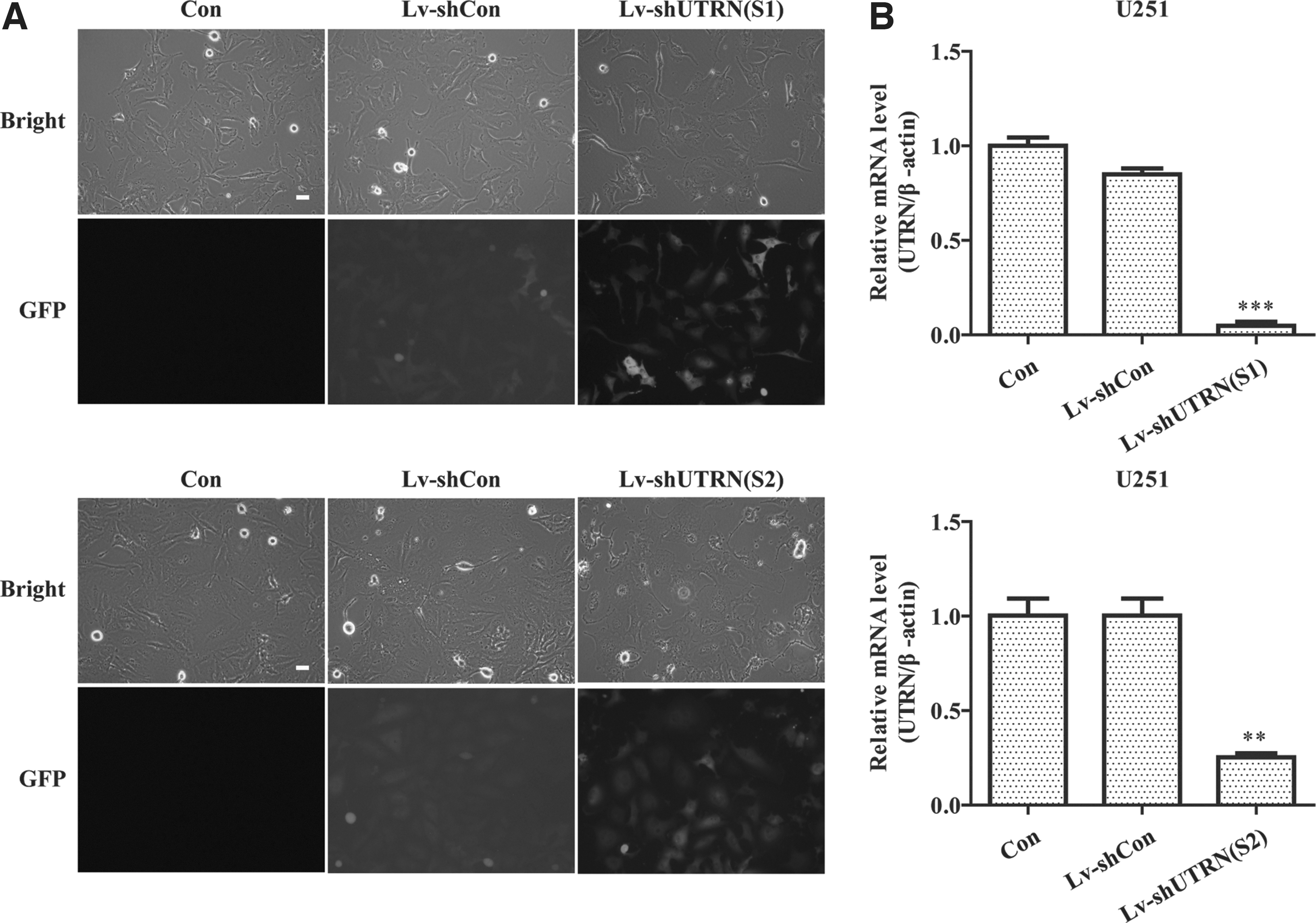
Analysis of knockdown efficiency of UTRN gene in U251 cells.
Knockdown of shUTRN (S1) and shUTRN (S2) suppressed the cell proliferation
The MTT assay was used to compare the proliferation difference between Lv-shUTRN and shCon group. As shown in Figure 2A, the growth curve was sharply increased in Lv-shCon and Con groups, but relatively smooth in Lv-shUTRN (S1) and Lv-shUTRN (S2). Statistical analysis showed that the rate of proliferation was significantly decreased in Lv-shUTRN (S1) and Lv-shUTRN (S2) compared to that in Lv-shCon group (***p < 0.001).
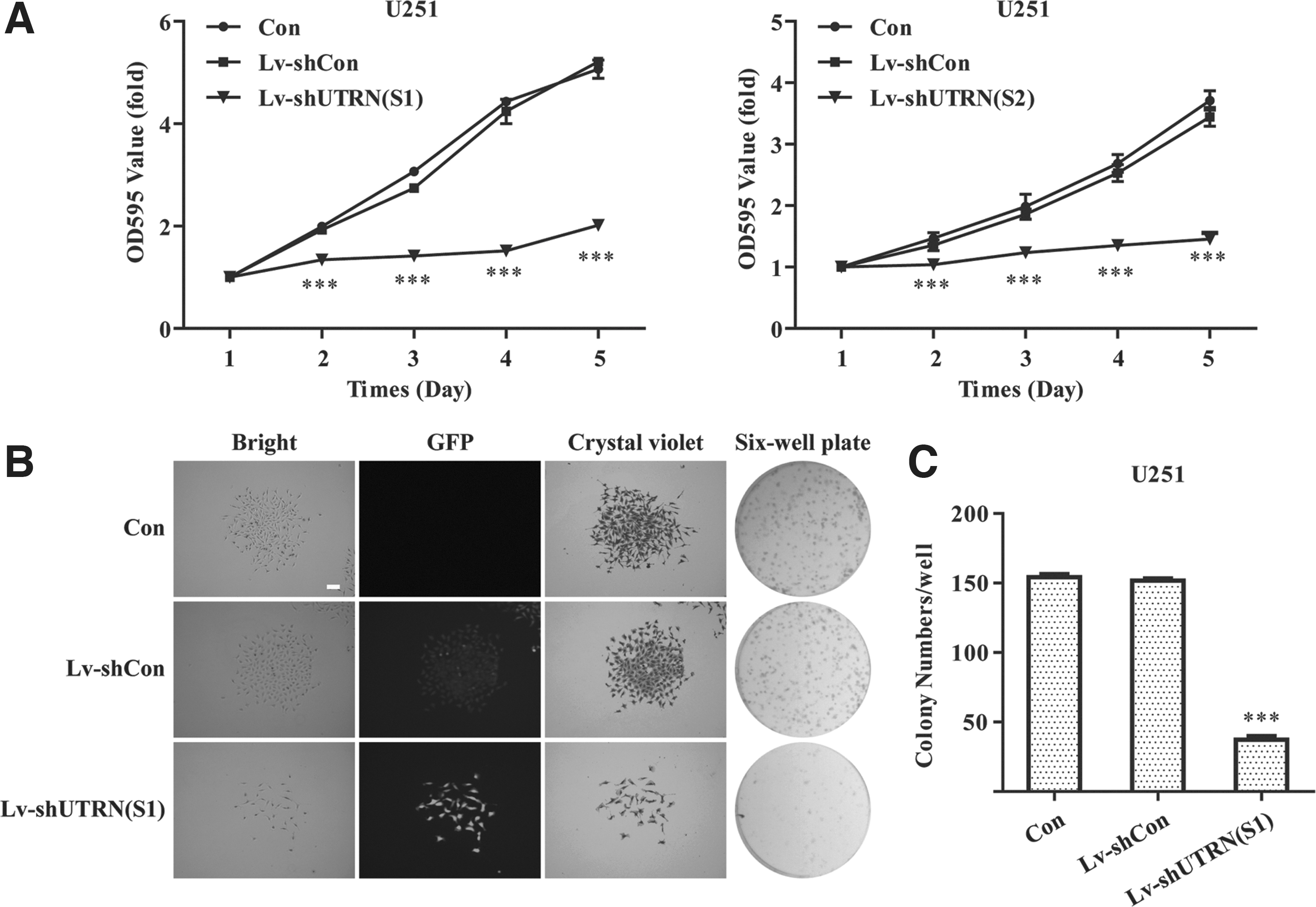
UTRN involves in the proliferation process in U251 cells.
Knockdown of shUTRN (S1) weakened colony formation ability
To further confirm the effect of UTRN knockdown on U251 cell growth, colony formation assay was conducted on U251 cell growth analysis. From Figure 2B, the representative image of cell colony formation indicated that there were smaller and fewer colonies in Lv-shUTRN (S1) group than that in Lv-shCon group, and this result was verified by statistical analysis in Figure 2C (***p < 0.001).
Knockdown of shUTRN (S1) arrested cell cycle
As known to all, cell cycle progression is closely related with abnormal cell proliferation. The flow cytometry was used to investigate the effect of UTRN knockdown on cell cycle distribution of U251 cells (Fig. 3A). According to their results (Fig. 3B), the cell percentage of S phase was remarkably decreased in Lv-shUTRN (S1). In addition, UTRN knockdown of U251 cells caused a significant increase in the cell percentage of G0/G1 (*p < 0.05) and G2/M phases (***p < 0.001) compared with that in the Lv-shCon group.
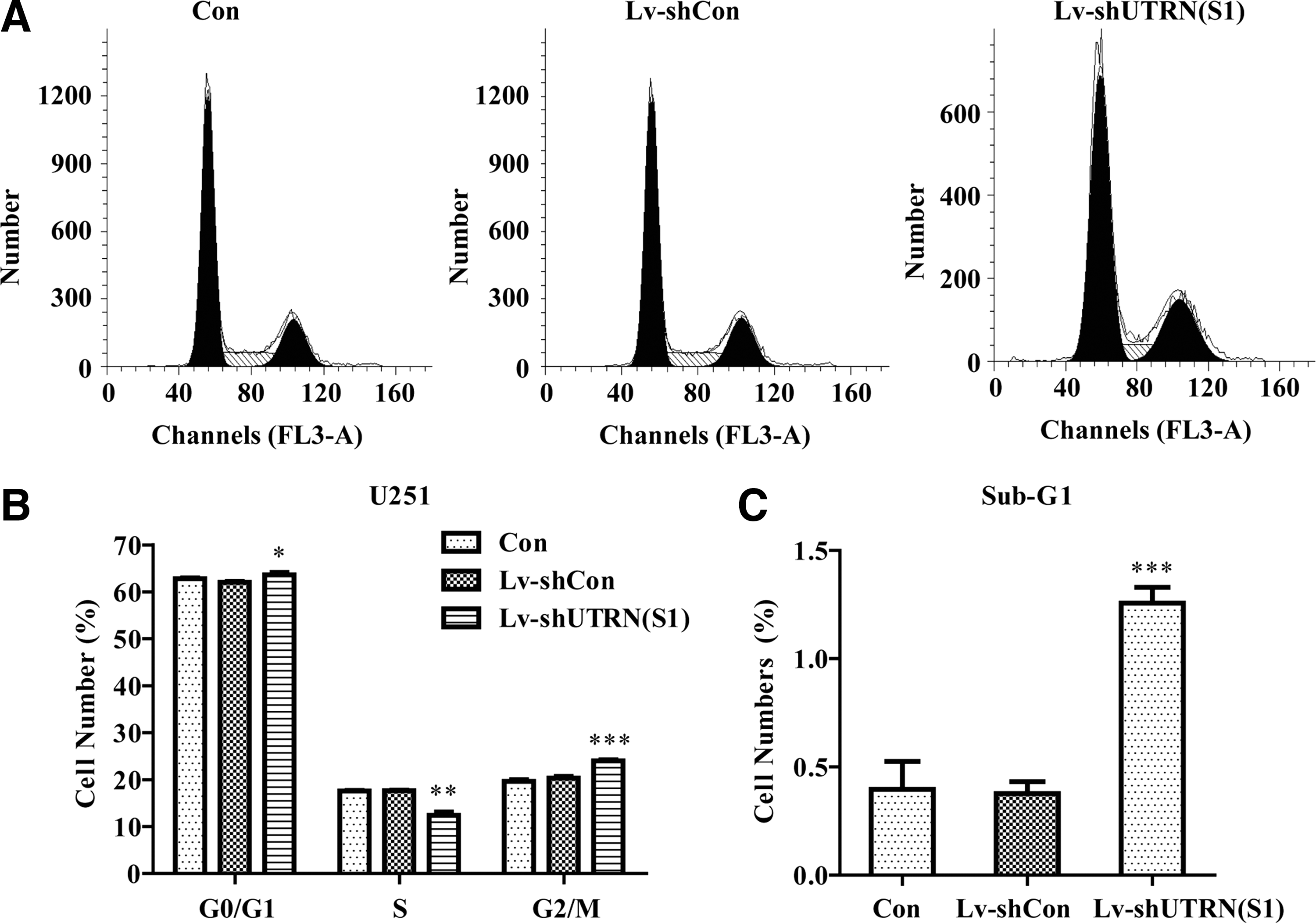
The silence of UTRN induced cell cycle arrest.
Knockdown of shUTRN (S1) increased cell death
Cell cycle analysis showed that there was an accumulation of cell percentage in sub-G1 phase (***p < 0.001) after UTRN knockdown, and the increased sub-G1 phase cells represent apoptotic cells, suggesting that UTRN knockdown could affect the U251 cell apoptosis (Fig. 3C). To determine its apoptosis effects, flow cytometric apoptosis assay was applied. The result (Fig. 4A, B) indicated that both early-stage apoptosis and late-stage apoptosis cells were significantly increased in Lv-shUTRN (S1) group compared with that in the Lv-shCon group (***p < 0.001).
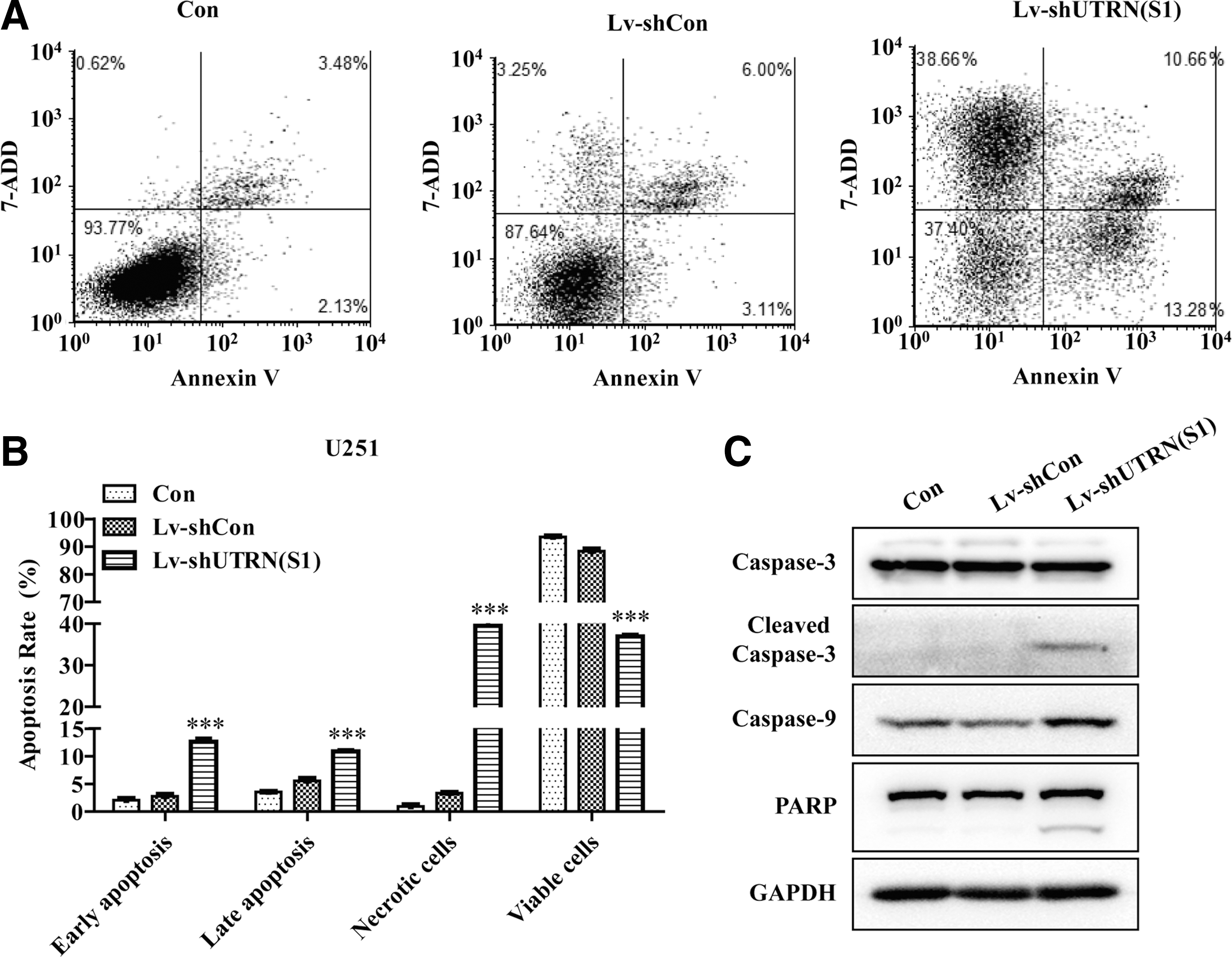
The silence of UTRN induced apoptosis through caspase pathway.
Expression of caspases and PARP in U251 cells after infected with Lv-shUTRN
To further explore the molecular mechanism of UTRN knockdown on cell proliferation and apoptosis, Western blot was applied to analyze the protein expression level of caspase-3, caspase-9, and PARP on UTRN knockdown cells. As is shown in Figure 4C, the caspase-9, cleaved caspase-3, and cleaved PARP expression of Lv-shUTRN (S1) were markedly higher than those of Lv-shCon group.
Discussion
Gliomas with poor survival median are considered as the most devastating brain tumor and make up 80% of malignant brain tumors. 3 It is necessary to find diagnostic markers for gliomas because of ineffective therapies. UTRN has been reported to express in rat C6 glioma cells 18 and promote tumor cell growth in breast cancer cells. 19 However, its role in human malignant glioma cells remains unclear.
In this study, the authors successfully constructed silenced UTRN cell model using the lentiviral transfection technology. The results indicate that U251 cell proliferation is significantly suppressed after UTRN gene knockdown, and UTRN plays an important role in regulating glioma cell cycle and apoptosis progression. To further confirm these results, apoptotic-related markers were detected by Western blot assay. There is mounting evidences suggesting cleaved PARP and caspase-3, as reliable indicators of apoptosis, are increased during cell apoptosis. 25 Cleaved PARP, a downstream target of cpp32 (casein phosphopeptide–amorphous calcium phosphate), has been activated in the procedure of promoting glioma cells' apoptosis. 26 Caspase-3, as cysteine proteases, has been reported to be effectively increased in promoting glioma cells' apoptosis. 2 In addition, caspase-3 can cause sequential activation of cell apoptosis by interacting with caspase-9 proteins. 27 What's more, the combination of PARP and caspase-3 pathways has been demonstrated to arrest cell cycle and cells' apoptosis in colon cancer. 28 UTRN has been shown to be concentrated in astrocyte membrane. 15 The occurrence of astrocytomas, as gliomas, may be associated with UTRN overexpression based on these data.
In summary, the authors have provided some evidences that UTRN could regulate glioma cell proliferation and apoptosis by cleaved caspases and PARP to some degree. UTRN may be a therapeutic target for glioma occurrence, which is closely related with cell cycle arrest and apoptosis progression.
Footnotes
Acknowledgment
This work was supported by the Natural Science Foundation of China (81271332).
Disclosure Statement
No competing financial interests exist.