
Abstract
Objective:
The authors have conjugated chelating agents (DOTA and NODAGA) with a peptide (pituitary adenylate cyclase-activating peptide [PACAP] analogue) that has a high affinity for VPAC1 receptors expressed on cancer cells. To determine a suitable chelating agent for labeling with 68Ga, they have compared the labeling kinetics and stability of these peptide conjugates.
Methods:
For labeling, 68GaCl3 was eluted in 0.1 M HCl from a [68Ge-68Ga] generator. The influences of peptide concentration, pH, and temperature on the radiolabeling efficiency were studied. The stability was evaluated in saline, human serum, DTPA, transferrin, and metallic ions (FeCl3, CaCl2, and ZnCl2). Cell binding assay was performed using human breast cancer cells (T47D). Tissue biodistribution was studied in normal athymic nude mice.
Results:
Optimal radiolabeling (>95.0%) of the DOTA-peptide conjugates required a higher (50°C–90°C) temperature and 10 minutes of incubation at pH 2–5. The NODAGA-peptide conjugate needed incubation only at 25°C for 10 minutes. Both radiocomplexes were stable in saline, serum, as well as against transchelation and transmetallation. Cell binding at 37°C for 15 minutes of incubation with 68Ga-NODAGA-peptide was 34.0% compared to 24.5% for 68Ga-DOTA-peptide. Tissue biodistribution at 1 hour postinjection of both 68Ga-labeled peptide conjugates showed clearance through the kidneys.
Conclusions:
NODAGA-peptide showed more convenient radiolabeling features than that of DOTA-peptide.
Introduction
Positron emission tomography (PET) has become a prominent imaging modality in the field of oncology. 1 PET has several technical advantages over the single-photon emission computed tomography, such as attenuation correction, and a higher spatial resolution. 2,3 Small molecules such as sugars, amino acids, nucleic acids, or receptor-binding ligands are labeled with positron-emitting radionuclides for PET imaging to study in vivo visualization of physiological processes on a molecular level. 4 –7 There are several positron-emitting radionuclides like fluorine-18 [18F] (t1/2 ≈ 110 minutes), copper-64 [64Cu] (t1/2 ≈ 12.7 hours), carbon-11 [11C] (t1/2 ≈ 20 minutes), and oxygen-15 [15O] (t1/2 ≈ 2 minutes). However, their production requires in-house cyclotron, not available at every academic institution and hospital.
In recent years, Gallium-68 [68Ga]—a positron-emitting radionuclide conveniently available in the form of Germanium-68 (t1/2 ≈ 270 days)-Gallium-68 (t1/2 ≈ 68 minutes) [68Ge-68Ga] generator, has drawn considerable attention for oncologic diagnostic applications. The short physical half-life of 68Ga induces low radiation burden on patients and makes it an ideal radionuclide for diagnostic use. 8 –10 The labeling of biomolecules with 68Ga is achieved by using bifunctional chelators (BFCs) such as DTPA (diethylene triamine pentaacetic acid), DOTA (1,4,7,10-tetraazacyclododecane-1,4,7,10-tetraacetic acid), NOTA (1,4,7-triazacyclononane-1,4,7-triacetic acid), or NODAGA (1,4,7-triazacyclononane-1-glutamic acid-4,7-diacetic acid). BFCs differ from each other in their physiochemical properties, which can influence radiolabeling kinetics. They can also influence overall charge of the complex biodistribution and more importantly uptake in the tumor site. 11 –13
In recent years, there has been increasing interest in the receptor targeted scintigraphy in which peptides have emerged as promising biomolecules. 14 Their properties such as fast clearance, rapid tissue penetration, low antigenicity, and relatively easy synthesis have added to their applicability. 15 In the past few years, this laboratory has designed, synthesized, and radiolabeled peptide conjugates that have high affinity for VPAC1 receptors, overexpressed in many malignant tumors, including those of the breast and prostate. 16 –20 The peptides are analogues of pituitary adenylate cyclase-activating peptide (PACAP)—a 27 amino acid peptide and vasoactive intestinal peptide (VIP)-28 amino acid. Both have similar biochemical properties and bind to VPAC1 receptors expressed in high density on the surface of certain cancers such as of the breast, prostate, and urinary bladder (100%), colon (96%), pancreas (65%), lung (58%), stomach (54%), and liver (49%). 21 –24
The present study was aimed to investigate the influence of these two BFC agents on the PACAP analogue, which differs in terms of their radiolabeling kinetics, biological activity, and stability of the resultant product.
Materials and Methods
Reagents
Fmoc amino acid, solvents and reagents for peptide chelator synthesis, was purchased from Fluka Chemicals (St. Louis, MO). The BFC DOTA was purchased from macrocyclics (Dallas, TX) and NODAGA was purchased from Chematech (Dijon, France). Sodium acetate, diethylenetriamine pentaacetic acid (DTPA), calcium chloride (CaCl2), zinc chloride (ZnCl2), Ferric chloride (FeCl3), hydrochloric acid solutions were purchased from Fisher Scientific, Inc. (Waltham, MA). Transferrin was purchased from Sigma-Aldrich (St. Louis, MO). A 370 MBq [68Ge-68Ga] generator was purchased from Eckert and Zeigler (Berlin, Germany). Sodium chloride solution (0.9%) was prepared in this laboratory using deionized water. Human serum was prepared from whole blood drawn from a volunteer. All chemicals were used without further purification.
Instruments
The peptide was purified using a preparative column on a high-pressure liquid chromatography (HPLC) system (Shimadzu Corporation, Kyoto, Japan) equipped with gradient pumps and UV/VIS detector, an NaI(Tl) radioactivity monitor, and a rate meter. The reverse-phase C18 microbound column (4.6 × 250 mm) served as the stationary phase, and two solvents, 0.1% trifluoroacetic acid (TFA) in H2O and 0.1% TFA in acetonitrile, as mobile phase. The gradient was such that 10% CH3CN in aqueous 0.1% CF3COOH to 100% CH3CN in 0.1% CF3COOH at a flow rate of 1 mL/min over 28 minutes at 22°C. For thin layer chromatography (TLC), precoated aluminum sheets with silica gel were purchased from Merck (Kenilworth, NJ) and radioactivity was measured on a Perkin Elmer 2480 wizard 2 automatic gamma counter (Waltham, MA). The pH of the solutions was measured using either a pH meter (Sartorius, Gottingen, Germany) or pH strips (Fisher Scientific, Inc.).
Synthesis of peptide conjugate
Briefly, the PACAP analogue with C-terminal NODAGA/DOTA chelators was synthesized on a Wang resin using an ABI 341A peptide synthesizer (Applied Biosystems, Foster City, CA). Fmoc-Lys (ivDde) was first introduced at the C terminus of the peptide, followed by 4-aminobutyric acid (y-Aba). The 27-amino-acid-long PACAP sequence was then assembled by standard Fmoc coupling with the final histidyl residue, being a t-Boc–protected His(Trt) derivative. The capping t-Boc function was necessary to ensure that the N-terminal amino group remained protected during subsequent deprotection and coupling cycles performed at the γ-amino group of the C-terminal lysine. The ivDde group at the C-terminal lysine was then selectively removed with 2% hydrazine, followed by coupling with DOTA or NODAGA. The peptide was cleaved from the resin using TFA/water/phenol/thioanisole/ethanedithiol (82.5:5:5:5:2.5) and precipitated with diethyl ether. The crude peptide was purified to homogeneity by reversed phase HPLC using a preparative column. The peptide was characterized by matrix-assisted laser desorption/ionization- time of flight (MALDI-TOF) mass spectrometry.
Radiolabeling
For radiolabeling, 300 μL (14.5–18.5 MBq) of 68GaCl3 was added in a clean glass test tube to which were added varying amounts of acetate buffer to study the influence of pH (1.0–6.0) and varying amounts of a 4 mg/mL peptide-chelator water solution (1–20 μg). The mixture was heated at predetermined temperatures (25°C, 50°C, 70°C, and 90°C) for prechosen time periods (10, 15, 20, and 30 minutes) to study the kinetics of the radiolabeling reactions with respect to temperature and time. The radiolabeling was evaluated by radio-HPLC.
Preparation of free 68GaCl3, 68Ga-hydrolyzed, 68Ga-DTPA, and 68Ga-Tf as reference standard
To identify HPLC peaks of the 68Ga species, HPLC peaks, which may have formed during the chelation experiments, free 68GaCl3 (as eluted from generator with 0.1 M HCl), 68Ga-hydrolyzed products, 68Ga-DTPA, and 68Ga-Transferrin (Tf) were prepared as described 25,26,13 and their retention times were recorded on radio-HPLC.
Stability (in vitro), transchelation, and transmetallation studies
For the stability, transchelation, and transmetallation studies, 68Ga-peptide-chelators were prepared by mixing 300 μL (14.5–18.5 MBq) of 68GaCl3 in 0.1 M HCl, 120 μL of 1.0 M sodium acetate, and 5 μL (20 μg) of peptide-chelators. The preparation was incubated at 90°C for 30 minutes. Aliquots of 68Ga-peptide-chelator solutions (0.3 mL, ∼15 MBq) were alternatively mixed with (1) 0.3 mL of 0.9% NaCl, (2) 0.3 mL of 100 nM Fe3+/Zn2+/Ca2+(for transmetallation study), (3) 0.3 mL of a 200 μM DTPA solution, (4) 0.3 mL of a 50 μM transferrin solution (for transchelation study), and (5) 0.3 mL of human serum solution. The mixtures were incubated at 37°C and analyzed after 0, 10, 30, 60, and 120 minutes of incubation, respectively. Ten microliters of (in 100 μL of normal saline) aliquot preparations of (1), (2), and (3) were injected in the radio-HPLC while preparations (4, 5) were analyzed by instant thin layer chromatography-silica gel (ITLC-SG) using 0.1 M citric acid as a solvent.
Cell binding (in vitro) assay
Estrogen receptor-positive T47D human breast tumor cells from American Type Culture Collection were maintained in the RPMI-1640 medium with 2 mM
Biodistribution studies
The biodistribution of 68Ga-labeled peptide was studied in normal (n = 10), athymic nude, female mice (20–25 g). The resultant optimal preparation procedure was used, which contained 300 μL of 68GaCl3 in 0.1 N HCl, 120 μL of 1.0 M sodium acetate solution, and 20 μg of peptide conjugate, incubated for 10 minutes at room temperature (for NODAGA-peptide) and 90°C (for DOTA-peptide). Following determination of radiochemical purity (>95%) by radio-HPLC analysis, 68Ga-peptide (3.7–5.5 MBq) was administered intravenously through a lateral tail vein in 200 μL of the preparation (pH ≈5.5). Radioactivity in the syringe before and after administration was measured in an energy-calibrated dose calibrator (CRC-15; Capintec, Ramssey, NJ), and the exact quantity received by each animal was determined. A standard 68GaCl3 solution of a known quantity was prepared at the time of injection. The animals (n = 5, each study) were sacrificed at 1 hour postinjection by CO2 inhalation. Various organs were removed, washed free of blood, blotted dry, and weighed. The associated radioactivity was counted in the γ-counter and results were calculated as percentage injected dose per gram (% ID/g) of tissue.
Results
Purity of peptide-chelator conjugate
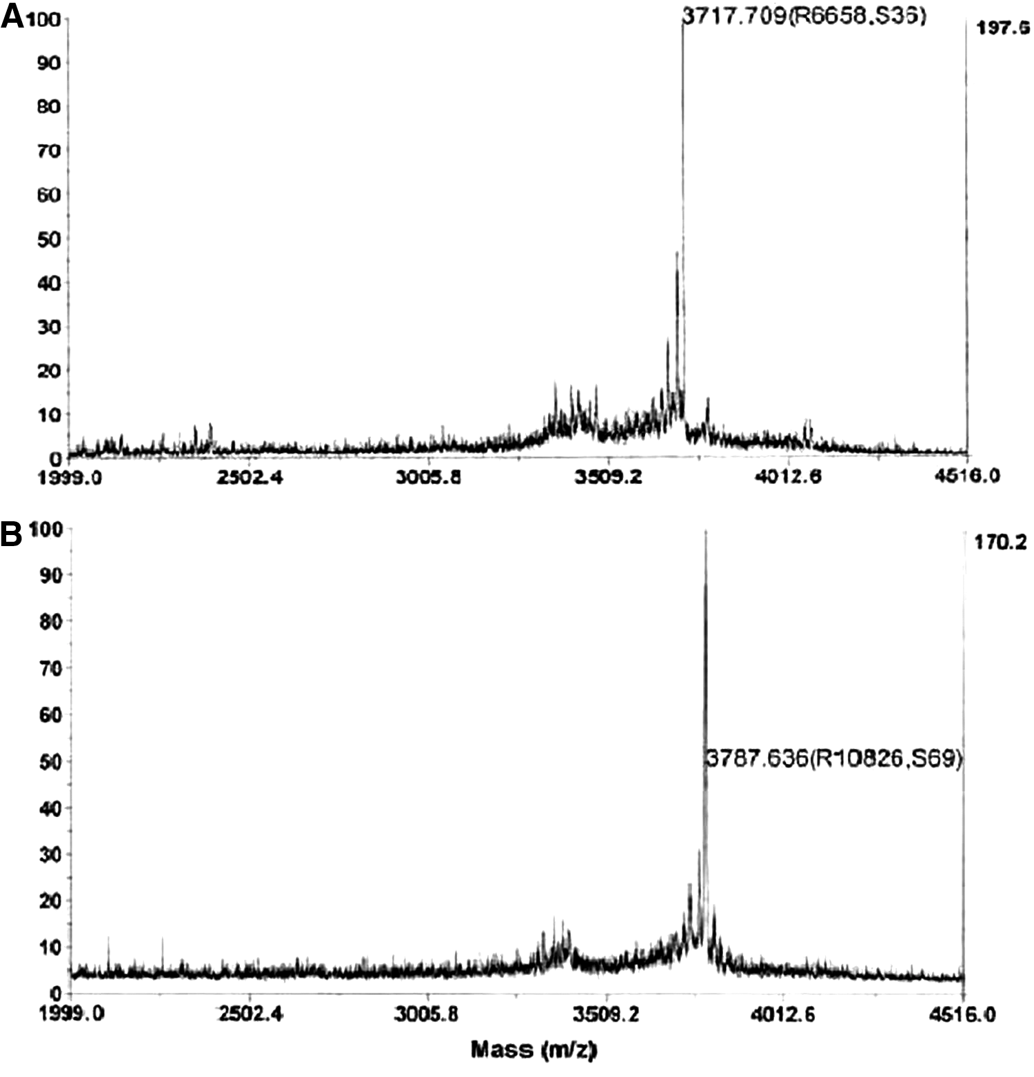
The NODAGA- and DOTA-peptide purity as determined by HPLC was 99%. The MALDI-TOF demonstrated that for NODAGA-peptide, the observed molecular mass was 3716 (calculated molecular mass of 3718), whereas for DOTA-peptide, the observed molecular weight was 3787 (calculated molecular mass of 3785). The MALDI peaks for the peptide conjugates are given in Figure 1A and B.
Mass spectrometry of
Radiolabeling kinetics
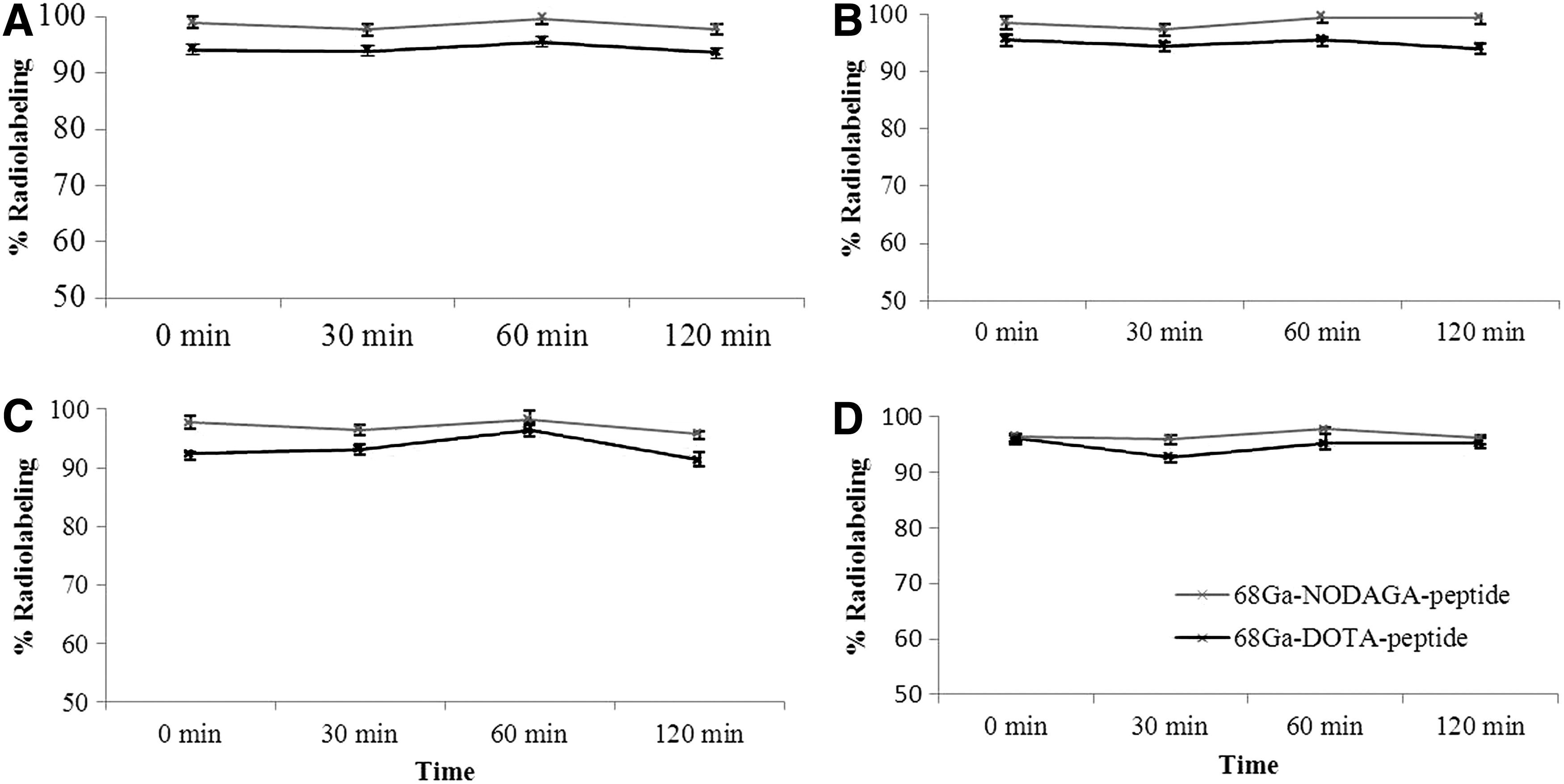
Optimal radiolabeling (>95.0%) of the DOTA-conjugated peptide required pH 2–5 and a higher (50°C–90°C) temperature, while the NODAGA-conjugated peptide needed incubation only at room temperature at the same pH range. At higher temperature (90°C), the radiolabeling was not affected by pH for both peptide conjugates (Fig. 2A, B) and can be labeled at a wide range of pH (1.0–6.0). NODAGA-peptide can bind 68Ga at a minimal quantity of 1 μg, but DOTA-peptide required a minimum of 20 μg to yield optimal (≥95%) radiolabeling (Fig. 2C). The minimum incubation time needed for optimal (>95%) radiolabeling was 10 minutes at 90°C (Fig. 2D). Radio-HPLC retention time for both radiocomplexes was 9.9 ± 0.3 minutes compared to 3.4 ± 0.2 minutes for free 68Ga, as shown in Figure 3B. The retention time was also determined for the reference standard such as hydrolyzed 68GaCl3, 68Ga-DTPA, and 68Ga-transferrin and found to be 3.5 ± 0.2 minutes.

Influence of temperature and pH on

Stability studies
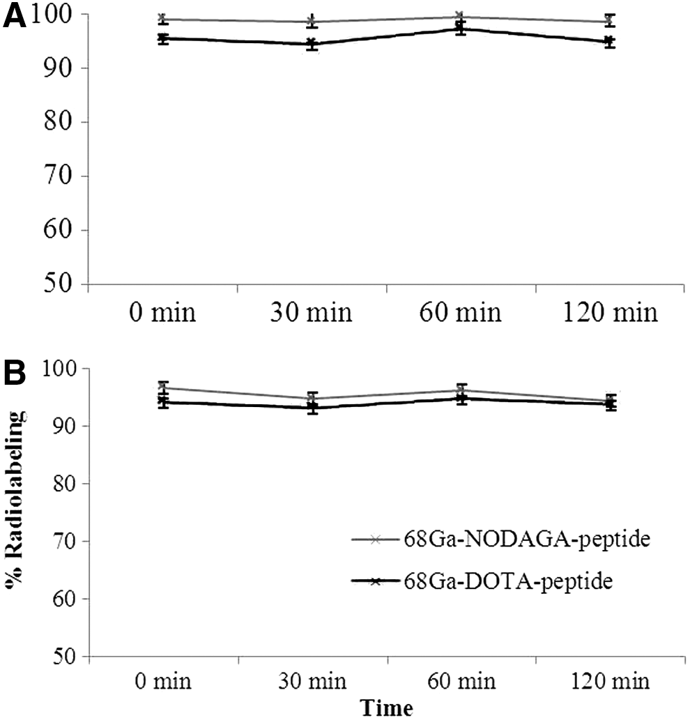
Both 68Ga-labeled peptide conjugates were stable in 0.9% normal saline (Fig. 4A) and showed minimal (1%–2%) degradation against (FeCl3, CaCl2, and ZnCl2) transmetallation (Fig. 4B–D) up to 2 hours. The transchelation study with DTPA (Fig. 5A) and transferrin (Fig. 5B) solution showed that both radiocomplexes remained intact over 2 hours. Radio-TLC showed a ratio factor value of 0.0–0.1 for 68Ga-labeled peptide and 0.9–1.0 for free 68GaCl3.
Stability in
Transchelation study with
Cell binding (in vitro) assay
Cell binding assay showed a higher uptake for 68Ga-NODAGA-peptide, 34.0% ± 0.8%, compared with 24.5% ± 0.9% for 68Ga-DOTA-peptide at 15 minutes. However, at 2 hours of incubation at 37°C, the uptake was nearly the same, 48.9% ± 3.9% for 68Ga-NODAGA-peptide and 45.2% ± 2.1% for 68Ga-DOTA-peptide (Table 1).
Biodistribution studies
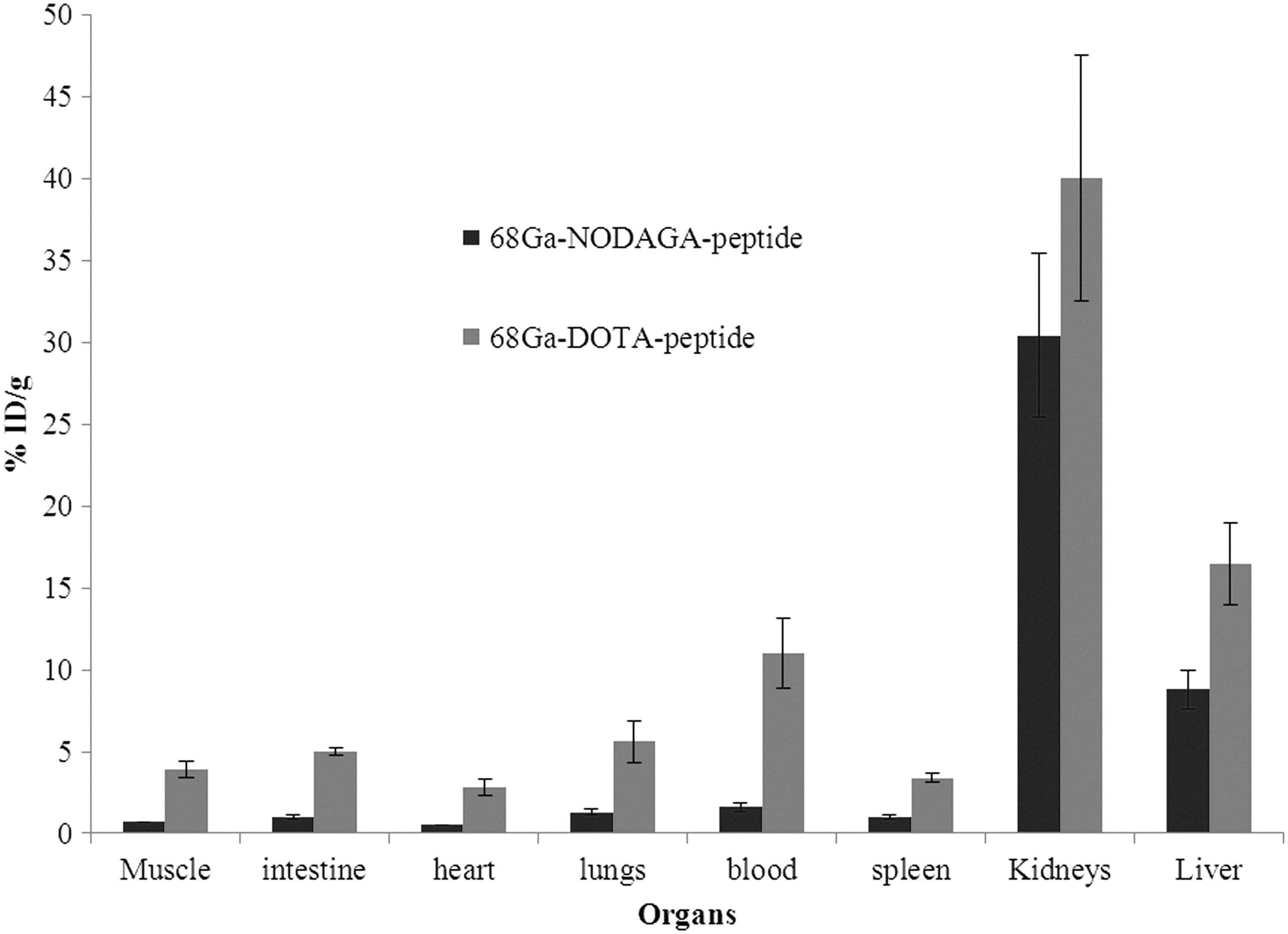
The biodistribution pattern of the both 68Ga-labeled peptide conjugates is shown in Figure 6. In both cases, a higher uptake is seen in the kidneys followed by the liver, blood, and lungs. It was evident that the 68Ga-NODAGA-peptide showed rapid blood clearance compared to the 68Ga-DOTA-peptide. Liver uptake was 8.8% for 68Ga-NODAGA-peptide, lower (p = 0.01) compared to 16.5% for 68Ga-DOTA-peptide. Most of the activity was excreted through the kidneys.
Tissue biodistribution of 68Ga-NODAGA-peptide (n = 5) and 68Ga-DOTA-peptide conjugate (n = 5) at 1 hour.
Discussion
With its several advantages such as the availability in a generator form, cyclotron independency, multielution ability in a single day, simplified radiolabeling chemistry with several biomolecules using bifunctional chelating agents, the use of 68GaCl3 is rapidly growing for diversified clinical applications. 27 The presence of even μg levels of metallic impurities (with aging of generator) in 68GaCl3 or reagent used may hamper the radiolabeling complexation. The removal of radionuclide impurities, however, has helped expansion of its use for clinical application. 28
The eluate of 68GaCl3 from the generator may contain metal ion impurities such as Fe3+, Cu2+, Al3+, Zn2+, and Sn4+, which could hamper the radiolabeling of biomolecules. It is important therefore to identify the suitable chelating agent for the radiolabeling, which is least affected by these impurities as well the stability of the labeled compound. 29,30 DOTA is the most commonly used chelating agent and 68Ga-DOTATOC is accepted in the European Pharmacopeia. 31 68Ga-labeled DOTA biomolecule conjugates (like peptides, antibodies, and small molecules) have been successfully developed and used for many clinical applications. The disadvantage of DOTA conjugate is its slow reaction kinetics for complex formation with 68Ga. 32 These limitations prompted us to investigate the use of NODAGA for comparison with DOTA.
Chelator-peptide conjugates were synthesized by the solid-phase peptide synthesis method. Both peptide conjugates were synthesized with high purity (∼99.0%). The NODAGA-peptide conjugate showed faster kinetics compared to DOTA-peptide conjugate and less influenced by the pH variation. The NODAGA-peptide conjugate yielded a higher radiolabeling efficiency at minimal quantity of peptide (1 μg), at room temperature, and within a short incubation (5 minutes) time. For the preparations with short lived radionuclides like 68Ga, these characteristics of NODAGA are advantageous. The faster radiolabeling kinetics may be attributable to its ionic size compatibility with cavity size of the nine-membered triazomacrocycle of NODAGA and therefore yielding a high radiolabeling in 5 minutes at room temperature. 33 A low radiolabeling was observed in acidic (pH 1.0) conditions for both peptide conjugates when incubated at temperature (25°C–70°C). It may attribute to a strongly acidic environment that might have protonated the donor atoms of the ligand and destabilized the 68Ga complex. 34 At high temperature (∼90°C), the radiolabeling was found to be independent of pH variation (Fig. 2A, B). However, at pH 7.0 and above, labeling decreases (data not included). This may be due to a fact that at acidic pH, 68Ga remains predominant in cation Ga3+ form (>90%), although soluble complex ions Ga(OH)2+ and Ga(OH)+ 2 may be present in the solution. As the pH of the solution shifts toward basic (>7.0), the hydrolyzed form of Gallium, Ga(OH)− 4, increases and decreases the radiolabeling yield. 35 Once the peptide conjugate is radiolabeled, then adjusting the pH to physiological pH 7.0 does not adversely influence the chelated Ga-68.
The use of sodium acetate buffer maintained the pH and allowed a strong formation BFC-68Ga complex. The other advantage of using the sodium acetate buffer is its biocompatibility for human use. 36 The DOTA-peptide conjugate showed labeling at high temperature. Therefore, DOTA may not be useful for those peptides that can denature at higher temperature and become unsuitable for molecular imaging. In contrast, radiolabeling of NODAGA-peptide at room temperature ruled out major thermal denaturation. Radio-HPLC showed that the formation of the 68Ga-NODAGA- and DOTA-peptide conjugate has retention time of around 9.9 minutes, whereas free 68Ga eluted at 3.6 minutes (Fig. 3B) and the complex formation between 68Ga and NODAGA-peptide conjugate is shown in Figure 3A. The HPLC UV detector also detected the UV peak for NODAGA- and DOTA-peptide conjugates at around 9.9 minutes. Therefore, it confirmed the complexation of the 68Ga with peptide conjugate, which leads to the retention of the 68Ga-labeled peptide approximately at the same time.
Both radiocomplexes were found to be stable in normal saline (Fig. 4A) and human serum over 120 minutes. The stability in human serum (at 37°C) indicated its suitability for in vivo applications and showed inertness toward the metal ion/species that may be present in the serum. The transmetallation study was important not because these ions are present as impurities in the eluate but because these ions are also present in serum in appreciable quantity. 37 68Ga-NODAGA- and DOTA-peptide conjugates showed minimal (1%–2%) degradation in radiolabeling after 120 minutes of incubation with the respective ion solution at 37°C (Fig. 4B–D). This could also provide an indication about the stability of these radiocomplexes in an in vivo environment. Transferrin is the major protein in the plasma that is known to form a strong complex with 67Ga-citrate. 38 The minimal transchelation (∼1% to 2%) of 68Ga-peptide conjugates with transferrin showed the stability of radiocomplexation (Fig. 5A, B).
The cell binding assay was performed on T47D breast cancer lines that express VPAC1 receptors on their surface. 19 68Ga-NODAGA-peptide showed a higher uptake in 15 minutes compared to 68Ga-DOTA-peptide, but the uptake was almost the same at 2 hours (Table 1), which may be due to saturation of the receptor site. The biodistribution data (Fig. 6) showed that both radiocomplexes were cleared through the kidneys. Hence, the renal system was the major route of clearance from the body. 68Ga-NODAGA-peptide was cleared rapidly from the blood and had significantly less liver uptake (p < 0.05) compared to 68Ga-DOTA-peptide at 1 hour. The fast clearance from the blood may be advantageous in reducing the background radioactivity for PET imaging, but it could also limit the accumulation of radioactivity at the target (tumor) area. Hence, a detailed in vivo investigation will be required further for any conclusion.
In the present study, the authors have shown that 68Ga-NODAGA-peptide exhibited the most favorable features from a radiochemistry point of view. Chelator NODAGA has flexibility over the influence of temperature and acidic environment. 13 Labeling at room temperature provides an advantage and could prevent any deleterious effect on peptide conjugate due to heating. Both peptide conjugates were found to be stable in serum against transchelation and transmetallation experimentations. A detailed in vivo study is in progress to investigate the influence of chelators on the biodistribution and tumor targeting.
Conclusions
A comparison between NODAGA and DOTA revealed that NODAGA-peptide conjugate could be efficiently radiolabeled with 68Ga at room temperature with a high yield. 68Ga-NODAGA-peptide conjugate remained stable in serum, in the presence of major metal ion impurities, against transchelation with DTPA or transferrin. The biodistribution data showed clearance from the kidneys, which minimizes the radiation burden on the organs. The clearance of 68Ga-NODAGA-peptide from blood was faster compared to 68Ga-DOTA-peptide and would be advantageous for PET imaging.
Footnotes
Acknowledgments
The research, in part, was supported by NIH/NCI RO1 CA 157372 (M.L.T.), NIH/NCI 1S10OD012406 (M.L.T.), and NIH/NCI S10RR23709 (M.L.T.).
Disclosure Statement
M.L.T. is consultant to NuView and Zevacor, Inc. No other potential conflicts of interest relevant to this article are reported.