
Abstract
The members of the integrin αv (ITGAV) family are widely expressed on many types of tumors and have been reported to be involved into angiogenesis, tumor metastases, and multicellular radioresistance. Osteosarcoma (OS) is the most common primary malignant bone tumor and the role of ITGAV in OS needs to be further elucidated. MicroRNAs are aberrantly expressed in a variety of cancers. Thus, the authors collected OS tissues (n = 15) and corresponding paracancerous tissues (n = 15) and found that the expression of miR-548c-3p was significantly downregulated in OS tissues and cell lines 143B, SaoS2, and HOS when compared to the corresponding paracancerous tissues and human osteoblast cell line hFOB (OB3), respectively. In addition, the authors identified that miR-548c-3p could directly target the 3′-untranslated region of ITGAV, and miR-548c-3p overexpression inhibits the mRNA and protein levels of ITGAV, which were confirmed by the luciferase reporter assays. Interestingly, they also uncovered that miR-548c-3p overexpression or knockdown of ITGAV remarkably suppressed cell vitality and promoted apoptosis and G2/M cell cycle arrest, leading to abrogating the ability of colony formation. The results indicated that the miR-548c-3p, similar to the target agents against integrin αv in clinical trials, could negatively regulate the ITGAV and be a promising tumor therapeutic target.
Introduction
Osteosarcoma (OS) is an aggressive bone cancer and ∼60% of cases are pediatric patients between 10 and 20 years of age. The patients with OS exhibit a predilection to occur in the metaphysis of long bones, including distal femur, proximal tibia, or humerus. Most metastatic diseases occur in the lung and bone. 1 –3 The current standard treatment for newly diagnosed OS combined the neoadjuvant chemotherapy and surgical removal. 4 New therapies have been investigated to improve overall survival in patients with OS. 5
As the cellular adhesion molecules, integrins consist of a large family of cell surface glycoproteins and function as mediators for the interactions between cells and the extracellular matrix (ECM) components, as well as cell-to-cell interactions. 6,7 The integrin family contains 18 larger α subunits and 8 smaller β subunits, which combine to form 24 α/β heterodimer receptors. The combination of α/β heterodimers allows cells to sense and respond to the tissue microenvironment through the transduction exogenous signals from the ECM into intracellular signals in cell-type and tissue microenvironment-dependent manners, which contribute to several cellular functions, such as cell apoptosis, proliferation, angiogenesis, migration, invasion, and tumor progression. 8 –10 Integrin αv subunits, which are encoded by integrin αv (ITGAV) genes, are found to be involved in tumor angiogenesis, tumor metastases, and multicellular radioresistance in various types of cancers, including colorectal cancer, 11 human epidermal cancer, 12 medulloblastoma, 13 breast cancer, 14 and human nasopharyngeal carcinoma. 15 In colorectal cancer, overexpression of integrin αv had been found to be associated with advanced T and N stage and could be an independent prognostic factor in colorectal cancer. 11 Importantly, target agents against integrin αv have been tested on clinical trial in many cancers, and these target agents include CNTO 95, a fully human monoclonal antibody against integrin αv, MEDI-522 (LM609; a humanized monoclonal antibody against integrin αv β3), and cilengitide, a small cyclic (Arg-Gly-Asp) RGD peptide against αv β3 and αv β5. 8,16 Therefore, the identification of the role of ITGAV in OS would benefit the appropriate application of these target agents.
MicroRNAs (miRNAs) are small noncoding regulatory RNAs ranging in size from 17 to 25 nucleotides and play pivotal roles in the manipulation of gene expression at the post-transcriptional level, and miRNAs act by imperfectly base-pairing with the 3′-untranslated region (3′-UTR) of target messenger RNAs to prevent mRNA translation or degradation and repress protein accumulation. 17,18 The abnormal expression of miRNAs has been investigated in diverse cellular activities, including immune response, viral replication, carcinogenesis, metastasis, and response to anticancer treatments. 19 In medulloblastoma, miR-367 was significantly overexpressed, which correlated with poor prognosis, and ITGAV as its target was significantly inhibited. 13 miR-142-3p in breast cancer cells was reported to target ITGAV, which in turn resulted in a significant inhibition of cellular invasiveness. 8 In this study, the authors found that the expression of miR-548c-3p was notably downregulated in OS tissues and OS cell lines 143B, SaoS2, and HOS. Meanwhile, they identified that miR-548c-3p could directly target the 3′-UTR of ITGAV and control the levels of ITGAV through the luciferase reporter assays. Furthermore, miR-548c-3p overexpression or knockdown of ITGAV remarkably suppressed the cell proliferation and colony formation, which could be a promising and potential tumor therapeutic strategy for the treatment of OS.
Materials and Methods
Cell culture, tissue collection, and reagents
The human OS cell lines 143B, SaoS2, and HOS and the human osteoblast cell line hFOB (OB3) were cultured in Dulbecco's modified Eagle's medium (DMEM) supplemented with 10% fetal bovine serum (FBS; Life Technologies), ampicillin, and streptomycin at 37°C, 5% CO2 conditions. OS tissues (n = 15) and corresponding paracancerous tissues (n = 15) were collected to assess the expression of miR-548c-3p. All the patients diagnosed with primary OS were confirmed by hematoxylin and eosin staining by experienced pathologists. Patients who were diagnosed with autoimmune or other malignant diseases and pregnant or lactating individuals were excluded from this experimental group. No patients underwent preoperative chemotherapy and/or radiotherapy. This study was approved by the Research Ethics Committee of Southern Medical University, Guangzhou, China. Written informed consent was obtained from all the patients. According to the WHO classifications, all the slides of OS tissues were evaluated by two pathologists. SiRNA of ITGAV and miRNA mimics or inhibitors for miR-548c-3p were purchased from RiboBio (siRNA-ITGAV: 5′-GGCUGUCGGAGAUUUCAAUTT-3′). Reporter plasmid of full-length 3′-UTR (wild type or mutant) of ITGA2 mRNA was conducted by GenePharma. Anti-ITGAV and GAPDH antibodies were obtained from Cell Signaling Technology.
RNA isolation and qRT-PCR
According to the standard RNA isolation protocol, total RNA from tissues or cells was extracted using the TRIzol reagent (Invitrogen). Quantitative real-time RT-PCR (qRT-PCR) was performed by Applied Biosystems™ (Life Technologies), and the expression levels of ITGAV were normalized to GAPDH for gene expression. The primers were purchased from GenePharma and listed below: ITGAV-F: 5′-GACCCCTTA CCCCAACTTTAT-3′, ITGAV-R: 5′-TGACAGCCGAGACT GATTTTA-3′; GAPDH-F: 5′-TGTTCGTCATGGGTGT GAA-3′, GAPDH-R: 5′-ATGGCATGGACTGTGGTCAT-3′; miR-548c-3p -F: 5′-ACACTCCAGCTGGGCAAAAATCTC AAT-3′, miR-548c-3p -R: 5′-CTCAACTGGTGTCGTGGA-3′; U6-F: 5′-CTCGCTTCGGCAGCACA-3′, U6-R: 5′-AACGCTT CACGAATTTGCGT-3′.
Western blots
Cells for western blots were collected and total protein was isolated from the cell samples according to the manufacturer's protocol. Detailed procedures for immunoblotting are described elsewhere. 14
Cell transfection
According to the manufacturer's protocol, 2 × 10⁵/well cells were cultured in 12-well plates and transfected with siRNA-NC/ITGAV or miR-548c-3p by Lipofectamine2000 (Invitrogen) and cultured for 48 hours.
CCK-8 assay
After the transfection, cells were harvested and washed with phosphate buffered saline, then the cell counting kit-8 (Dojindo) mixed with DMEM was used for cell viability assay, and the absorbance was measured at 450 nm by a microplate reader.
Colony formation assay
Cells were exposed to indicate treatments of RNA oligonucleotides or plasmids after 48 hours of transfection and harvested. The cells were resuspended in a complete medium containing 10% FBS and were seeded into six-well plates for 10 days. Using 0.1% crystal violet, cells fixed with methanol for 15 minutes were visualized under a dissection microscope (Olympus) and colonies consisting of 50 cells or more were counted.
Flow cytometry assay
For the apoptosis analysis, the cells were fixed in cold 70% ethanol at −20°C for 2 hours. Then, cells were treated with 10 mg/mL RNase and stained with 2 μL of Annexin V mixed with 2 μL of propidium iodide (PI; eBioscience) according to the manufacturer's instructions and quantified by flow cytometry on a FACS Calibur instrument. Annexin-V(+)/PI(−) was identified as early apoptosis and Annexin-V(+)/PI(+) was identified as late apoptosis.
Statistical analyses
Using the Statistical Package for Social Sciences version 16.0 (SPSS 16.0; SPSS, Inc.) and the Prism statistical software package (version 5.0; GraphPad Software, Inc.), the statistical analyses were performed. Unpaired t-tests or Mann–Whitney U tests were used to compare the two groups, and multiple group comparisons were analyzed with one-way analysis of variance. p < 0.05 was considered statistically significant. All experiments were performed at least three times.
Results
The expression of miR-548c-3p is decreased during carcinogenesis
miRNAs have demonstrated to be aberrantly expressed in a variety of cancers. OS tissues (n = 15) and corresponding paracancerous tissues (n = 15) were collected and the Q-PCR analysis showed that the expression of miR-548c-3p was significantly lower in OS tissues than that in the corresponding paracancerous tissues (Fig. 1A). To confirm the results, the authors performed the analysis of the level of miR-548c-3p in related cell lines. Similar results showed that the human osteoblast cell line hFOB (OB3) harbored a higher expression of miR-548c-3p than that in OS cell lines 143B, SaoS2, and HOS (Fig. 1B). These results indicated that the decreased expression of miR-548c-3p in OS could be involved into tumor progression.
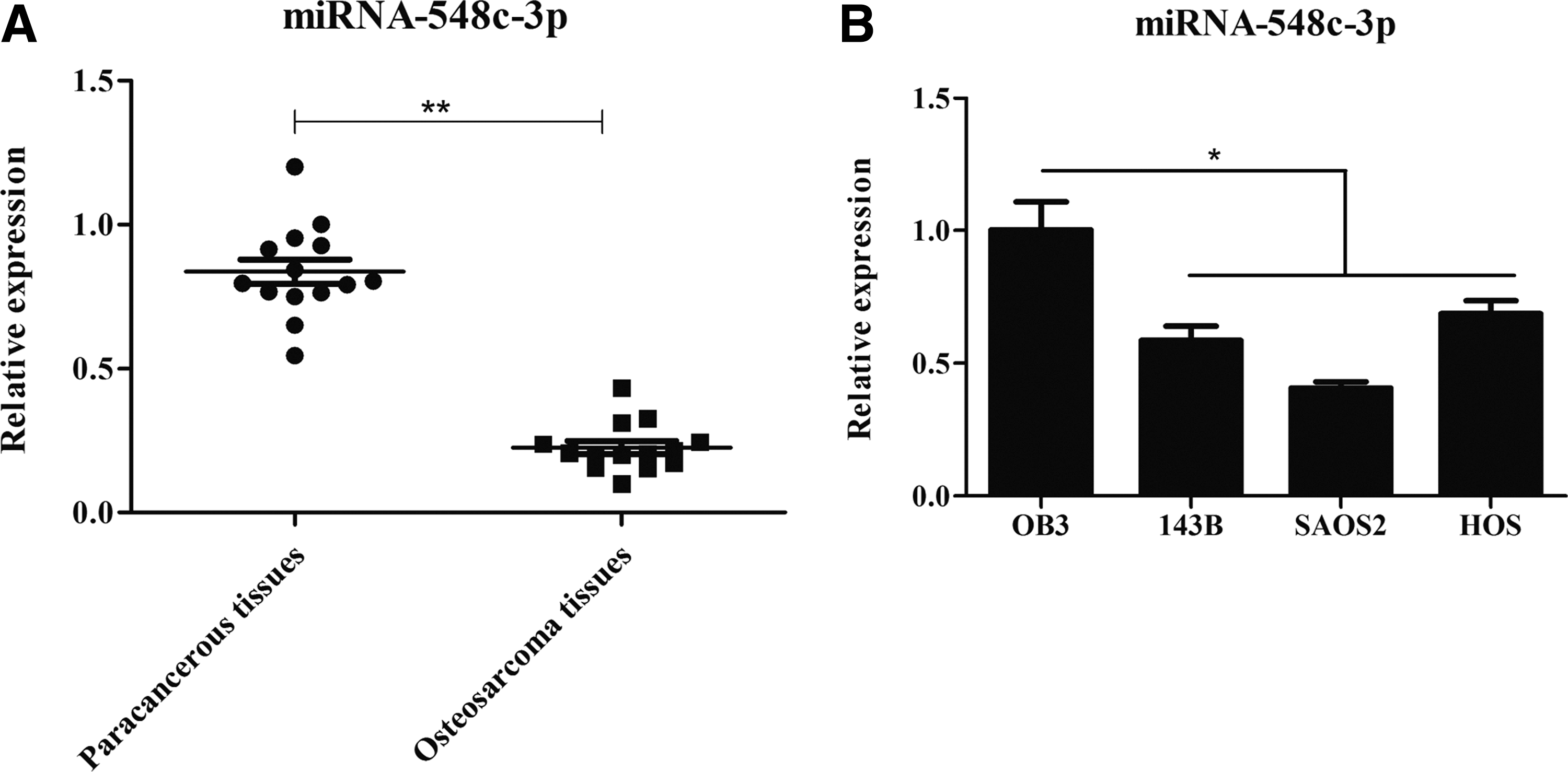
The miRNA expression of miR-548c-3p in osteosarcoma (OS) tissues
miR-548c-3p could directly target and inhibit the expression of ITGAV
The mechanism of miR-548c-3p in carcinogenesis of OS needed to be explored. Thus, the predicted genes that could be targeted by miR-548c-3p were screened by miRWalk (
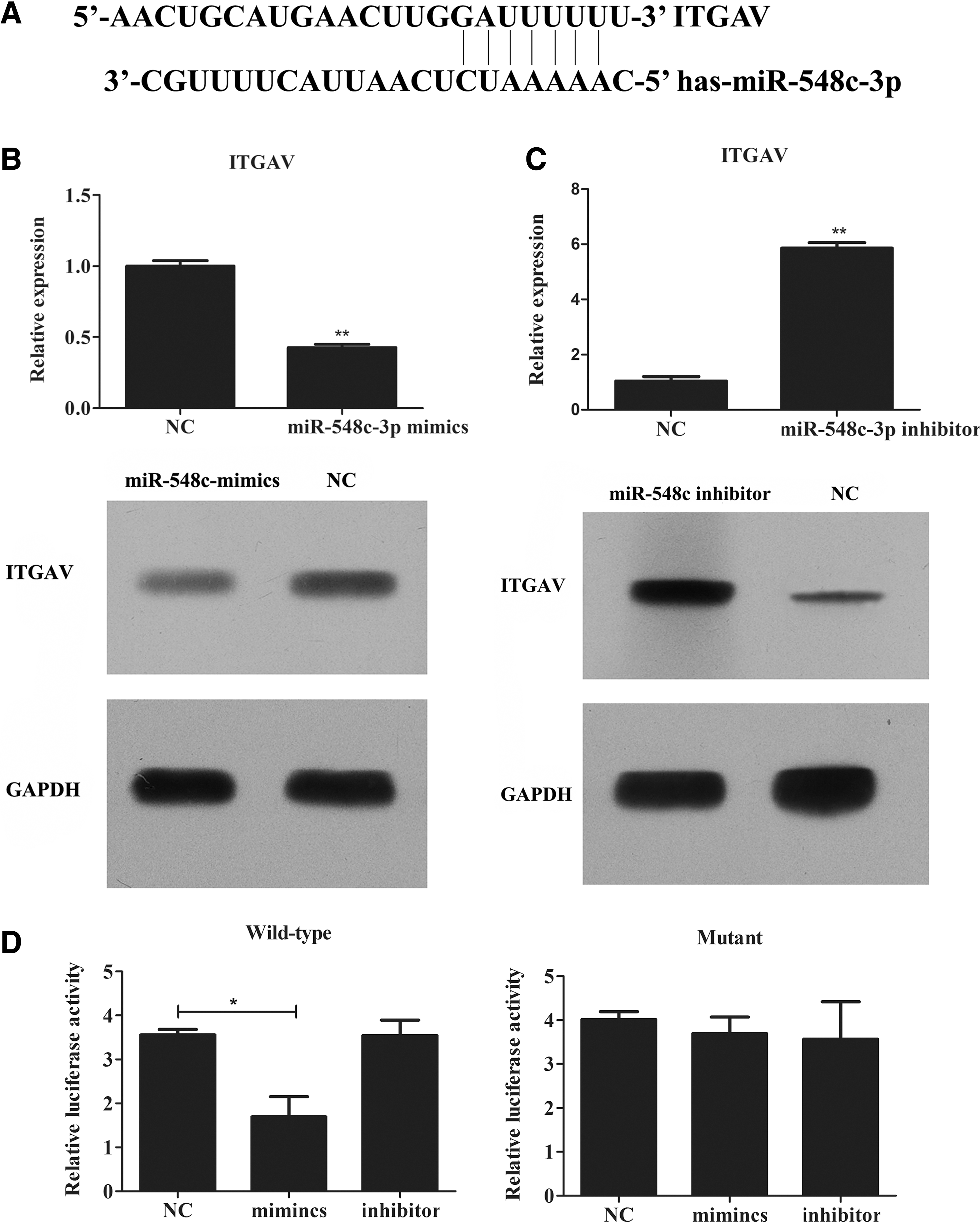
miR-548c-3p could directly target ITGAV
miR-548c-3p negatively regulates the cell vitality and colony formation via ITGAV
Since the expression of miR-548c-3p was downregulated in OS tissues and ITGAV mRNA was its direct target, miR-548c-3p could participate in tumor progression via ITGAV. The authors transfected the miR-548c-3p mimics or siRNA of ITGAV into the SaoS2 cell line for indicated times. The CCK-8 assay indicated that transfection of miR-548c-3p mimics notably decreased cell vitality and siRNA-ITGAV showed similar effects on cell vitality (Fig. 3A). Colony formation assay also showed that miR-548c-3p mimics or siRNA of ITGAV could abrogate the ability of colony formation of SaoS2 cell line (Fig. 3B), suggesting negative correlation between miR-548c-3p and ITGAV on regulating cell vitality and colony formation.
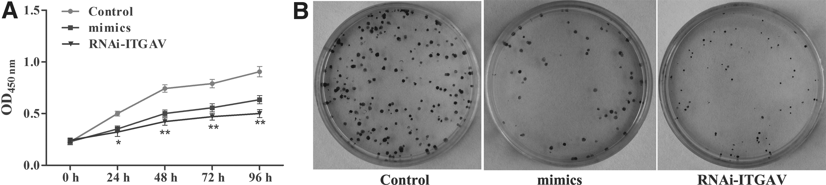
Overexpressed miR-548c-3p mimics or siRNA-ITGAV inhibit cell vitality
miR-548c-3p promotes apoptosis and cell cycle arrest via ITGAV
To investigate the mechanism of decreased cell vitality and colony formation, the authors hypothesized that miR-548c-3p mimics or siRNA of ITGAV might affect the apoptosis and cell cycle of SaoS2 cell line. Therefore, they overexpressed the miR-548c-3p mimics or siRNA-ITGAV and the results showed that the apoptosis rate was significantly higher when compared to the negative control (Fig. 4A) and the cell cycle was found to be arrested in G2/M, leading to decreased cell vitality and colony formation (Fig. 4B).
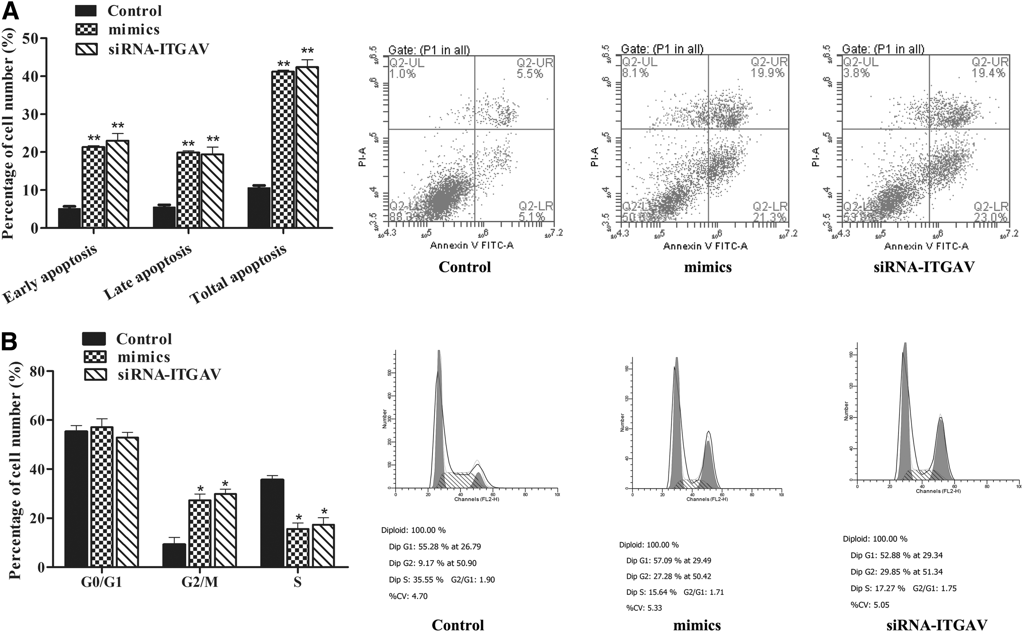
Apoptosis
Discussion
Despite chemotherapy and surgical excision of the primary tumor, OS is a highly malignant bone tumor that frequently leads to poor clinical outcomes due to pulmonary metastasis, and the molecular mechanism of carcinogenesis remains to be understood. 7 The authors here reported that the expression of miR-548c-3p was downregulated in OS tissues and OS cell lines, which in turn negatively regulated the expression of ITGAV by directly targeting its 3′-UTR and contributed to elevated cell vitality, decreased apoptosis, G2/M arrest, and enhanced ability of colony formation, which shed light on the role of miR-548c-3p/ITGAV in the progression of OS.
Integrins play pivotal roles in epithelial adhesion, angiogenesis, proliferation, wound healing, and carcinoma. The αv subunit binds to five different β subunits (β1, β3, β5, β6, and β8), but β4, β5, β6, and β8 each bind to only 1 α subunit. 20,21 α and β subunits share the similar structure 4 consisting of an extracellular domain, a single transmembrane domain, and a short, noncatalytic intracellular tail. Although the knockout of the αv gene was lethal, mice showed extensive brain and intestinal blood vessel abnormalities, resulting in death before or soon after birth, which indicated that the αv subunit is essential for normal development. 22 In addition, integrin αv subunits have been demonstrated to be involved in tumor angiogenesis, metastasis, and cancer progression in various types. He et al had reported that decreased expression of αv integrin reduced multicellular radioresistance to oxaliplatin via the diminished of phosphorylated NF-κB p65 and enhanced phosphorylated JNK2 in multicellular spheroids in the HT29 cell line. 23 In human epidermal cancer, acute loss of αvβ5 and αvβ6 resulted in the loss of de novo epidermal tissue generation and broke the tissue maintenance that promoted tumor invasion. 12 A humanized monoclonal antibody against integrin αvβ3, LM609, could block the growth of human melanomas in vivo, suggesting a possible therapeutic potential for αvβ3 antagonists. 24 CNTO 95, the fully human monoclonal antibody against integrin αv, plus Dasatinib, was found to effectively inhibit adhesion, migration, and integrin signal transduction in the colorectal cancer cell line, 25 indicating that integrin αv could be a diagnostic and therapeutic target of these cancers.
miRNAs are strongly associated with carcinogenesis, affecting nodal points in immune response, genome integrity, stress responses, apoptosis, and metastasis. 18 To date, it was found that miRNAs were directly involved in human cancers, including the lung, breast, brain, liver, colon cancer, and leukemia. 15 miR-192 could decrease ITGAV, ITGB1, ITGB3, and CD47 expression to decrease cellular anchoring for the dissemination in medulloblastoma tissues. 26 ITGAV was also identified as a regulatory target for miR-142-3p in MCF-7 and MDA-MB-468 cells for modulating breast cancer metastasis. 14 The hsa-mir-548 expression was identified to be upregulated in colorectal cancer tissue and many genes encoding cell division cycle protein involved in cellular proliferation were identified as its potential targets and repressed in colorectal cancer tissue. 27 miR-548c-3p had been investigated in other cancers, including breast cancer and ovarian cancer. miR-548c was reported to be downregulated in breast cancer and ovarian cancer tissues when compared to normal tissues, 28,29 and in ovarian cancer, decreased miR-548c expression correlated with poor prognosis in endometrial cancer patients. 29
In sum, in this study, the authors found that the expression of miR-548c-3p was decreased in OS tissues and OS cell lines and they identified that the ITGAV was the direct target. Thus, this study implicated that the decreased miR-548c-3p could elevate the expression of ITGAV for tumor progression, including facilitating cell proliferation and inhibiting apoptosis, inducing G2/M arrest.
Footnotes
Disclosure Statement
No competing financial interests exist.