
Abstract
The biodistribution of extracellular vesicles (EVs) is a fundamental question in the field of circulating biomarkers, which has recently gained attention. Despite the capabilities of nuclear imaging methods, such as single-photon emission computed tomography, radioisotope labeling of EVs and the use of the aforementioned methods for in vivo studies hardly can be found in the literature. In this article, the authors describe a novel method for the radioisotope labeling of erythrocyte-derived EVs using the 99mTc-tricarbonyl complex. Moreover, the capability of the developed labeling method for in vivo biodistribution studies is demonstrated in a mouse model. The authors found that the intravenously administered 99mTc-labeled EVs mostly accumulated in the liver and spleen. The in vivo stability of the labeled EVs was assessed by the comparison of the obtained biodistribution of EVs with that of the free 99mTc-tricarbonyl. According to the authors' data, only a minor fraction of the radioactive label became detached from the EVs.
Introduction
Extracellular vesicles (EVs) are recently recognized as key players in many physiological and pathological conditions. 1 –6 EVs are not only present in all human body fluids but also can be found, for example, in ocean water 7 and beer 8 due to the fact that yeasts and other microorganisms also release EVs. The clinical relevance of EVs was first recognized for their diagnostic use as biomarkers of different diseases. 5 Recently, the therapeutic application of EVs is also emerging 9,10 ; for example, the application of dendritic cell (DC)-derived exosomes for cancer treatment was recently under investigation in a clinical trial (NCT01159288). Understanding the role of these membrane vesicles in intercellular communication and also their applicability as vehicles for drug delivery requires the investigation of their biodistribution. The in vivo fate of EVs has only been addressed in the last few years, and most studies used fluorescent imaging for this purpose. 11 –18 In these studies, EVs were either labeled with a membrane dye 13,18 or engineered to display a membrane reporter (e.g., Gaussia luciferase) and administered exogenously 15 or used endogenously produced, genetically modified EVs (such as GFP-tagged CD63 bearing EVs). 19 In contrast, the application of nuclear imaging techniques, such as single-photon emission computed tomography (SPECT) or positron emission tomography (PET), using isotopically labeled EVs hardly can be found in the literature, despite the fact that these techniques have indisputable advantages over fluorescent imaging regarding quantitative measurement of the biodistribution of the labeled compounds. 20
In this article, the authors report a novel method for radioisotope labeling of EVs using 99mTc-tricarbonyl complex and demonstrate the applicability of this method for the noninvasive assessment of the biodistribution of erythrocyte-derived EVs using SPECT/CT.
Materials and Methods
Preparation of erythrocyte vesicles
Erythrocyte vesicles were isolated from freshly outdated erythrocyte concentrates (Hungarian National Transfusion Service). The erythrocyte concentrate was twofold diluted in phosphate-buffered saline (PBS, P4417; Sigma-Aldrich), and the red blood cells were removed by two centrifugation steps at 1500g for 20 minutes at 4°C. EVs in the erythrocyte-free supernatant were concentrated by ultracentrifugation (T-1270 fixed-angle rotor; Thermo Sorvall WX Ultracentrifuge) at 130,000g for 30 minutes and washed once with PBS using the same parameters. After resuspending the EV pellet in PBS, the sample was filtered through a 600-nm polycarbonate filter (Whatman® Nuclepore™) and finally snap frozen in 100 μL aliquots using liquid nitrogen and stored at −20°C until use.
Freeze-fracture transmission electron microscopy
The vesicle sample (1–2 μL) was pipetted onto a gold sample holder, frozen by plunging it immediately into partially solidified freon for 20 seconds, and stored in liquid nitrogen. Fracturing was performed at −100°C in a Balzers freeze-fracture device (Balzers BAF 400D; Balzers AG). Replicas of the fractured faces etched at −100°C were made by platinum–carbon shadowing and then cleaned with a water solution of surfactant and washed with distilled water. The replicas were placed on 200 mesh copper grids and examined in a Morgagni 268D (FEI) transmission electron microscope.
Size-exclusion chromatography combined with dynamic light scattering
Size-exclusion chromatography combined with dynamic light scattering (SEC-DLS) analysis was performed with a Jasco HPLC system (Jasco) consisting of a PU-2089 pump, a UV-2075 UV/Vis detector, and a W130i online DLS detector (Avid Nano Ltd.) controlled by ChromNAV software v. 1.17.02. Sepharose CL-2B gel was used as a stationary phase filled in a Tricorn 5/200 glass column (GE Healthcare Life Sciences). CL-2B is a cross-linked agarose gel with a fractionation range of 70–40,000 kDa for dextran and was found to be suitable for purification of EV samples. 21 The eluent was PBS, and the elution speed was 0.25 mL/min. The scattering intensity at 90° and the autocorrelation function accumulated for 3 seconds were measured with the DLS setup equipped with a 660-nm laser. The online DLS data were processed with iSize 3.0 software (Avid Nano Ltd.).
Purification and concentration of erythrocyte-derived EVs
Before labeling of the erythrocyte-derived EVs obtained by differential centrifugation, the EV sample was purified using a 10-mL plastic gravity column filled with the same Sepharose CL-2B gel used for the SEC-DLS investigation. 0.5 mL EV sample was introduced onto the column, and PBS was used as the eluent. The EV-containing fraction corresponding to the void volume of the column was collected (1 mL) and further concentrated to 0.6 mL using Vivaspin 500 centrifugal filters with 100 kDa MWCO (Sartorius Stedim Biotech GmbH).
99mTc labeling of the purified erythrocyte-derived EVs
Radiolabeling of the erythrocyte-derived EVs was performed with the 99mTc-tricarbonyl complex [99mTc(CO)3(H2O)3]+ using a commercial kit (Isolink®; Mallinckrodt Medical B.V.), according to the manufacturer's instructions. 1.2 GBq [99mTcO4]− was eluted in 1 mL saline and added to the kit, followed by placing the vial into boiling water for 20 minutes. The basic solution was neutralized by the addition of ∼200 μL 1 M HCl solution. 0.6 mL of the EV sample was added to 0.8 mL 99mTc-tricarbonyl complex solution (430 MBq) and incubated for 30 minutes at room temperature. Separation of the free 99mTc-tricarbonyl was performed using a desalting column (Zeba™ Spin Desalting Column, 0.5 mL) applying the manufacturer's instructions.
In vivo SPECT/CT imaging
In vivo imaging was carried out on male BALB/c mice (n = 3; Charles River Hungary). The body mass of the experimental animals was 28 ± 5 g, and they were 10–12 weeks old. Animal experiments were carried out at the Nanobiotechnology and In Vivo Imaging Center, Semmelweis University, with permission from the local institutional animal ethics committee no. XIV-I-001/29-7/2012 and in compliance with the relevant European Union and Hungarian regulations. Images were acquired with a NanoSPECT/CT Silver Upgrade (Mediso Ltd.) sequential animal SPECT/CT imaging system. In the SPECT/CT experiment, 15 ± 2 MBq of 99mTc-labeled erythrocyte EVs in 200 μL volume was injected into the tail vein of the mice. Control measurements (n = 3) were performed by injecting 99mTc-tricarbonyl complex only. During the scans, the animals were continuously anesthetized using a mixture of 1%–1.5% isoflurane and medical oxygen. Their body temperature was maintained at 37°C throughout the scanning. The SPECT scans started 1 hour after the time of administration, and the acquisition lasted 45 minutes.
To prevent movements, the animals were immobilized in a MultiCell™ Imaging Chamber (Mediso Ltd.) and positioned in the center of field of view. The reconstructed voxel size was 300 μm in 120 × 120 × 328 pixel matrix both in SPECT and CT modalities. The image acquisitions were started with CT imaging after intravenous injection of 99mTc-labeled EVs. The CT and subsequent SPECT imaging lasted 10.5 and 30 minutes, respectively. Reconstructed, reoriented, and coregistered images were further analyzed with Fusion (Mediso Ltd.) and VivoQuant (inviCRO LLC, US) dedicated image analysis softwares by placing appropriate volume of interests (VOIs) on the organs. The VOI was delineated manually on each CT scan. Radioactivity concentrations in MBq/cm3 were determined for each VOI and corrected for scattering and isotopic decay in the reconstruction algorithm. The uptake values were measured in the following organs: the heart, lungs, kidneys, liver, spleen, and bladder.
Results and Discussion
Figure 1A shows the TEM images of the platinum–carbon replicas of the freeze-fractured EV sample. Spherical vesicles within the size range of 80–300 nm can be observed on these images, clearly demonstrating the applicability of the used isolation protocol. SEC-DLS analysis was used to characterize the purity of the EV sample as well as determine the mean hydrodynamic diameter of the vesicles (Fig. 1B). During the SEC analysis, the EVs were excluded from the pores of the used gel (Sepharose CL-2B), and consequently, they are eluted at shorter elution times (at 4.6 minutes in this case). The online DLS detector enabled the characterization of the mean size of the vesicle fraction, which was found to be 188 ± 11 nm (standard deviation). Smaller objects, such as lipoproteins and soluble plasma proteins, enter the pores of the gel, which results in longer elution times (at 14.3 minutes in this case). Due to the smaller size of the plasma proteins, their contribution cannot be detected based on the light scattering signal, but the UV absorption clearly indicates their presence in the studied sample.
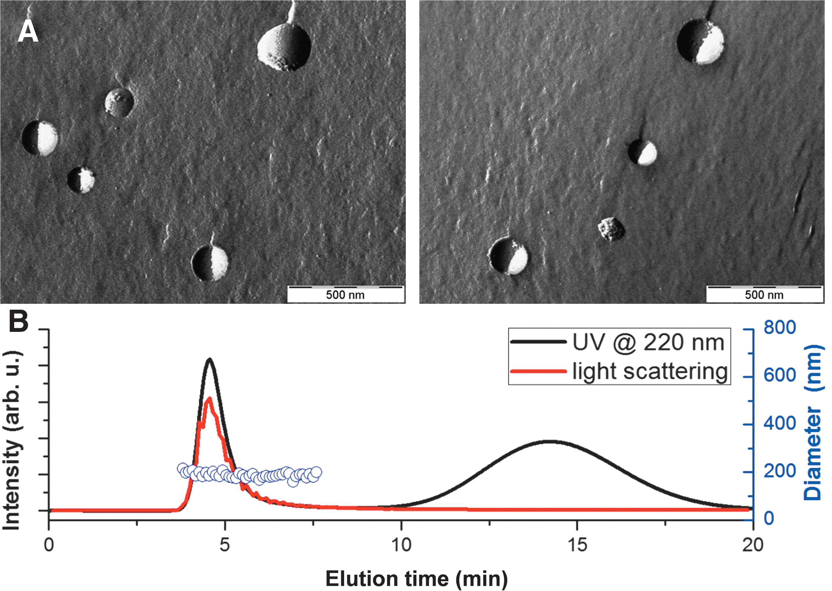
Contamination by plasma proteins in EV samples is common when using isolation by differential centrifugation only. Therefore, our sample was further purified before the radioisotope labeling. The purification was performed using the same Sepharose CL-2B gel that was used for the SEC-DLS analysis but in a preparative manner using a 10-mL plastic tube and a gravity separation protocol. It was recently shown by Böing et al. that the contamination from high-density lipoprotein is less than 5% and from plasma proteins is less than 1% in the purified EV fraction using this protocol. 21 The latter also agrees with our previous experience using the Sepharose CL-2B gel.
To the best of the authors' knowledge, no literature data are available for the radioisotope labeling of EVs. In contrast, several approaches exist for the labeling of liposomes, which can be treated as model systems of the EVs due to the fact that the phospholipid bilayer represents the basic structural building block for both. The most commonly used radionuclide for liposome labeling is 99mTc. There are several (afterloading and surface) radiolabeling techniques of liposomes in the literature. 22 The afterloading labeling technique is based on the application of lipid-soluble chelating agents. 23 One of the most frequently applied lipophilic chelator molecules is HMPAO (hexamethylpropyleneamineoxime). It is able to carry 99mTc inside the preformed vesicle, where the lipophilic HMPAO is transformed into its hydrophilic form in the presence of glutathione, and HMPAO is trapped. 22,24 –32 Another afterloading method for labeling liposomes uses 99mTc-BMEDA. This lipophilic radionuclide complex has very good in vitro and in vivo stability. 22 The direct surface labeling with 99mTc-pertechnetate is an easy manner of liposome labeling, but the radiochemical yield, the specific activity, and the stability of the forming complex seem to be very low. 33
Radiolabeling of artificial exosome-mimetic nanovesicles (ENVs) prepared by extrusion of macrophage cells was recently reported using the 99mTc-HMPAO method, which is an established technique in liposome research. 34 The in vivo biodistribution of ENVs used as model system for real EVs was investigated using SPECT/CT. Although this method can be used in principle to label real EVs, the low labeling efficiency of the HMPAO method at low concentrations in the case of liposomes might hinder the successful labeling of EVs.
In our experiment, the novel organometallic aquaion, 99mTc-tricarbonyl, was used for the radioisotope labeling of the purified EVs. This technique combines the advantages of high specific activity and small size of the labeling compound. 35 Hence, it retains the biological activity of the labeled object. The 99mTc-tricarbonyl has been used for the labeling of a wide range of biomolecules from small tracer molecules, peptides, and antibodies to liposomes containing a DTPA chelater. 33,36 The 99mTc-tricarbonyl binds to several amino acids, such as histidine, methionine, and cysteine, 37 and consequently, it was assumed that this aquaion will inherently bind to the surface of EVs due to the presence of membrane proteins in the vesicles. After the labeling procedure, the unbound 99mTc-tricarbonyl was removed using desalting columns. The used desalting column removes more than 98% of the free 99mTc-tricarbonyl, according to our previous experience, which also agrees with the specification from the manufacturer. By measuring the activities of the elutes and the columns, a labeling efficiency of 38.8% ± 6.2% was obtained for the erythrocyte-derived EVs, which is reasonable, especially if the generally low concentration of EVs is considered.
Figure 2 shows a typical SPECT/CT image with the biodistribution of the labeled erythrocyte-derived EVs. High accumulation of the injected EVs can be observed in the liver and spleen in accordance with previous fluorescent imaging studies. The activity appearing in the bladder can be attributed to the free 99mTc-tricarbonyl, as demonstrated by a control measurement with the labeling compound. The real advantage of modern nuclear imaging methods is that the distribution of the labeled objects can be quantified without the need for removing the specific organs. To determine the percentage of the radioactivity in the different organs, appropriate VOIs were used. Figure 3 shows the distribution of the labeled EVs within the different organs as a percentage of the total injected radioactivity (Fig. 3A) and also as standardized uptake value, known as “SUV” (Fig. 3B). SUV is defined as “the ratio of the tissue radioactivity concentration and the injected activity divided by the body weight” (as often used in PET). A major advantage of preclinical SPECT imaging is its ability to produce quantitative results for biodistribution, 38 just like PET imaging. SUV takes into account the volume of the segmented organ and so highlights organs with higher tissue uptake values.
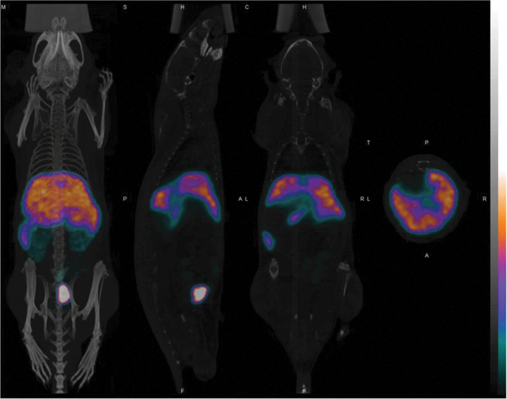
In vivo SPECT-CT images of the 99mTc-labeled erythrocyte-derived EVs. The three-dimensional, reconstructed, and coregistered SPECT and CT images are shown together with sagittal, coronal, and axial images (from left to right). Uptake of the EVs by the liver and spleen is clearly visible on the images, while the activity detected in the bladder corresponds to the free 99mTc-tricarbonyl complex. EVs, extracellular vesicles; SPECT, single-photon emission computed tomography. Color image available online at
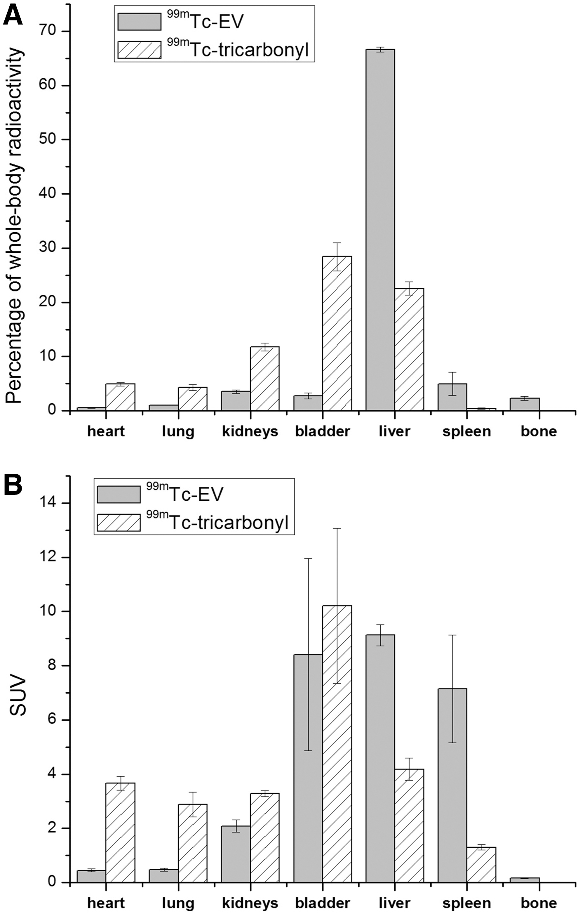
Quantitative distribution of 99mTc-labeled erythrocyte-derived EVs together with the results of the control measurement performed using the 99mTc-tricarbonyl complex alone. The activities measured in the specified organs in the percentage of the whole-body radioactivity are shown
The distribution values for the labeling compound alone are also presented in Figure 3. The distributions of the free 99mTc-tricarbonyl and that of the labeled EVs differ significantly, which is an evidence of the successful labeling. The radioactivity detected in the bladder (3% of the injected radioactivity) originates from the free 99mTc-tricarbonyl that corresponds to the radiochemical purity of the labeled EV sample (taking into account ∼40% labeling efficiency and 98% efficiency of the desalting columns used to remove the remaining free 99mTc-tricarbonyl after labeling), which also indicates a good in vivo stability of the labeling.
The main aims of the SPECT/CT investigation of the 99mTc-labeled erythrocyte-derived EVs were to demonstrate the applicability of the developed labeling procedure for in vivo experiments and assess the in vivo stability of the labeling. The deep analysis of the biodistribution of erythrocyte-derived EVs is out of the scope of this article; however, it can be compared to recent studies using fluorescently labeled EVs 13 and using 99mTc-labeled ENVs. 34 Wiklander et al. used a lipophilic near-infrared fluorescent dye (DiR: 1,1-dioctadecyl-3,3,3,3-tetramethy-lindotricarbocyanine iodide) that only fluoresces intensely when inserted into a lipid membrane to assess the biodistribution of EVs isolated from different cell lines. The obtained distribution of HEK293T cell-derived EVs agrees well with our observation, namely high uptake of the EVs in the liver (60%–80% of the total fluorescence, depending on the dose and way of administration) and the spleen (10%–20% of the total fluorescence). Wiklander et al. also investigated EVs from different sources, such as C2C12 mouse muscle cell line, B16F10 melanoma cells, and primary immature bone marrow-derived DCs. The liver uptake was the highest among the organs in all cases, but interestingly, they have found differences for the extent of liver accumulation being the highest for the C2C12 cell-derived EVs (71% ± 1.5%) and the lowest for DC-derived EVs (46% ± 0.9%). Confirmation of these observations using radiolabeled EVs and using SPECT/CT (which is superior to fluorescent imaging regarding quantitative analysis) would be an interesting application of the 99mTc-tricarbonyl EV labeling procedure presented in this study.
The biodistribution obtained by Hwang et al. for 99mTc-HMPAO-labeled ENVs from murine macrophage Raw 264.7 cell line also shows great similarity with the results presented in this article. 34 Hwang et al. prepared ENVs by extrusion of whole cells and subsequent purification by gradient ultracentrifugation. High concentration of ENVs can be achieved by this method compared to natural EVs, which enabled the 99mTc-HMPAO labeling. In contrast, the authors also mention in their article that labeling of natural EVs with the 99mTc-HMPAO method is challenging due to the low radiochemical yield of the method at low EV concentrations. 34
In summary, a novel method for the radioisotope labeling of erythrocyte-derived EVs using 99mTc-tricarbonyl complex is presented. Special attention was paid to the purity of the EV sample that was achieved by SEC purification. This procedure assured that only EVs were labeled since the used radiolabel could have also bound to free serum proteins. Acceptable radiochemical yield was achieved by the use of the novel 99mTc-tricarbonyl complex, which has high affinity to biomolecules. The applicability of the presented radiochemical labeling procedure was demonstrated by in vivo SPECT/CT imaging experiments using a mouse model, which also confirmed the in vivo stability of the labeling. The presented method might pave the way for further studies addressing the in vivo fate of EVs using nuclear imaging methods.
Footnotes
Acknowledgments
Part of this work was funded by the János Bolyai Research Scholarship, OTKA 111958, COST BM1202 ME HAD, and MEDINPROT grants.
Disclosure Statement
No competing financial interests exist.