
Abstract
This study aimed to radiolabel finasteride, a novel 5α-reductase inhibitor, to evaluate its cancer targeting potential in experimental model of prostate carcinogenesis. Finasteride was effectively radiolabeled with 99mTc and showed >90% labeling efficiency. The radiopharmaceutical was found to be stable up to 6 hours in rat serum at 37°C. The blood kinetics of the 99mTc-finasteride followed a biphasic release pattern, whereby fast-release phase was observed at 15 seconds and a slow-release phase was observed after 30 minutes of administration. The plasma protein binding of the radio complex observed was 83.89%. For biodistribution studies, the rats were divided into two groups. Group I served as normal controls, while group II was subjected to carcinogen N-methyl-N-nitrosourea (MNU) and hormone testosterone propionate (T) for induction of prostate carcinogenesis, which was confirmed histopathologically. The biodistribution studies on control and carcinogen-treated rats revealed a significant percent-specific uptake in prostate, which was found to be increased significantly as a function of time. The most significant finding of the study was an increase in the percent-specific uptake in prostate of carcinogen-treated animals when compared to the percent-specific uptake in prostate of normal rats after 2 and 4 hours postinjection. The study concludes that 99mTc-finasteride possesses selectively toward prostate cancer tissue and can be explored further for its role in detection of prostate cancer.
Introduction
Prostate cancer is the second-leading cause of cancer death in men. 1 Prostate cancer is a heterogeneous and biologically complex disease, as a result of which it poses challenges in screening, diagnosis, and disease characterization. Once an early and accurate diagnosis is made, a large number of treatment options are available for management of prostate cancer viz; radical prostatectomy, radiotherapy, or drug treatment. 2 Thus the major goal of prostate cancer management is more accurate disease characterization through various anatomic, functional, and molecular imaging information. 3
There is limited role of imaging modalities for prostate cancers. Ultrasonography is mainly used for biopsy guidance, while magnetic resonance (MR) imaging is helpful for evaluating local tumor extent and is useful in depicting lymph node metastases. Further computed tomography (CT) is reserved for the evaluation of advanced disease. 3
Imaging with radiopharmaceuticals is a noninvasive technique and has gained more importance because of the ability to detect the functional status of the disease and characterize the disease at the molecular level. Out of the conveniently used radionuclides in radionuclide imaging, 99mTc has the ideal characteristics and is cost-effective. It is easily available from generator in the form of pertechnetate (99mTcO4 −) and its gamma energy of 140 keV makes it suitable for gamma scintillation imaging. Radionuclide bone scanning in prostate cancer is mainly used to detect metastatic spread of disease to bones using 99mTc-methylene diphosphonate (MDP). 3 Furthermore, the role of 18 F-fluorodeoxyglucose (FDG) positron emission tomography (PET)/CT in prostate cancer is limited. This is related to slow glycolysis of prostate cancer cells and low FDG avidity on PET imaging. 4 Other PET imaging agents, 11 C-choline and 18 F-choline, are used particularly in biochemical relapse and high-risk staging. 5 –7 However, the choline PET has low detection rates in cases of biochemical relapse with low prostate-specific antigen (PSA) rise. 8 Recently, Ga-68-labeled PSMA ligands for PET/CT imaging has shown promising detection rates for functional imaging of recurrent prostate cancer. 9 However, only few clinical studies to date have evaluated its role in clinical workflow of prostate cancer. 10
Finasteride was the first drug to be approved in United States for benign prostatic hyperplasia (BPH) 11 and male pattern baldness. 12 Finasteride, an inhibitor of 5a-reductase, inhibits the conversion of testosterone to dihydrotestosterone (DHT), the primary androgen in the prostate. The serum DHT levels get reduced by about 65%–70% and the prostate DHT levels by up to 85%–90%, by finasteride administration. 13,14 It is being reported that all forms of androgen deprivation therapy, including finasteride, induce distinctive histological changes in benign and neoplastic prostatic epithelial cells. 15 Also, the findings of prostate cancer prevention trial (PCPT) provide significant evidence of a role for 5αR inhibitors in the chemoprevention of prostate cancer. 16 –18 A study showed that finasteride was associated with a 24.8% reduction in the prevalence of prostate cancer compared with placebo over a 7-year period. 16 Recently, another study of PCPT suggested that finasteride exposure may reduce prostate cancer risk. 19
Taking into consideration the above-mentioned points, the aim of this study was to optimize the radiolabeling of finasteride with 99mTc and evaluate its role in the diagnosis of experimentally induced prostate carcinogenesis in rats.
Materials and Methods
Chemicals
Finasteride (100 mg) >98%, stannous chloride dihydrate (SnCl2.2H2O), n-octanol, methyl-N-nitrosourea (MNU), and testosterone propionate (T) were purchased from Sigma-Aldrich, Inc. Sodium pertechnetate Na99mTcO4 was obtained from Post-Graduate Institute of Medical Education and Research (PGIMER, Chandigarh, India). Sodium hydroxide (NaOH) and instant thin layer chromatography-silica gel (ITLC-SG) strips were purchased from MERCK. Trichloroacetic acid (TCA), sodium chloride (NaCl), hydrochloric acid (HCl), sodium dihydrogen phosphate, and disodium hydrogen phosphate were purchased from SRL. Primary antibody PSA polyclonal was obtained from Bioworld Technology, Inc. β-actin and secondary antibody were purchased from Sigma-Aldrich, Inc. All other chemicals were purchased from SRL.
Animals
Six- to seven-week-old healthy adult male Sprague Dawley rats of weight 200–250 g were procured from the Central Animal House, Panjab University (Chandigarh, India). The animals were housed in polypropylene cages under hygienic conditions in the departmental animal house. The rats were maintained on a standard laboratory pelleted feed (Ashirwad Industries, Tirpari, India) and water ad libitum throughout the period of experimentation. All the procedures were done in accordance with the standard guidelines for care and use of laboratory animals and the protocols followed were approved by the Institutional Animal Ethics Committee (IAEC), Panjab University.
Radiolabeling procedure
99mTc-finasteride was prepared by adding 7.4 MBq of 99mTcO4 − to a vial containing 20 μg of finasteride (1 mg/1 mL in 10% ethanol). To the mixture, 40 μg of SnCl2.2H2O (1 mg/mL in 0.1 N HCl) was added and the pH was adjusted to 7 using 0.01 N NaOH. The reaction mixture was vortexed and kept at ambient temperature for 30 minutes to complete the reaction. Optimization of amount of all the chemical constituents viz: SnCl2.2H2O, finasteride, and 99mTc in the reaction of radiolabeling of 99mTc-finasteride was carried out to obtain the maximum radiochemical yield. Furthermore, stability of the radio complex at different pH values and reaction time was also examined.
Radiochemical purity analysis
Radiochemical purity/percentage labeling of finasteride with 99mTc was determined by ascending chromatographic technique. Briefly, ITLC-SG strips were cut into appropriate length and width (0.5 × 10 cm) and were marked at the point of origin and end line (solvent front) from the base. A single spot of preparation was applied on the strip at the point of origin. Two such strips were prepared and then placed in tubes containing acetone and PAW (a mixture of pyridine: acetic acid: water in the ratio 3:5:1.5 v/v) as mobile phases to measure the amount of free-99mTc fraction and hydrolyzed-99mTc fraction, respectively. PAW as the solvent system was used to exclude percentage of hydrolyzed-99mTc fraction, which remains bound at the origin. 20 The strips were left undisturbed to allow movement of the solvent. The strips were then removed from the developing vials and counted for activity in well-type gamma-sensitive probe (ECIL, Hyderabad, India).
Paper electrophoresis
The charge of 99mTc-finasteride was determined by paper electrophoresis using sodium phosphate buffer solution of pH 6.8 and Whatman No.1 paper. Briefly, the sample was run at a constant voltage of 300 V for 1 hour. The Whatman paper strip was removed, dried, and cut into 1 cm pieces to be counted in a well-type gamma scintillation counter. For comparison, a sample of Na99mTcO4 − was also run with identical conditions. 21
Partition coefficient measurement Log Po/w
Partition coefficient P is defined as the ratio of the concentrations of a neutral compound in organic (Corg) and aqueous (Caq) phases under equilibrium conditions. 22 Briefly, to a mixture of 2.9 mL n-octanol and 3 mL normal saline, 100 μL of radiopharmaceutical was added and the mixture was incubated at room temperature for 30–40 minutes with constant stirring. The two fractions were counted separately using a gamma ray scintillation counter. The partition coefficient (Po/w) was calculated as the ratio of (activity in n-octanol layer)/(activity in the aqueous layer) and was expressed as log Po/w.
Serum stability and plasma protein binding
For the assessment of stability of 99mTc-finasteride in serum, blood samples were drawn from rats under light ether anesthesia by puncturing the retro-orbital plexus using sterilized glass capillaries. Serum was then collected for the serum stability analysis of the radio complex. Briefly, 100 μL of the radio complex was incubated with 900 μL of serum at 37°C up to 6 hours. Small aliquots from the above mixture were applied on ITLC-SG strips and developed in 100% acetone to check for any dissociation or degradation of labeled complex. 23
In vitro plasma protein binding of the radio complex was estimated in rat plasma by incubating 100 μL of the radio complex with 900 μL of plasma at 37°C up to 1 hour. One milliliter of 10% TCA was added to the complex and the mixture was centrifuged at 268 × g for 5 minutes. Supernatant was collected in a different tube, and the pellet was suspended in 1 mL of 5% TCA and centrifuged again. Radioactivity was measured in both the precipitate and supernatant fraction in a well-type gamma counter (ECIL). Plasma protein binding of the radio complex was expressed as the percentage of radioactivity bound to protein to the total activity.
Blood pharmacokinetics
In vivo pharmacokinetics of the radio complex was assessed by injecting 7.4 MBq of the radio complex into the penile vein of the rats. Blood was drawn at different time intervals from the ocular vein and counted for radioactivity. 23
Prostate cancer induction
Rats were subjected to carcinogen and hormone treatment by a modified protocol derived by Mccormick and Rao and Liao et al. 24,25 Briefly, each rat received daily intraperitoneal (i.p.) injections of testosterone propionate (50 mg/kg body weight) for 21 consecutive days. On day 23, rats received daily i.p. injections of 100 mg testosterone propionate/kg body weight in 0.3 mL propylene glycol for 3 days. On day 27, all the rats received a single intravenous (i.v.) dose (50 mg/kg body weight) of MNU (dissolved in saline at 10 mg/mL), through the tail vein. One week after MNU administration, rats received i.p. injection of 4 mg testosterone propionate/kg body weight alternatively for 120 days.
Serum prostatic acid phosphatase determination
Serum prostatic acid phosphatase (PAcP) has long been used as a biomarker in prostate cancer diagnosis. In this study, the serum PAcP activity was measured by the method of Tenniswood et al. 26 Briefly, 0.5 mL of sample was taken in a test tube and 0.5 mL of the substrate solution (p-nitrophenyl phosphate) and 0.5 mL of 0.1 N citrate buffer were added. Thereafter, the test tubes were kept in water bath maintained at 37°C for 30 minutes and the reaction was arrested by adding 3.8 mL of 0.1 N sodium hydroxide. The color formed at the end was read at 415 nm in ultraviolet (UV)-visible spectrophotometer (LABINDIA/UV 3000+).
Western blot analysis of PSA
PSA, a member of the kallikrein gene family has been known as the best biomarker for monitoring prostate cancer progression. 27 For the analysis of PSA expression levels in prostate tissue, 20% tissue homogenates were prepared in fresh ice-cold lysis buffer (10 mM Tris, 100 mM NaCl, 5 mM EDTA, 1% Triton X, 1 mM PMSF, and 2 mM DTT [pH 8.0]). The extracts were separated by centrifugation at 10,000 × g for 10 minutes at 4°C and the resultant supernatant was used for Western blot analysis of PSA. Protein (80 μg) was boiled for 7 minutes at 95°C in sodium dodecyl sulfate (SDS) sample buffer (Tris–HCl, pH 6.8, dithiothreitol, 10% glycerol, 2% SDS, and 0.1% bromophenol blue) and was subjected to 10%–15% SDS-polyacrylamide gel electrophoresis. Then, the proteins were electrotransferred to a polyvinylidene difluoride membrane (100 V, 120 minutes) and incubated at 25°C for 1 hour with a blocking buffer (1% bovine albumin and 0.01% tween-20 in TBS). The membrane was then probed using primary antibodies Rabbit Polyclonal PSA (L106) antibody (1:700) and β-actin for 12 hours at 4°C. The blot was then washed with Tween 20 Tris-buffered saline and Tris-buffered saline and the membrane was incubated in dark at 25°C with peroxidase-labeled secondary antibody (1:1000) for 3 hours. Finally, the membrane was developed in dark by adding diaminobenzidine (50 mg DAB, 20 mL TBS, and 200 μL H2O2). Bands obtained were analyzed densitometrically using Image J software and the density was expressed as gray values in the densitometric units.
Histopathology
The histological confirmation of prostate cancer in rats was evaluated by hematoxylin/eosin (H/E) staining. Briefly, following an overnight fixation in 10% formalin, prostate tissues were dehydrated using different grades of alcohol and finally embedded in paraffin wax. The tissues were then sliced into 5 μm sections and placed on glass slides to be stained with H/E stain. Stained transverse sections were permanently mounted with dibutyl phthalate xylene (DPX) mount and were examined under a light microscope for preneoplastic/neoplastic changes in MNU-/T-treated tissue sections. 23
Biodistribuion studies
For biodistribution studies, a total of 30 animals were divided into two groups. Group I served as normal controls, while group II was subjected to carcinogen MNU and hormone testosterone propionate treatment. Biodistribution studies of 99mTc-finasteride were carried out in normal control and in rats treated with MNU and T. Briefly, 7.4 MBq of the radio complex was injected intravenously through the penile vein in each animal. Animals were sacrificed after giving an overdose of ether
28
at different desired time intervals after the administration of the radiopharmaceutical. Organs were excised and counted in an automated well scintillation counter. The percentages of injected dose per gram of tissue (ID/g ± standard deviation [SD]) were calculated by comparing with the standard activity representing the injected dose per animal.
Statistical analysis
Each experimental parameter was repeated thrice and differences in the data were evaluated with one way analysis of variance (ANOVA) test. Results are reported as mean ± SD. The level of significance was set at p ≤ 0.05. Averages were compared using the student's t-test.
Results
Radiosynthesis and characterization of 99mTc-finasteride
The radiochemical purity of 99mTc-finasteride was determined by chromatography using acetone as mobile phase for the assessment of free pertechnetate fraction. Free pertechnetate moved to solvent front in acetone with a retardation factor (Rf) value of 1, while bound and hydrolyzed remained at the origin (Rf = 0). The mixture of pyridine: acetic acid water (3:5:1.5 v/v) was used as a mobile phase to assess the hydrolyzed fraction. In this mixture, bound and free pertechnetate moved to solvent front with an Rf value of 1, while hydrolyzed fraction remained at the application point. Percentage bound of finasteride with 99mTc was found to be >90%. The radio complex was found to be stable up to 6 hours in serum at 37°C temperature. Paper electrophoresis showed that the 99mTc-finasteride neither moved to cathode nor to anode, therefore exhibiting the neutral behavior of the radio complex. Partition coefficient, log P o/w, value of 99mTc-finasteride obtained was 0.257 ± 0.107, indicating that the radio complex is slightly lipophilic at pH 7.
Factors affecting the radiolabeling yield
Optimization of chemical constituents
Optimization of amount of SnCl2.2H2O as a chemical constituent in the reaction of radiolabeling of 99mTc-finasteride was carried out to prevent the formation of undesired radiochemical species; such as colloids and free pertechnetate that may affect the biodistribution of the radio complex. The procedure was carried out by varying the amount of SnCl2.2H2O from 10 to 100 μg. The results obtained from the analysis are shown in Figure 1A. The percent radiochemical yield observed at 10 μg was 79.092% and increased further as a function of concentration up to 40 μg, after which the percent radiochemical yield showed insignificant change. Furthermore, the optimization of amount of finasteride was carried out, in which the amount of finasteride was varied from 10 to 100 μg as shown in Figure 1B. The percent labeling efficiency at 10 μg of finasteride obtained was 96.97%. The labeling yield increased slightly to 97.74% by increasing the amount of finasteride to 20 μg. However, there was no significant increase in percent radiochemical yield with further increase in the amount of finasteride. Furthermore, the pertechnetate (99mTcO4 −) activity was also varied from 1.85 to 18.5 MBq and the highest percent radiochemical yield was observed at 7.4 MBq of activity as shown in Figure 1C.

Effect of pH
The effect of pH on the percent radiochemical yield was evaluated and the results from the analysis are shown in Figure 1D. The labeling efficiency at pH 2 observed was 89.92%, which increased to 98.33% at pH of 7, and decreased slightly at pH 10 to 96.61%. Therefore for further experimentation, pH of the formulation was kept at 7.
Effect of reaction time
The effect of reaction time on the labeling yield of 99mTc-finasteride is shown in Figure 2. The percent labeling efficiency after 10 minutes of incubation observed was 92.57%. The percent labeling efficiency increased from 92.57% to 97.15% after 30-minute incubation at room temperature and thereafter decreased gradually to 87.59% after 120 minutes of incubation at room temperature.
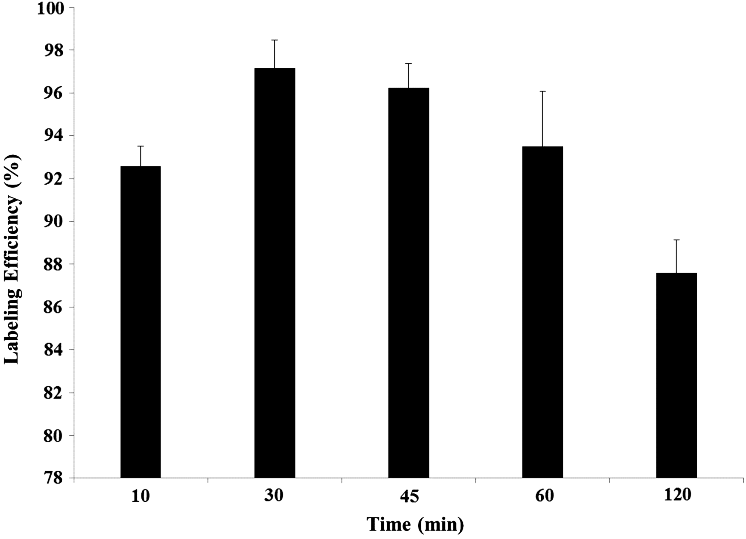
Effect of reaction time on labeling efficiency of 99mTc-finasteride (N = 3 per experiment, p < 0.05).
Quality control
In vitro plasma protein binding of 99mTc-finasteride observed was 83.89%. Furthermore, the blood kinetic profile results of the radio complex (Fig. 3) exhibited a two compartmental pharmacokinetic model of 99mTc-finasteride. An abrupt fall in radioactivity was observed after 15 seconds of administration of the radio complex followed by a slow clearance phase after 30 minutes postadministration.
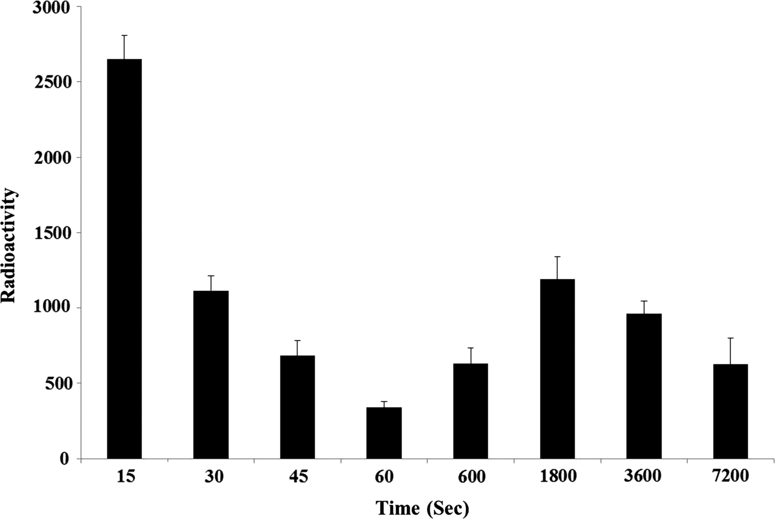
Blood pharmacokinetic profile of 99mTc-finasteride following i.v. injection to rats (N = 3). Higher uptake in blood was observed after 15 and 1800 seconds after i.v. injection. The statistical significance was considered at the level of p < 0.05 following ANOVA test. i.v., intravenous; ANOVA, analysis of variance.
Serum PAcP
The serum PAcP activity level was found to be significantly increased in MNU+T-treated rats compared with that of control rats and the results from the analysis are shown in Figure 4.

Serum prostatic acid phosphatase activity level of MNU+T-treated rats and control rats (N = 10). a p < 0.001. MNU, methyl-N-nitrosourea; T, testosterone propionate.
Protein levels of PSA
There was a significant increase in PSA levels in prostate of MNU+T-treated rats when compared to control rats. The results from the analysis are shown in Figure 5
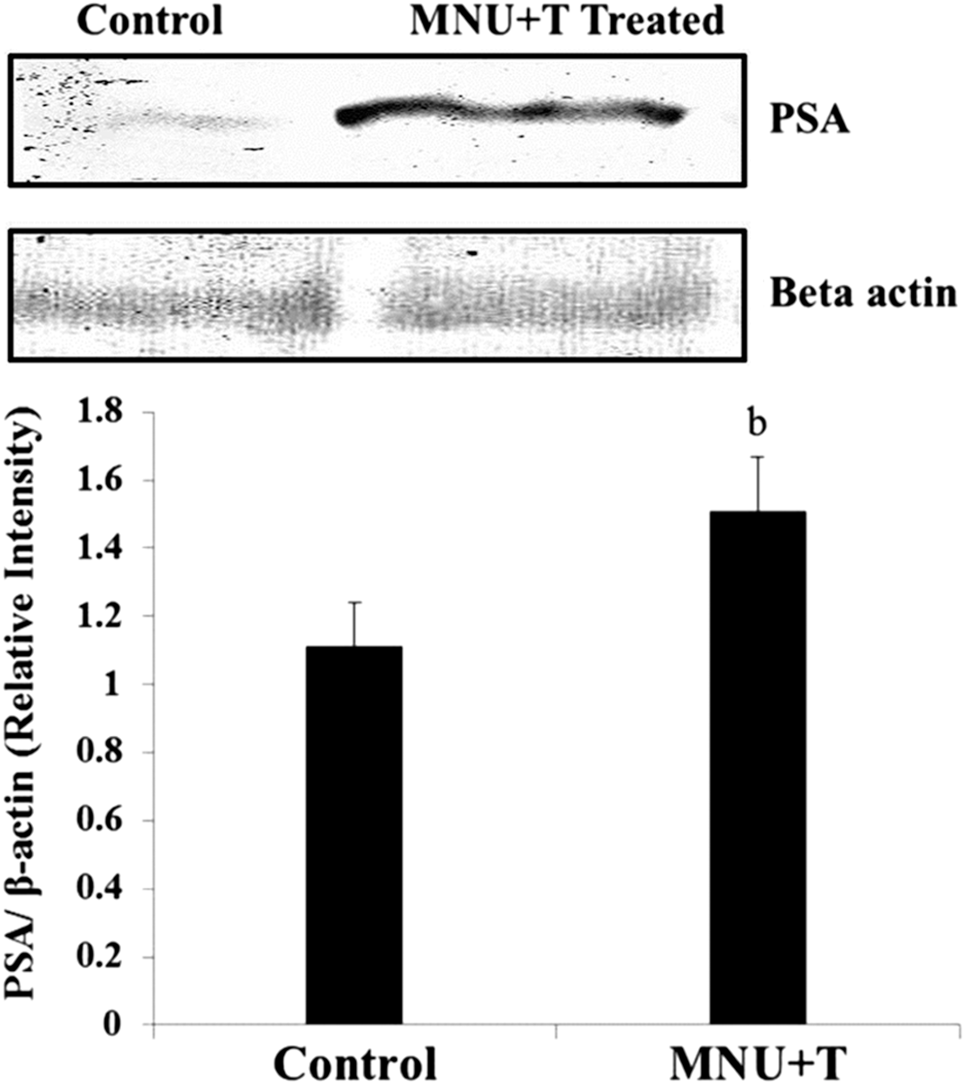
Western blot and densitometric analysis of PSA in prostate of control and MNU+T-treated rats. Each value is expressed as mean ± SD of three independent observations. b p = 0.028 (N = 3) following student's t-test when relative intensity of PSA with that of β-actin in prostate of MNU+T-treated rats is significantly different compared with control rats. PSA, prostate-specific antigen; MNU, methyl-N-nitrosourea; T, testosterone propionate; SD, standard deviation.
Histological evaluation of prostate cancer
A preneoplastic change, characterized by an increase in the number of epithelial cells called prostatic intraepithelial neoplasia (PIN), was observed in rats treated with MNU and T when compared with control. Furthermore, cellular pleomorphism in adenocarcinoma was also observed in treated rats as shown in Figure 6.
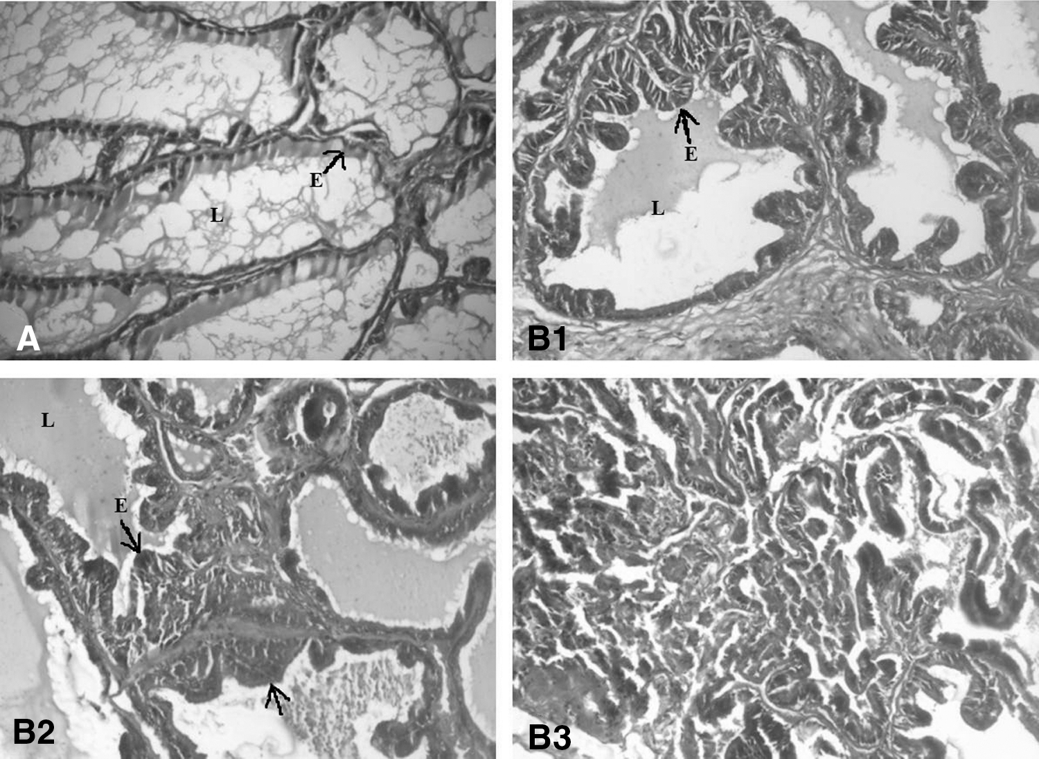
Histology of prostate 20 × magnification treated with N- MNU+T.
Tumor incidence
PIN was found in 7 (70%) of 10 animals of the prostate of MNU+T-treated rats. Further hyperplasia and dysplasia of about 70% and 60%, respectively, were observed in rats treated with MNU+T. The mean survival of rats treated with MNU+T was limited to 20 weeks from the start of treatment.
The percent-specific uptake values expressed as % ID/g ± SD of in vivo biodistribution of 99mTc-finasteride in normal and rats treated with MNU+T at different time intervals are shown in Tables 1 and 2, respectively. From the biodistribution study, the highest percent-specific uptake value was observed in the liver followed by the spleen, kidney, and lung. Furthermore, the percent-specific uptake of 99mTc-finasteride observed in prostate increased significantly from 1 to 6 hours with time in MNU+T-treated rats. The most significant finding of the study was an increase in uptake in prostate of treated rats after 2 and 4 hours postadministration when compared with uptake in prostate of normal rats as shown in Figure 7.
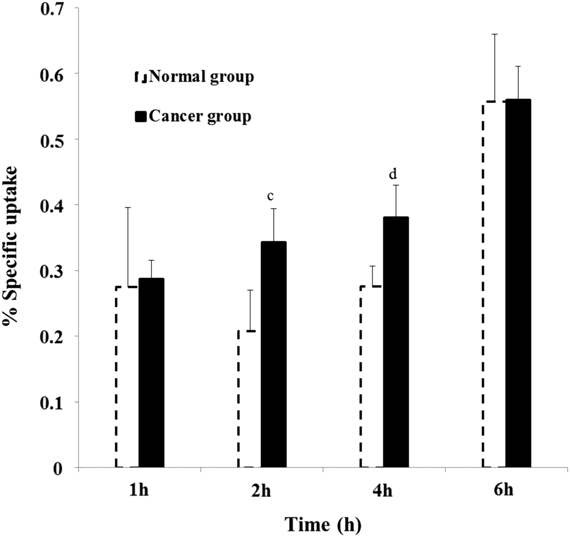
Comparative percent-specific uptake of 99mTc-finasteride in prostate of normal control group and in rats treated with MNU+T. c p = 0.043 (N = 3), d p = 0.035 (N = 3) following student's t-test when percent-specific uptake in prostate of MNU+T treated rats is significantly different compared with control rats. MNU, methyl-N-nitrosourea; T, testosterone propionate.
Each value represents the mean ± SD (n = 3) of injected dose per gram of tissue (ID/g ± SD) of 99mTc-finasteride in normal control rats. The statistical significance was considered at the level of p < 0.05 following ANOVA test.
SD, standard deviation; ID, injected dose; ANOVA, analysis of variance.
Each value represents the mean ± SD (n = 3) of injected dose per gram of tissue (ID/g ± SD) of 99mTc-finasteride in rats treated with MNU+T. The statistical significance was considered at the level of p < 0.05 following ANOVA test.
SD, standard deviation; ID, injected dose; MNU, methyl-N-nitrosourea; T, testosterone propionate; ANOVA, analysis of variance.
Discussion
This study radiolabeled finasteride with 99mTc with >90% labeling efficiency. Optimization of reaction constituents was carried out to obtain maximum percent radiolabeling yield. The radio complex was found to be stable in serum under physiological conditions for a period of 6 hours. The plasma protein binding of the radio complex had a direct effect on its uptake and rate of clearance in vivo. 29,30 The protein binding of 99mTc-finasteride in vitro was found to be 83.89%. The higher protein binding of 99mTc-finasteride depicts its slow clearance from the reticuloendothelial and urinary system in vivo. Furthermore, the in vivo blood pharmacokinetics of the radio complex showed a biphasic clearance of the radio complex. A fast clearance phase was observed after 15 seconds of administration of the radio complex followed by a slow clearance phase after 30 minutes postadministration. The first peak at 15 seconds indicates fast clearance from the blood suggesting that the radio complex is quickly taken up by different organs or is being eliminated from the body at a very fast rate. The slow clearance of tracer observed in the second phase of blood kinetics is suggestive of the slow release of the tracer from different organs into the systemic pool.
The study revealed a similar pattern of biodistribution of 99mTc-finasteride in the normal control group and in the MNU+T-treated group (Fig. 8). The maximum percentage activity per gram of tissue after 1 hour postinjection was observed in the liver followed by the spleen and kidneys. The activity in the liver was not reduced even after 4 hours. Finasteride gets extensively metabolized in the liver to inactive metabolites, which are eliminated through the bile and urine with elimination half-life (t1/2) of 4.7–7.1 hours, 31 resulting in an increased percent-specific uptake of radio complex in the liver. Kidneys also showed retention of the radio complex even after 4 hours, indicating the excretion of activity through the renal route. Furthermore, the delayed clearance of the radio complex can be well corroborated from this finding of high protein binding of the 99mTc-finasteride.
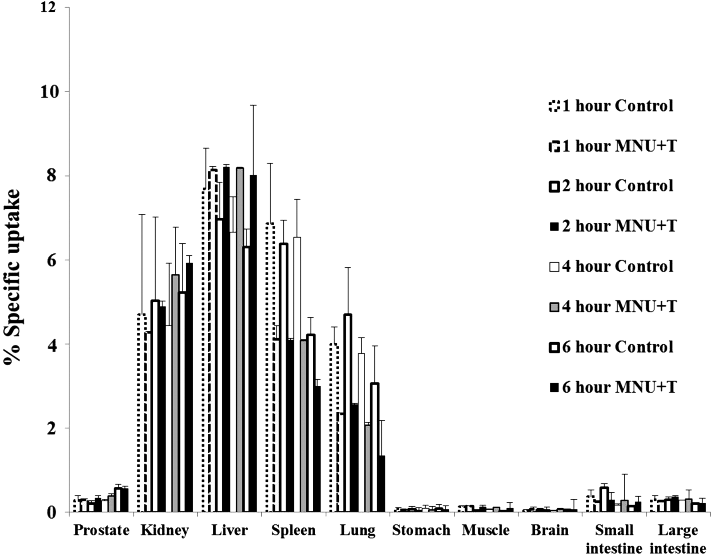
Bars and error bars represent mean ± SD values for percent-specific activity in various organs as a function of time (1–6 hours) after i.v. administration of 99mTc-finasteride in control and MNU+T-treated rats. Statistical significance was considered at the level of p ≤ 0.05 following ANOVA test. SD, standard deviation; i.v., intravenous; MNU, methyl-N-nitrosourea; T, testosterone propionate.
The percent-specific uptake of 99mTc-finasteride did not show significant change up to 4 hours in the prostate of control rats. However, the percent-specific uptake of 99mTc-finasteride increased significantly as a function of time and reached maximum at 6 hours in MNU+T-treated rats. The important finding of the study was a significantly higher uptake of 99mTc-finasteride in prostate of MNU+T-treated rats after 2 and 4 hours of administration, when compared with uptake in normal control group rats at the same time interval.
For biodistribution studies, the animals subjected to MNU+T treatment were analyzed histopathologically as well as for PSA levels. Histological evaluation in the treated group confirmed neoplastic changes with an increased cellular activity (hyperplasia) and PIN. Furthermore, prostate tissue PSA levels were significantly increased in MNU+T-treated rats compared to control rats, since there exists direct correlation between the increased 5α-reductase isozyme II activity and the development and progression of prostate cancer. 32 Therefore, the increased percent uptake of 99mTc-finasteride in prostate of MNU+T-treated rats can be due to the rise in level of the isozyme 5α-reductase type II.
Conclusions
Finasteride is successfully labeled with 99mTc with proven in vitro and in vivo stability. Furthermore, the biodistribution pattern suggests that 99mTc-finasteride possesses better selectivity toward the cancerous prostate tissue when compared to normal prostate tissue in rats. Clinically, the developed radio complex may prove to be an economically viable candidate with specificity for prostate cancer detection.
Footnotes
Acknowledgment
This work was supported and funded by Council of Scientific and Industrial Research (CSIR), India.
Disclosure Statement
There are no existing financial conflicts.