Improving the in vivo pharmacokinetics (PK) of positron emission tomography (PET) radiotracers is of critical importance to tumor diagnosis and therapy. In the case of peptide-based radiotracers, the modification and addition of a linker or spacer functional group often offer faster in vivo pharmacokinetic behavior. In this study, the authors introduced two new PEGlyated dimeric c(RGD-ACH-K) conjugates, in which an aminocyclohexane carboxylic acid (ACH) is inserted into the ring chain of the cyclic RGD peptides, with a common bifunctional chelator (DOTA or NOTA) used for labeling with radiometals (including 68Ga and 64Cu). The addition of polyethylene glycol (PEG) and dimerization of c(RGD-ACH-K) affected the PK of the renal system and the tumor-targeting ability, relative to unmodified molecule. As a result, both 64Cu-DOTA-E[c(RGD-ACH-K)]2 (complex 1) and 64Cu-NOTA-E[c(RGD-ACH-K)]2 (complex 2) exhibited specific tumor-targeting properties relative to tumor-blocking control group, most likely resulting from improved in vivo tumor imaging. The in vivo tumor-to-blood ratio of the 64Cu(NOTA) complex shows better PET imaging than that of the 64Cu(DOTA) complex, which should lead to improved dosimetry and increased suitability for noninvasive monitoring of tumor growth or tumor-targeted radionuclide therapy.
Introduction
Over the last two decades, many radiolabeled cyclic RGD peptides (c(RGD)) have been evaluated as potential radiotracers for imaging and targeted therapy of integrin ανβ3-positive tumors, using either single-photon emission computed tomography or positron emission tomography (PET).1,2 Several 18F- and 68Ga-labeled RGD peptide tracers, such as [18F]Galacto-RGD, [18F]Fluciclatide, [18F]RGD-K5, [18F]FPPRGD2, [18F]Alfatide, [68Ga]NOTA-RGD, and [68Ga]NOTA-PRGD2, have been used in clinical investigation.3 Both monomeric and multimeric c(RGD) peptides have been modified to improve the overall tumor accumulation and retention. Dimeric and tetrameric c(RGD) peptides have higher ανβ3-binding affinities and better tumor-targeting capabilities than monomeric analogs.4 Bivalency and enhanced local c(RGD) concentrations contribute to the ανβ3-binding affinity of multimeric c(RGD)s.
The bivalency, the distance between two c(RGD) motifs in a multimeric c(RGD), must be sufficiently long to achieve simultaneous integrin ανβ3 binding.5 As expected, local c(RGD) concentrations increased as the number of c(RGD) moieties increased. However, these strategies inadvertently resulted in increased uptake of the multimeric c(RGD) peptides in nontarget organs with increasing peptide multiplicity.6
To improve the pharmacokinetics (PK) of peptide radiopharmaceuticals, several studies have demonstrated that modifying RGD peptides using polyethylene glycol (PEG) or galactose linkers is faster their PK and PD properties.3,7 For instance, galacto sugar amino acid (SAA) and PEG linkers have been used to minimize the accumulation of radioactivity in the liver and increase the target-to-nontarget ratios of radioisotope-labeled c(RGD).8–12 The linker or spacer function is generally used to increase the hydrophilicity of the imaging probe and thus decrease nontarget organ accumulation. In addition, linkers have been proposed to increase the distance between two c(RGD) moieties, resulting in effective bivalency.
Based on this finding, the authors developed a new PEGylated dimeric c(RGD) peptide, E[c(RGD-ACH-K)]2, by derivatizing c(RGD-ACH-K) with the hydrophilic linker PEG to improve the tumor imaging capability and to effectively reduce nonspecific organ uptake. The derivative was conjugated to DOTA or NOTA for 64Cu (t1/2 = 12.7 hours; β+ 19%, EC 41%)13 labeling (Fig. 1) before use as integrin ανβ3-targeted PET radiotracers. In this study, the authors used cyclic RGD peptides incorporating aminocyclohexane carboxylic acid (ACH), c(RGD-ACH-K), as the tumor-targeting peptides. This work builds on the previous studies of the design and synthesis of a new series of RGD peptide-based radiopharmaceuticals.14–16
Chemical structure of 64Cu-DOTA-PEG[c(RGD-ACH-K)]2 (complex 1), 64Cu-NOTA-PEG[c(RGD-ACH-K)]2 (complex 2), and 64Cu-DOTA-c(RGD-ACH-K). Scheme of complexes 1 and 2 is shown in the Supplementary Data.
Materials and Methods
64Cu-DOTA-PEG[c(RGD-ACH-K)]2 (complex 1) and 64Cu-NOTA-PEG[c(RGD-ACH-K)]2 (complex 2)
The syntheses of DOTA-PEG-[c(RGD-ACH-K)]2 (7) and NOTA-PEG-[c(RGD-ACH-K)]2 (10) are described in Schemes S1 and S2 in the Supporting Information. 64CuCl2 (∼5.0 mCi in 10 mL) was completely dried in a vial by purging with nitrogen gas and heating to over 100°C before being adjusted to pH 5–6 with NaOAc buffer solution (500 μL, 1 M) and adding compound 7 or 10 (100 μg). The reaction mixture was heated at 50°C for 30 minutes. After labeling with 64Cu, the product (complex 1 or complex 2) was used without further purification. The radiochemical purity was analyzed by a radio-TLC scanner (AR-2000, Eckert & Ziegler co.) with instant thin-layer chromatography (ITLC) using a mobile phase of citrate buffer (pH 5.0, 0.1 M).
Partition coefficient determination
The octanol/water partition coefficients of complexes 1 and 2 were determined according to the following protocol. Solutions of complexes 1 and 2 (0.1 μCi) in PBS (3.0 mL, pH 7.4) were added to neat octanol (4.0 mL) and then stirred vigorously for 5 minutes and centrifuged (3000 rpm) for an additional 5 minutes. The radioactivities of both the PBS and octanol phases were measured with a gamma counter, and the log p-values were calculated (n = 3).
In vitro serum stability
Complexes 1 and 2 (500 μCi/50 μL) were incubated at 37°C into each solution (1 mL) of human serum, mouse serum, and PBS buffer (pH 7.0) for different time intervals (30 minutes, 1 hour, 3 hours, 24 hours, and 48 hours). After incubation, the sample solution (5 μL) of each mixture was directly spotted on ITLC paper, which was eluented by a mobile phase of citrate buffer solution (pH 3.0, 0.1 M) into a sealed bottle. Each radiochemical stability value of above samples was analyzed by Radio-TLC scanner (AR-2000; Eckert & Ziegler co.).
Cell culture and tumor xenograft model
A human glioma cell line, U87MG, was obtained from the ATCC (American Type Culture Collection) and was grown in Dulbecco's modified Eagle's medium (DMEM) supplemented with 10% fetal bovine serum and 1% penicillin–streptomycin in 5% CO2 at 37°C.
Animal procedures were performed according to a protocol approved by the animal research committee of the Korea Institute of Radiological and Medical Sciences (KIRAMS). Female BALB/c nude mice (SLC, Hamamatsu, Japan) at 4–6 weeks of age were injected subcutaneously in the left arm with 5 × 10⁶ U87MG cells suspended in 100 μL DMEM. The mice were subsequently used for biodistribution and PET studies when the tumor volumes reached 0.7–0.9 cm.
Biodistribution
Mice were injected with complex 1 or 2 (10 μCi/100 μL). Mice (n = 4 per time point) were sacrificed by exsanguination at different time points (30 minutes, 1 hour, 4 hours, and 18 hours) postinjection (p.i.). Organs of interest (such as blood, muscle, heart, lung, liver, spleen, stomach, intestine, kidney, bone, brain, and tumor) were harvested and measured for radioactivity using a 1480 WIZARD (WALLAC) gamma counter. The organ uptake was calculated and expressed as a percentage of the injected dose per gram (% ID/g).
In vivo PET imaging
Whole-body PET images of the mice were obtained using a dedicated small animal PET scanner (INVEON, Siemens Medical Solutions). The U87MG glioma-bearing mice (n = 3) were anesthetized with 1.5% isoflurane, and complex 1 or 2 (200 μCi/100 μL) was injected through a tail vein. Thirty-minute static scans were acquired at 30 minutes, 1 hour, 3 hours, and 18 hours after injection. PET scans were reconstructed to images using a Fourier rebinning and 2-dimensional ordered-subset expectation maximization algorithm with no correction of attenuation or scatter. On the plane of the reconstructed images showing the tumor region, circular 3D regions of interest were drawn around the tumor area, using analysis software (INVEON Research Workplace, Siemens). The pixel values of the reconstructed images were converted to % ID/g values by means of a previously obtained cross-calibration factor. For the blocking experiment, a mouse bearing a U87MG tumor was injected with complex 1 or 2 (200 μCi/100 μL) along with c(RGDyK) (10 mg/kg). Thirty-minute static PET images were then acquired at 1 hour p.i.
Statistical analysis
All data are represented as mean ± standard deviation. Statistical analysis was performed by one-way analysis of variance with Tukey's post hoc test using GraphPad Prism 5 (GraphPad Software, Inc., La Jolla, CA). p-Values of less than 0.05 were regarded as statistically significant.
Results and Discussion
A variety of radiolabeled RGD peptides as a PET tracer have been developed and showed an attractive diagnostic and therapeutic approach. Good results have already been achieved and especially introduce multimerization of peptides to elevate tumor specificity and insertion of PEG or SAA for faster PK.3 Although the multimeric RGD peptides showed incremental enhancement on the receptor specificity, the relatively high accumulation in nontarget organs could be an unwanted consequence for clinical study. In the case of 64Cu, moreover, demetalation of Cu ions from chelators under physiological conditions results in high uptake of liver, therefore choosing the chelator is very important as well as selection of linkers.
This study describes the synthesis of PEGylated dimeric c(RGD-ACH-K) peptides and evaluates a comparison of two chelators, DOTA and NOTA, in in vivo PET imaging. Product (compounds 1–10) formation was confirmed by MALDI-mass spectroscopy and the chemical purity was confirmed by HPLC. The radiochemical yields and purities of 64Cu-DOTA-PEG-[c(RGD-ACH-K)]2 (complex 1) and 64Cu-NOTA-PEG-[c(RGD-ACH-K)]2 (complex 2) were 99.2% ± 0.2% (n = 10) and 99.4% ± 0.3% (n = 10), as confirmed by radio-TLC scanning (free 64Cu: Rf = 0.3–0.5, complex 1 or 2: Rf = 0.7–1.0), respectively. The specific activities of complexes 1 and 2 after labeling (n = 10) were 121.0 ± 10.9 and 125.4 ± 7.7 mCi/μmol, respectively.
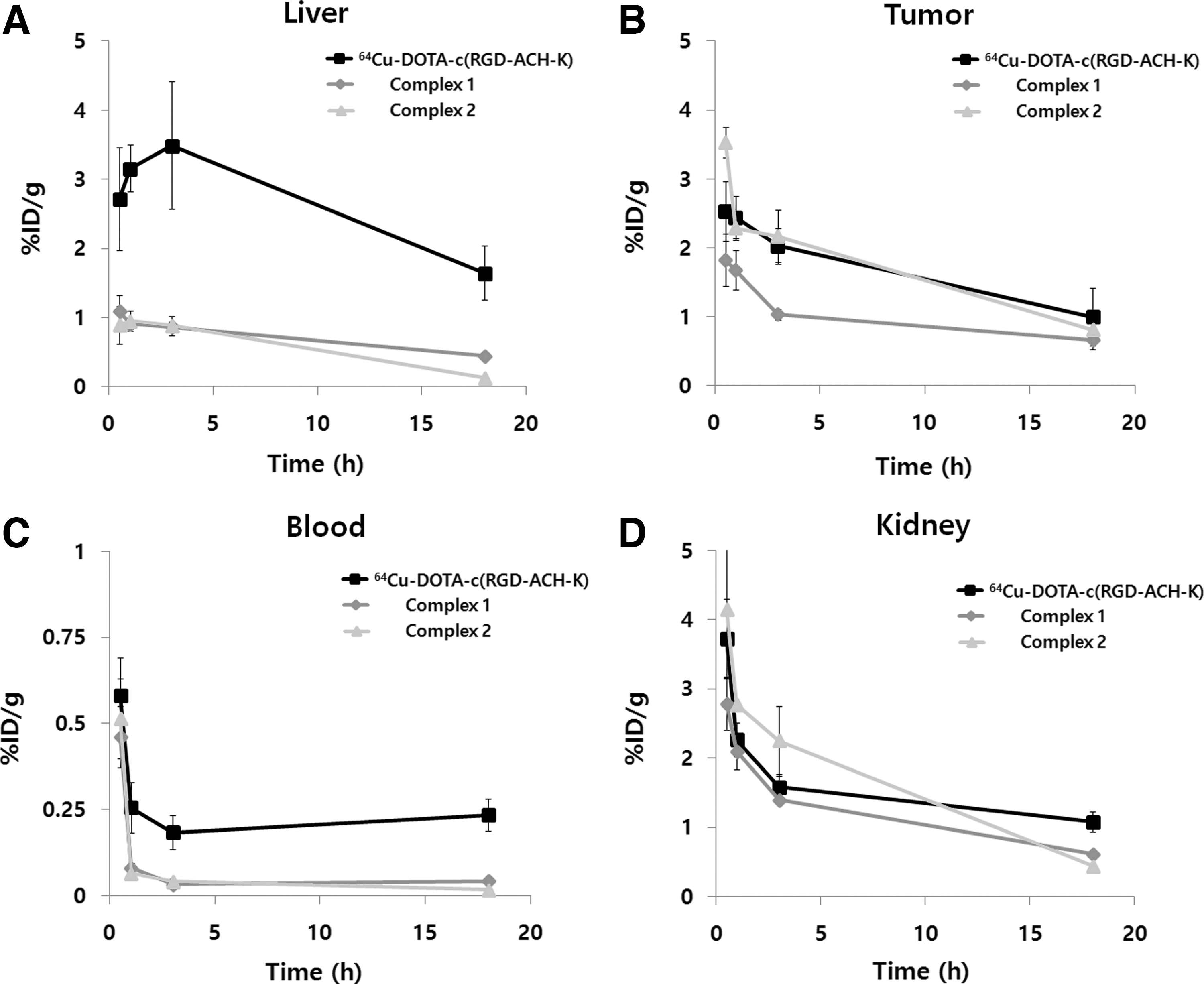
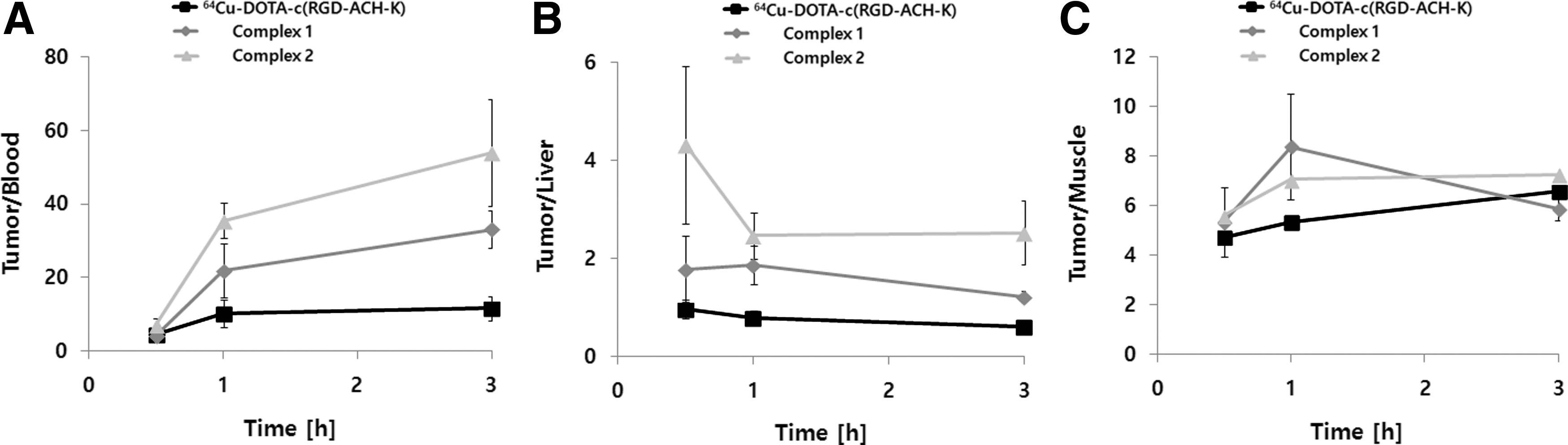
The hydrophilicity of complexes 1 and 2 was determined using an octanol–water partition assay. The recorded logP values for complexes 1 and 2 were −1.13 ± 0.04 and −1.27 ± 0.01, respectively. Although both compounds are more lipophilic than 64Cu-DOTA-c(RGD-ACH-K) (logP = −4.41 ± 0.05),15 the liver uptake of complex 1 or 2 (0.86 ± 0.07 or 0.88% ± 0.13% ID/g at 4 hours p.i., respectively) was significantly lower compared with that of 64Cu-DOTA-c(RGD-ACH-K) (3.49% ± 0.92% ID/g at 4 hours p.i.). The biodistribution data are summarized in Tables 1 and 2 and Fig. 2. Low liver uptake of these complexes may also be a consequence of the compound's stability. As shown in Supplementary Table S1, both PEGylated dimeric RGD conjugates showed good stability in plasma for up to 48 hours (radiochemical purity >99%), whereas 64Cu-DOTA-c(RGD-ACH-K) showed some radiochemical degradation (radiochemical purity >97%).15 The liver uptake of 64Cu-DOTA-c(RGD-ACH-K) was relatively high (0.5 hour, p < 0.01; from 1 hour to 18 hours, p < 0.001) compared with both complexes 1 and 2, which was most likely caused by its instability in a complex in vivo environment (Fig. 2A). The in vivo behavior of 64Cu like transmetalation was chemically influenced by the type of used chelator, unlike 111In and 68Ga. Roosenburg et al. reported a comparative study with 68Ga-, 111In-, and 64Cu-labeled PP-F11 peptides conjugated with the chelators, DOTA, NOTA, and NODAGA, and found that the chelator did not change the in vivo behavior of 68Ga- and 111In-labeled PP-F11, but that the 64Cu-labeled peptides showed different in vivo behaviors depending on the chelators.17 Several articles have addressed the relationship between 64Cu and chelators. For example, Hoffman et al. reported that 64Cu-labeled bombesin analogs conjugated with NOTA showed more ideal in vivo PK than those conjugated with DOTA and TETA chelators.18 Similarly, Prasanphanich et al. showed that 64Cu-NOTA-8-Aoc-BBN (7–14)NH2 has lower liver uptake than 64Cu-DOTA-8-Aoc-BBN(7–14)NH2.19 Most recently, Dumont et al. reported that replacing DOTA with either CB-TE2A or NODAGA in 64Cu-labeled RGD peptides improved their biodistribution profiles.20 In this study, however, the liver uptakes of complexes 1 and 2 were not statistically significant (p > 0.05, from 0.5 to 18 hours), as shown in Figure 2A. This finding indicates that the in vivo behavior of the 64Cu-peptide conjugates may not be dependent on the chelator when PEG is attached to the peptides. It is well known that PEG addition influences the PK of PEGylated molecules and their fast blood clearance.7,21 This is in line with the results for complexes 1 and 2. The PEGylated dimeric RGD compounds showed surprisingly low liver uptake (<1% ID/g until 4 hours p.i.) compared with that of 64Cu(DOTA-3PEG4 dimer) (2–3% ID/g until 4 hours p.i.).22 Overall, they showed more rapid blood clearance than 64Cu-DOTA-c(RGD-ACH-K) (Fig. 2B). The kidney uptake of complex 1 was lower compared with both complex 2 and 64Cu-DOTA-c(RGD-ACH-K). Although complex 2 showed slightly higher kidney uptake than complex 1 and 64Cu-DOTA-c(RGD-ACH-K) at 4 hours p.i., the kidney uptake of complex 2 was the lowest at 18 hours p.i. (Fig. 2C). The highest tumor uptake was observed at 0.5 hour p.i. and some washout was noted (Fig. 2D). Because of its fast blood clearance, tumor accumulation of complex 1 was lower compared with 64Cu-DOTA-c(RGD-ACH-K) (1 hour and 3 hours, p < 0.01), although complex 1 has dimeric c(RGD-ACH-K). However, the tumor/blood ratio of complex 1 was higher compared with 64Cu-DOTA-c(RGD-ACH-K), although the uptake of complex 1 in tumors was less than half observed for 64Cu-DOTA-c(RGD-ACH-K), as shown in Figure 3A. Interestingly, the tumor uptake of complex 2 was two times higher compared with complex 1 (0.5 hour and 3 hours, p < 0.001) (Fig. 2D). As a result, its tumor/blood ratio increased from 7.1 ± 1.7 at 30 minutes p.i. to 53 ± 14.4 at 3 hours p.i. and remained at this level until 18 hours p.i (Fig. 3A). The tumor/liver ratio of complex 2 was larger compared with complex 1 and 64Cu-DOTA-c(RGD-ACH-K) (Fig. 3B) and the tumor/muscle ratio of complex 2 was significantly higher compared with 64Cu-DOTA-c(RGD-ACH-K) (Fig. 3C). Recently, an elegant study compared the effect of several bifunctional chelators for 64Cu-labeled peptides and concluded that NOTA was more stable than DOTA, especially in an acidic environment, such as that found in a tumor.17–19,23 In summary, the data suggest that complex 2 might be useful for imaging integrin ανβ3-positive tumors. To the best of the authors' knowledge, this is the first comparative experimental study on conjugation of PEGylated dimeric c(RGD-ACH-K) peptides with the bifunctional chelators, DOTA and NOTA.
Comparison of biodistribution data for complex 1, complex 2, and 64Cu-DOTA-c(RGD-ACH-K) in the liver (A), blood (B), kidney (C), and tumor (D). Error bars denote standard deviations (SDs) (n = 4).
Comparison of biodistribution of the tumor/blood ratio (A) and tumor/liver ratio (B) and tumor/muscle ratio (C) of complex 1, complex 2, and 64Cu-DOTA-c(RGD-ACH-K). Error bars denote SDs (n = 4).
Biodistribution Results of64Cu-DOTA-PEG[c(RGD-ACH-K)]2(Complex 1) in U87MG Tumor-Bearing Nude Mice at 30 Minutes, 1 Hour, 3 Hours, and 18 Hours Postinjection
Organ
30 minutes
1 hour
3 hours
18 hours
Blood
0.46 ± 0.09
0.08 ± 0.01
0.03 ± 0.01
0.04 ± 0.01
Muscle
0.65 ± 0.64
0.20 ± 0.03
0.17 ± 0.01
0.06 ± 0.03
Heart
0.43 ± 0.10
0.28 ± 0.04
0.16 ± 0.02
0.13 ± 0.01
Lung
1.33 ± 0.26
0.89 ± 0.08
0.44 ± 0.06
0.37 ± 0.04
Liver
1.09 ± 0.24
0.91 ± 0.07
0.86 ± 0.07
0.44 ± 0.04
Spleen
0.70 ± 0.09
0.79 ± 0.11
0.52 ± 0.05
0.45 ± 0.15
Stomach
1.31 ± 0.23
0.84 ± 0.32
0.59 ± 0.02
0.24 ± 0.04
Intestine
0.98 ± 0.20
0.77 ± 0.03
0.89 ± 0.19
0.28 ± 0.03
Kidney
2.78 ± 0.37
2.09 ± 0.25
1.39 ± 0.05
0.61 ± 0.03
Bone
0.59 ± 0.34
0.27 ± 0.07
0.30 ± 0.09
0.35 ± 0.22
Tumor
1.82 ± 0.37
1.67 ± 0.28
1.03 ± 0.08
0.66 ± 0.14
Tumor/blood
4.17 ± 1.48
21.84 ± 7.36
33.11 ± 5.18
16.74 ± 5.29
Tumor/liver
1.78 ± 0.68
1.85 ± 0.39
1.21 ± 0.11
1.52 ± 0.38
The data are expressed as % ID/g [mean ± SD] (n = 4).
% ID/g, percent of injected dose per gram; SD, standard deviation.
Biodistribution Results of64Cu-NOTA-PEG[c(RGD-ACH-K)]2(Complex 2) in U87MG Tumor-Bearing Nude Mice at 30 Minutes, 1 Hour, 3 Hours, and 18 Hours Postinjection
Organ
30 minutes
1 hour
3 hours
18 hours
Blood
0.51 ± 0.11
0.06 ± 0.01
0.04 ± 0.01
0.01 ± 0.01
Muscle
0.64 ± 0.09
0.33 ± 0.06
0.31 ± 0.06
0.18 ± 0.07
Heart
0.56 ± 0.13
0.27 ± 0.02
0.20 ± 0.06
0.10 ± 0.02
Lung
1.57 ± 0.39
0.95 ± 0.13
0.53 ± 0.12
0.21 ± 0.03
Liver
0.89 ± 0.26
0.95 ± 0.14
0.88 ± 0.13
0.13 ± 0.01
Spleen
1.14 ± 0.04
0.95 ± 0.10
0.84 ± 0.19
0.27 ± 0.03
Stomach
1.84 ± 0.46
0.76 ± 0.18
0.79 ± 0.23
0.09 ± 0.03
Intestine
1.25 ± 0.23
0.89 ± 0.09
1.04 ± 0.18
0.17 ± 0.02
Kidney
4.15 ± 0.97
2.77 ± 0.01
2.24 ± 0.50
0.44 ± 0.04
Bone
1.26 ± 0.26
1.17 ± 0.23
0.81 ± 0.21
0.93 ± 0.54
Tumor
3.52 ± 0.21
2.28 ± 0.17
2.17 ± 0.38
0.80 ± 0.09
Tumor/blood
7.13 ± 1.76
35.52 ± 4.85
53.93 ± 14.44
51.94 ± 12.07
Tumor/liver
4.30 ± 1.60
2.45 ± 0.47
2.52 ± 0.65
6.13 ± 0.94
The data are expressed as %ID/g [mean ± SD] (n = 4).
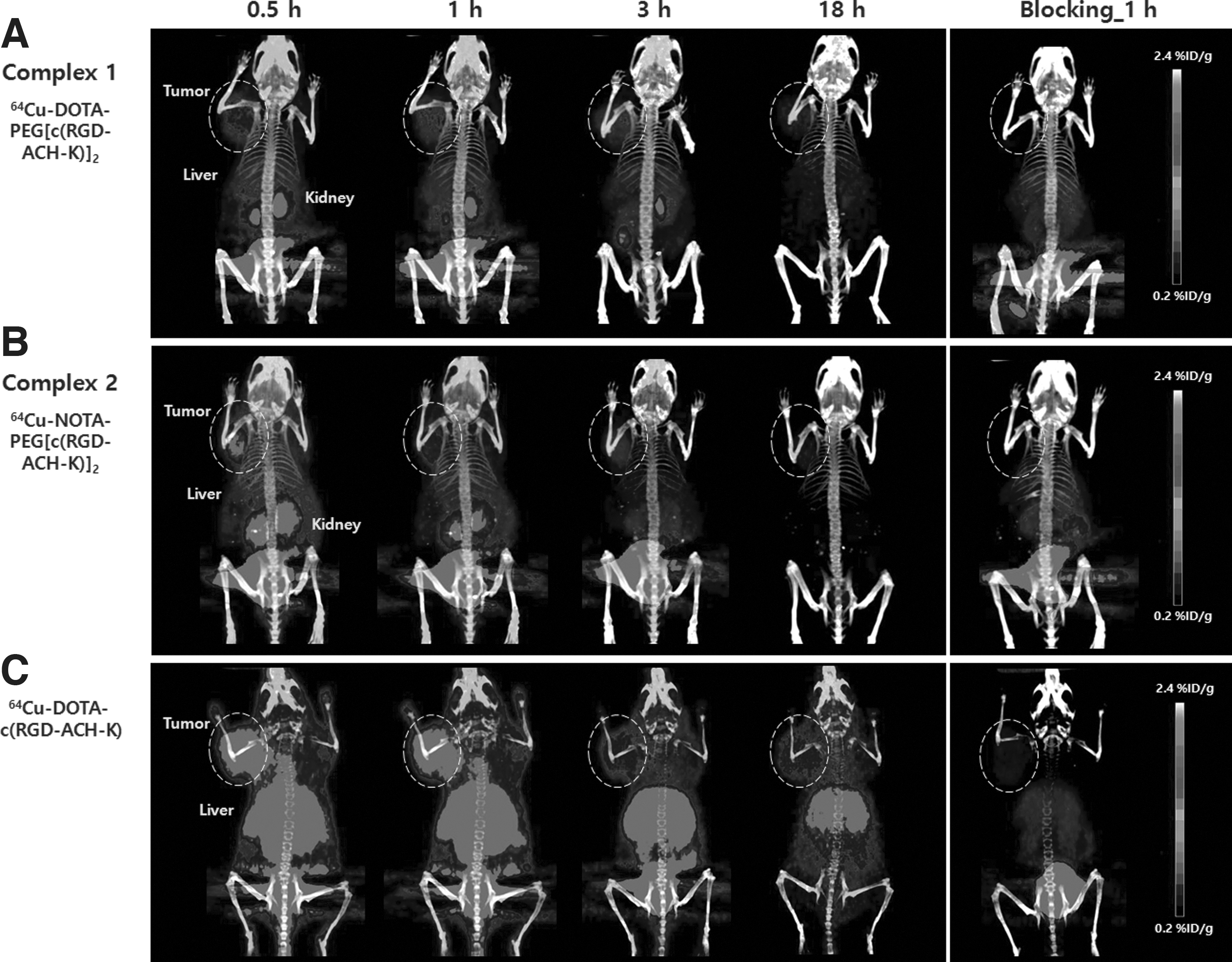
The in vivo quantitative results obtained from biodistribution studies and animal PET scans were in good agreement, confirming that the quantitative analysis of noninvasive PET images accurately reflected the distribution of PET tracers in vivo. With all three complexes employed, the tumor regions of U87MG-bearing mice are clearly visualized in the PET images (Fig. 4). As expected, complexes 1 and 2 showed good tumor-to-background images and low liver uptake. 64Cu-DOTA-c(RGD-ACH-K) not only exhibited the highest tumor uptake with % ID/g values at 1 hour p.i. as follows: 1.66 ± 0.52 (64Cu-DOTA-c(RGD-ACH-K)), 1.18 ± 0.21 (complex 2), and 0.86 ± 0.10 (complex 1), but 64Cu-DOTA-c(RGD-ACH-K) also showed the highest liver uptake with % ID/g values at 1 hour p.i. as follows: 2.66 ± 0.62 (64Cu-DOTA-c(RGD-ACH-K)), 0.69 ± 0.12 (complex 2), and 0.64 ± 0.02 (complex 1). Effective blocking of ανβ3 integrin receptors with an excessive amount of c(RGDyK) demonstrated the tumor specificity of PET tracers in U87MG xenograft tumors (Fig. 4). Uptake in tumors was reduced dramatically in the blocking study; the values at 1 hour p.i. were as follows: 0.04 ± 0.01 (complex 1), 0.01 ± 0.01(complex 2), and 0.05 ± 0.01 (64Cu-DOTA-c(RGD-ACH-K).
Animal positron emission tomography/CT images (maximum intensity projection, MIP) of U87MG tumor-bearing nude mice injection with complex 1 (A), complex 2 (B), and 64Cu-DOTA-c(RGD-ACH-K) (C). Decay-corrected whole-body coronal images were acquired at 0.5 hour, 1 hour, 3 hours, 18 hours, and 1 hour coinjection with excess amount of c(RGDyK) after intravenous injection with ∼200 μCi of each radiotracer (n = 3). Circles indicate tumor location, and radioactivity is presented as percent of injected dose per gram (% ID/g).
In conclusion, the authors designed and synthesized two PEGlyated dimeric RGD peptides, complexes 1 and 2, for use as PET radiopharmaceuticals. Both complexes 1 and 2 exhibited specific tumor-targeting properties with significantly lower uptake by nontarget organs, most likely resulting from better in vivo kinetics. Complex 2 shows a better tumor-to-nontumor ratio than complex 1, which could lead to lower toxicity, suggesting that complex 2 is more suitable for noninvasive monitoring of tumor growth or targeted radionuclide therapy.
Footnotes
Acknowledgments
This research was supported by the Basic Science Research Program through the National Research Foundation of Korea (NRF) funded by the Ministry of Science, ICT and Future Planning (No. 2011-0030161 and 2011-0030162). FutureChem (Korea) is acknowledged for the synthesis of peptides and mass measurements.
Disclosure Statement
No competing financial interests exist.
References
1.
FaniM, MaeckeHR, OkarviSM. Radiolabeled peptides: Valuable tools for the detection and treatment of cancer. Theranostics, 2012; 2:481.
2.
ZhouY, ChakrabortyS, LiuS. Radiolabeled cyclic RGD peptides as radiotracers for imaging tumors and thrombosis by SPECT. Theranostics, 2011; 1:58.
3.
LiuS, LiuZ, ChenK, et al.18F-labeled galacto and PEGylated RGD dimers for PET imaging of αVβ3 integrin expression. Mol Imaging Biol, 2010; 12:530.
LiuS. Radiolabeled cyclic RGD peptides as integrin alpha(v)beta(3)-targeted radiotracers: Maximizing binding affinity via bivalency. Bioconjugate Chem, 2009; 20:2199.
6.
LiZB, CaiW, CaoQ, et al.64Cu-labeled tetrameric and octameric RGD peptides for small-animal PET of tumor αVβ3 integrin expression. J Nucl Med, 2007; 48:1162.
HaubnerR, WesterHJ, WeverWA, et al.Noninvasive imaging of alpha(v)beta(3) integrin expression using 18F-labeled RGD-containing glycopeptide and positron emission tomography. Cancer Res, 2001; 61:1781.
9.
HaubnerR, KuhnastB, MangC, et al.[18F]Galacto-RGD: Synthesis, radiolabeling, metabolic stability, and Radiation dose estimates. Bioconjugate Chem, 2004; 15:61.
10.
ChenX, ParkR, ShahinianAH, et al.18F-labeled RGD peptide: Initial evaluation for imaging brain tumor angiogenesis. Nucl Med Biol, 2004; 31:179.
11.
HaubnerR, FinsingerD, KesslerH. Stereoisomeric peptide libraries and peptidomimetics for designing selective inhibitors of the αvβ3 integrin for a new cancer therapy. Angew Chem Int Ed Engl, 1997; 36:1374.
12.
ChenX, ParkR, ShahinianAH, et al.Pharmacokinetics and tumor retention of 125I-labeled RGD peptide are improved by PEGylation. Nucl Med Biol, 2004; 31:11.
13.
KimJY, ParkH, LeeJC, et al.A simple Cu-64 production and its application of Cu-64 ATSM. Appl Radiat Isot, 2009; 67:1190.
14.
ParkJA, KimJY, LeeYJ, et al.Gadolinium complex of (125)I/(127)I-RGD-DOTA conjugate as a tumor-targeting SPECT/MR bimodal imaging probe. ACS Med Chem Lett, 2012; 4:216.
15.
ParkJA, LeeYJ, LeeJW, et al.Cyclic RGD peptides incorporating cycloalkances: Synthesis and evaluation as PET radiotracers for tumor imaging. ACS Med Chem Lett, 2014; 5:979.
16.
ParkJA, LeeYJ, KoIO, et al.Improved tumor-targeting MRI contrast agents: Gd(DOTA) conjugates of cycloalkane-based RGD peptides. Biochem Biophys Res Commun, 2014; 455:246.
17.
RoosenburgS, LavermanP, JoostenL, et al.PET and SPECT imaging of a radiolabeled minigastrin analogue conjugated with DOTA, NOTA, and NODAGA and labeled with (64)Cu, (68)Ga, and (111)In. Mol Pharm, 2014; 11:3930.
18.
HoffmanTJ, SmithCJ. Cu-64 targeting vectors based upon bombesin peptide. Nucl Med Biol, 2009; 36:579.
19.
PrasanphanichAF, NandaPK, RoldTL, et al.[64Cu-NOTA-8-Aoc-BBN(7-14)NH2] targeting vector for positron-emission tomography imaging of gastrin-releasing peptide receptor-expressing tissues. Proc Natl Acad Sci U S A, 2007; 104:12462.
20.
DumontRA, DeiningerF, HaubnerR, et al.Novel 64Cu and 68Ga-labeled RGD conjugates show improved PET imaging of αvβ3 integrin expression and facile radiosynthesis. J Nucl Med, 2001; 52:1276.
21.
ChenX, SieversE, HouY, et al.Integrin alpha v beta 3-targeted imaging of lung cancer. Neoplasia, 2005; 7:271.
22.
ShiJ, KimYS, ZhaiS, et al.Improving tumor uptake and pharmacokinetics of 64Cu-labeled cyclic RGD peptide dimers with Gly(3) and PEG(4) linkers. Bioconjugate Chem, 2009; 20:750.
23.
DearlingJL, VossSD, DunningP, et al.Imaging cancer using PET-the effect of the bifunctional chelator on the biodistribution of a (64)Cu-labeled antibody. Nucl Med Biol, 2011; 38:29.
Supplementary Material
Please find the following supplemental material available below.
For Open Access articles published under a Creative Commons License, all supplemental material carries the same license as the article it is associated with.
For non-Open Access articles published, all supplemental material carries a non-exclusive license, and permission requests for re-use of supplemental material or any part of supplemental material shall be sent directly to the copyright owner as specified in the copyright notice associated with the article.