
Abstract
Objective:
To explore the effects of recombinant human endostatin (endostar, ES) and cisplatin on the growth of gastric cancer-transplanted tumor in nude mice and the expression of microvessel density (MVD).
Methods:
Human gastric cancer SGC-7901 cells were subcutaneously injected into the armpit of nude mice to prepare cancer-bearing nude mice. A total of 32 cancer-bearing nude mice were divided into four groups (each group with 8 mice). The four groups included control group and other three groups in which mice were treated with 5 mg/kg of ES (group E), 5 mg/kg of cisplatin (group Ci), and 5 mg/kg of ES combined with 5 mg/kg of cisplatin (group C), respectively. MVD was determined by immunohistochemistry, and the expressions of mRNA and protein of inhibitor of differentiation-1 (ID1) and vascular endothelial growth factor (VEGF) were detected with reverse transcription polymerase chain reaction (RT-PCR) and western blot, respectively. Apoptosis was observed with transmission electron microscope.
Results:
Compared with control group, the sizes and weights of tumors were significantly decreased in other three groups (all p < 0.05). MVD was significantly lower in groups E, Ci, and C than in control group, and in groups E and C than in group Ci (all p < 0.05). Compared with control group, the expressions of mRNA and protein of ID1 and VEGF significantly decreased in groups E and C (all p < 0.05). There were no significant differences in the expressions of mRNA and protein of ID1 and VEGF between group Ci and control group. There was apoptosis in groups E and C, but no apoptosis was found in group Ci and control group.
Conclusion:
ES can inhibit the growth of gastric cancer cells through suppressing angiogenesis and promoting apoptosis of tumor cell. This study provides a new idea for the treatment of gastric cancer.
Introduction
Gastric cancer, a solid tumor, is one of the most common malignant tumors. The death rate of gastric cancer has taken the lead in malignant tumors because it is often in advanced stage once it is discovered or diagnosed. At present, surgery is only one radical treatment for gastric cancer. However, for advanced gastric cancer, the 5-year survival rate after surgery is about 50%. The poor prognosis of advanced gastric cancer is mainly caused by early metastasis of gastric cancer. Recombinant human endostatin (endostar, ES) has depressant effects on angiogenesis, 1 and has widely been used in the treatment of many kinds of malignant tumors. 2 ES has reliable therapeutic effects, slight side-effects, and enhancement effects on some antitumor drugs. 3 Cisplatin, a cytotoxic drug, is widely accepted in the treatment for malignant tumors, especially in postoperative chemotherapy for gastric cancer because it can inhibit gastric cancer cell growth and promote gastric cancer apoptosis. 4 In this study, the effects of ES or cisplatin alone and ES combined with cisplatin on gastric cancer were observed to explore the inhibitory effects of ES on gastric cancer and its mechanism, providing a new approach for treatment of gastric cancer.
Materials and Methods
All study methods were approved by the Ethics Committee of the First Affiliated Hospital, Liaoning Medical University.
Animals and materials
Balb/c nude mice were purchased from the Experimental Animal Center of Dalian Medical University (Dalian, China). The human gastric cancer cell line SGC-7901 was purchased from Xiehe Cell Bank (Beijing, China). RPMI1640 medium and fetal bovine serum (FBS) were purchased from Gibco (Carlsbad, California, China). TRIzol, RT-PCR kit, and Marker were purchased from TaKaRa (Dalian, China). The PV-two-step immunohistochemistry kit was purchased from Zhongshan Jinqiao Company (Beijing, China). Rabbit anti-human inhibitor of differentiation-1 (ID1), vascular endothelial growth factor (VEGF), CD34 monoclonal antibodies, and IgG-HRP and β-actin secondary antibodies were purchased from Santa Cruz (California). Other reagents were analytical pure. ES was from Xianshengmaide Company (Yantai, China). Cisplatin was from Jiutai Pharmaceutical (Jinzhou, China). The sequences of primer ID1 including positive strand 5′-CAAGTGGCCAGAGGCATGCACTT-3′ and negative strand 5′-GATGTAGTC TTTACCATCC TGTTG-3′; VEGF including positive strand 5′-GAAGTGGTGAAGTTCATGGATGTC-3′ and negative strand 5′-CGATCGTTCTGTATCACTCTTTCC-3′ were designed and synthesized by TaKaRa (Dalian, China).
Preparation of cancer-bearing nude mice
Human gastric cancer SGC-7901 cells were incubated in the RPMI 1640 medium containing 10% FBS in an atmosphere of 5% CO2 at 37°C. When cell growth reached 80% of area of the culture bottle, the cells were digested with 0.25% pancreatin at 37°C for 1 minute. After removal of pancreatin, cells were made into cell suspension with a fresh culture medium followed by passage. After passage was performed three times, SGC-7901 cells were subcutaneously injected into the armpit of nude mice.
Grouping
Balb/c cancer-bearing nude mice were randomly divided into four groups (each group with 8 mice). The four groups included control group and other three groups in which mice were treated with 5 mg/kg of ES (group E), 5 mg/kg of cisplatin (group Ci), and 5 mg/kg of ES combined with 5 mg/kg of cisplatin (group C), respectively. 5 ES was subcutaneously given every 2 days and cisplatin was intraperitoneally given every 4 days for 4 weeks. After administration, mice activity, ingestion, and drinking were observed and recorded every day, and the weights of mice were measured every week.
Samples
Five weeks after administration, mice in each group were anesthetized, and then, tumor was taken followed by measurement of volume and weight of tumors.
Immunohistochemistry
Samples were embedded in paraffin and then sectioned. The primary antibody, rabbit anti-human CD34, was diluted to 1:200 with PBS. At the same time, PBS was used as negative control. The secondary antibody, sheep anti-rabbit IgG-HRP, was diluted to 1:200. After visualization with DAB and sealing with gum, sections were observed. The cells with brown-yellow granules (expression of CD34 protein) in the cytoplasm and cell membrane were regarded as positive cells. Microvessel density (MVD) was evaluated with the microvessel counting method of Weidner. 6 In detail, sections were first placed under a microscope with 40-fold magnification to determine the areas of high-density blood vessels, and then, three areas of high-density blood vessels were selected to count the number of microvessels under a microscope with 200-fold magnification. The mean value of microvessels in the three areas was served as MVD.
Gene expressions of ID1 and VEGF determined by RT-PCR
The extraction of total RNA and RT-PCR were performed according to the instructions of the kit. PCR conditions were as follows: predenaturing at 95°C for 10 minutes, denaturing at 94°C for 1 minute, reannealing at 60°C for 1 minute, elongation at 72°C for 2 minutes, 30 cycles; and finally, elongation at 72°C for 8 minutes. PCR products underwent agarose gel electrophoresis. Image scanning was performed followed by analysis of imaging with Lab Works software. The level of gene expression was evaluated by calculating the ratio of average gray-level value of target gene to β-actin.
Protein expressions of ID1 and VEGF determined by western blot
Samples were washed with PBS three times, placed into 400 μL of lysate on ice for 30 minutes, and centrifuged at 14,000 rpm for 5 minutes at 4°C. Protein was extracted from the supernatant, underwent SDS-PAGE, and then was transferred onto nitrocellulose membrane followed by sealing using the TBST buffer containing 5% BSA for 1 hour. After washing the membrane, antibodies of ID1, VEGF, and β-actin were, respectively, added at 4°C overnight. Then, a secondary antibody was added followed by visualization.
Tumor cell apoptosis observed with transmission electron microscope
Samples were fixed with 4% of glutaral, washed with 0.1 mol/L PBS three times for 15 minutes of each time, fixed with osmic acid for 2 hours, and washed with 0.1 mol/L PBS three times for 15 minutes. Samples underwent gradient dehydration of acetone, including 30%, 50%, 70%, 80%, 90%, and 100%, respectively, for 20 minutes. The samples were placed in the mixture of embedding medium and acetone (1:1) at 37°C for 1 hour, in the mixture of embedding medium and acetone (3:1) at 37°C overnight, and in embedding medium alone for 24 hours, respectively. Forty-eight hours later at 60°C, samples were sectioned with the Leica UCT slicing machine, and then, apoptosis was observed with the Hitachi H-75000 transmission electron microscope.
Statistical analysis
Statistical treatment was performed with SPSS 17.0 software. Measurement data were expressed as x ± s. t-Test was used in the comparison between groups. The comparison of multiple groups was performed with one-factor analysis of variance. Numeration data were analyzed with χ2 test and fourfold table exact probability. Statistical significance was established at p < 0.05.
Results
General conditions of mice
Before administration, mice activity, ingestion, and drinking were similar in the four groups, and there were no significant differences in body weight between the four groups. After administration, mice ingestion and body weights continuously decreased until the end of this experiment in group Ci, but body weights continuously increased in control group, group E, and group C (Table 1). The body weight was significantly lower in group Ci than in control group, group E, and group C (all p < 0.05). There was not statistical significance in body weight between control group and group E (p = 0.53). However, the body weight was significantly lower in group C than in control group and group E (all p < 0.05).
Indicates p < 0.05 compared with group Ci.
Indicates p < 0.05 compared with group C. Group C: mice are treated with 5 mg/kg of endostar and 5 mg/kg of cisplatin; Group E: mice are treated with 5 mg/kg of endostar; Group Ci: mice are treated with 5 mg/kg of cisplatin.
Changes in tumor volume
Compared with control group, tumor growth was slow in groups E, Ci, and C (Table 2). Tumor volume was significantly larger in the control group than in groups E, Ci, and C (all p < 0.05). There were no statistical differences in tumor volume between groups E, Ci, and C (all p > 0.05).
Indicates p < 0.05 compared with control group. Group C: mice are treated with 5 mg/kg of endostar and 5 mg/kg of cisplatin; Group E: mice are treated with 5 mg/kg of endostar; Group Ci: mice are treated with 5 mg/kg of cisplatin.
Changes in tumor weight
Tumor weight was significantly higher in the control group than in groups E, Ci and C (all p < 0.05). There were no statistical differences in tumor weight between groups E, Ci, and C (all p > 0.05) (Table 2).
Immunohistochemistry
MVD was 11.38 ± 1.25 in group E, 16.17 ± 1.33 in group Ci, 10.93 ± 1.54 in group C, and 23.73 ± 2.40 in control group. MVD was significantly higher in control group than in groups E, Ci, and C (all p < 0.05), and in group Ci than in groups E and C (all p < 0.05). There was no significant difference in MVD between group E and group C (p > 0.05) (Fig. 1).
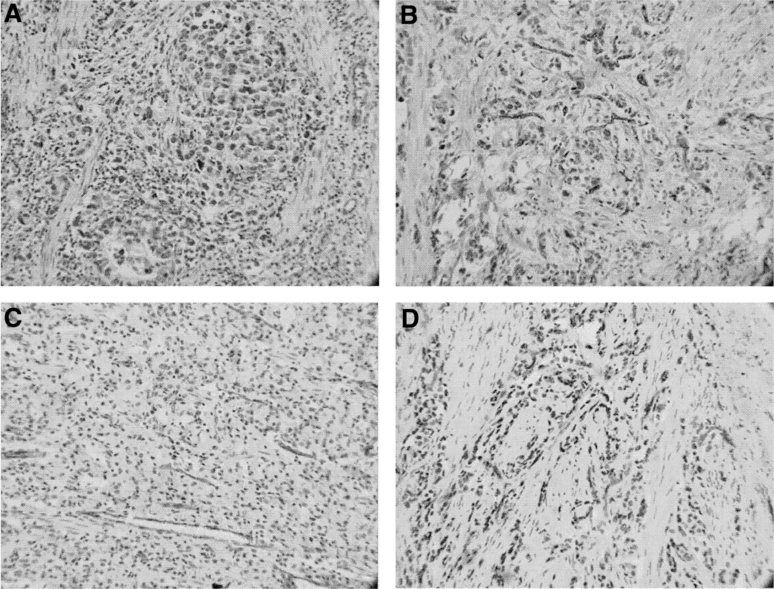
The brown granules are CD34 protein in cytoplasm ×100.
RT-PCR
mRNA expression of ID1 was 0.326 ± 0.083 in group E, 0.891 ± 0.020 in group Ci, 0.015 ± 0.002 in group C, and 0.954 ± 0.018 in control group. Compared with control group, mRNA expression of ID1 significantly decreased in groups E and C (all p < 0.05). However, there was no significant difference in mRNA expression of ID1 between control group and group Ci (p > 0.05). mRNA expression of VEGF was 0.257 ± 0.049 in group E, 0.799 ± 0.019 in group Ci, 0.031 ± 0.006 in group C, and 0.872 ± 0.014 in control group. Compared with control group, mRNA expression of VEGF significantly decreased in groups E and C (all p < 0.05). However, there was no significant difference in mRNA expression of VEGF between control group and group Ci (p > 0.05) (Fig. 2).

mRNA expressions of ID1 and VEGF determined by RT-PCR. Co, control group; Ci, cisplatin group; E, endostar group; C, combination group; ID1, inhibitor of differentiation-1; RT-PCR, reverse transcription polymerase chain reaction; VEGF, vascular endothelial growth factor.
Western blot
Protein expression of ID1 was 0.279 ± 0.051 in group E, 0.871 ± 0.096 in group Ci, 0.012 ± 0.003 in group C, and 0.922 ± 0.105 in control group. Compared with the control group, protein expression of ID1 significantly decreased in groups E and C (all p < 0.05). However, there was no significant difference in protein expression of ID1 between control group and group Ci (p > 0.05). Protein expression of VEGF was 0.236 ± 0.084 in group E, 0.980 ± 0.099 in group Ci, 0.026 ± 0.009 in group C, and 1.036 ± 0.122 in control group. Compared with the control group, protein expression of VEGF significantly decreased in groups E and C (all p < 0.05). However, there was no significant difference in protein expression of VEGF between the control group and group Ci (p > 0.05) (Fig. 3).
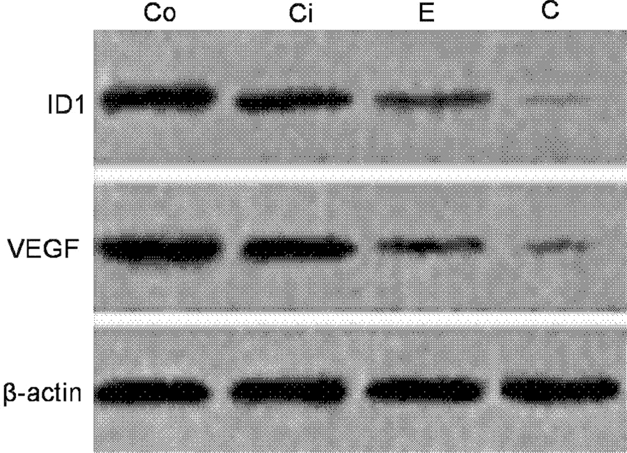
Protein expressions of ID1 and VEGF determined by western blot.
Ultrastructural changes in tumor cells
Under the transmission electron microscope, apoptotic cells were seen in groups E and C. The apoptotic cells exhibited nuclear fragmentation, slightly dilated rough endoplasmic reticulum, degranulation, and mitochondrial myelin (Fig. 4). However, in control group and group Ci, apoptotic cells were not found, but there were lots of necrotic cells. Necrotic cells exhibited disappearance of mitochondrial crista, injured mitochondrial membrane, severely dilated rough endoplasmic reticulum, degranulation, mitochondrial myelin, and degeneration and necrosis in the cytoplasm (Fig. 5).

Apoptotic cells in group E and C. Group E: endostar group; Group C: combination group.
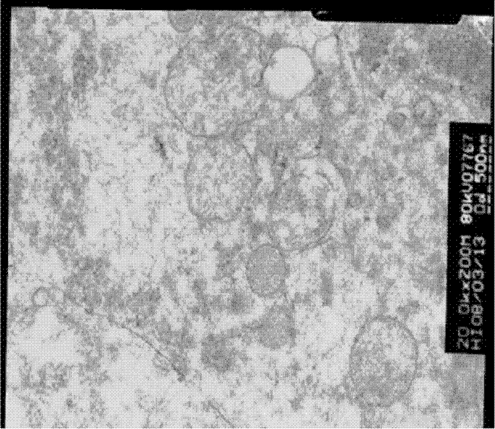
Necrotic cells in group Ci and control group. Group Ci: cisplatin group.
Discussion
Recombinant human endostatin (ES), an antiangiogenesis drug with multiple target points, was developed by China. Preclinical and clinical trials have confirmed the safety and efficacy of ES in tumor therapy. 7 The advantages of ES are that its target points directly expose to blood, which is conducive to play its role; gene expressions of target points are stable, which does not allow it to easily produce drug resistance, it has no specificity for tumors; it may inhibit tumor metastasis; it has downstream amplification effect and it has less adverse reactions. 8 –10 ES possesses antitumor synergistic effects with radiotherapy or chemotherapy, because (1) ES can make angiogenesis become normal in tumor, which reduces interstitial pressure and is conducive to drug transport; (2) radiotherapy- or chemotherapy-caused hypoxia in tumor can induce VEGF expression and inhibit apoptosis, but ES may prevent these secondary reactions.
Cisplatin, a cytotoxic drug, has marked antitumor effects. 11 In this study, ES and cisplatin were used in cancer-bearing nude mice to evaluate their therapeutic effects through observation of tumor size, and MVD, ID1, and VEGF expressions. In this study, compared with control group, weights and volumes of tumors were decreased in other experimental groups, demonstrating that both killing cells and inhibiting angiogenesis can suppress tumor growth. There were no significant differences in weights and volumes of tumors between the three experimental groups, which may be associated with a short observation period. Compared with the body weight before administration, weight loss was marked in group Ci after administration, but in the other three groups, the body weight was increased. This suggests that ES can relieve cisplatin-induced weight loss. Immunohistochemistry indicated that although MVD expression was significantly lower in group Ci than in control group, it was still significantly higher in group Ci than in group E and group C, demonstrating that ES has strong antiangiogenesis effects. It is reported that ES inhibits MVD through suppressing ID1 expression, a VEGF upstream factor. 12 In this study, the expressions of ID1 and VEGF were significantly decreased in group E and group C, which is consistent with the earlier result. In this study, the transmission electron microscope showed that there was apoptosis in both group E and group C, but in the control group and group Ci, necrotic cells were found instead of apoptosis. The authors further infer that ES inhibits tumor through both suppressing angiogenesis and promoting apoptosis.
In summary, the authors observed the effects of ES on human gastric cancer SGC7901 in cancer-bearing nude mouse models and found that ES inhibited tumor through both suppressing angiogenesis and promoting apoptosis. This study provides a new idea for treatment of gastric cancer.
Footnotes
Acknowledgment
This study was supported by a Project of the Young Science and Technology Foundation from the First Clinical College of Liaoning Medical University (No. FY2012-11).
Disclosure Statement
No competing financial interests exist.