
Abstract
Purpose:
Directly radioiodinated [131I]-rituximab has been developed as a radioimmunotherapeutic agent in patients with CD20-positive B cell non-Hodgkin's lymphoma. However, there are concerns over its in vivo catabolism and deiodination. A novel radioiodination linker, N-(4-isothiocyanatobenzyl)-2-(3-(tributylstannyl)phenyl) acetamide (IBPA), was synthesized for the preparation of stable radioiodinated proteins.
Methods:
The authors evaluated the potential of IBPA as a stable radioiodinated linker for rituximab. [125I]-IBPA was purified and conjugated with rituximab, and in vitro stability testing was performed in serum and liver microsomes. In vivo studies were performed after i.v. injection of [125I]-rituximab or [125I]-IBPA-rituximab to nude mice.
Results:
In in vitro studies, [125I]-IBPA-rituximab was stable in serum and liver microsomes. In static scans, high radioactivity was evident in the thyroid following injection of [125I]-rituximab, but low radioactivity was seen in the thyroid following injection of [125I]-IBPA-rituximab. In biodistribution studies, radioactivity uptake in thyroid glands of [125I]-IBPA-rituximab was decreased by approximately sevenfold compared to [125I]-rituximab. In pharmacokinetics, the half-life of [125I]-rituximab was shorter than that of [125I]-IBPA-rituximab in plasma of nude mice.
Conclusions:
The authors demonstrate that [125I]-IBPA-rituximab is more stable to metabolic deiodination in vivo than is [125I]-rituximab. Radioiodination of rituximab using IBPA is thus preferable to direct labeling in terms of in vivo stability.
Introduction
Bexxar® (131I-tositumomab; GlaxoSmithKline LLC) and Zevalin® (90Y-ibritumomab tiuxetan; Spectrum Pharmaceuticals) have been approved by the FDA for the therapy of relapsed or refractory CD20-positive B cell non-Hodgkin's lymphoma (NHL). 1 –3 Both agents are anti-CD20 mAbs labeled with a β-emitter. The crossfire effect of β-radiation permits cells devoid of radioactive-labeled antibody to be irradiated and killed. 4,5
Rituximab, a chimeric anti-CD20 antibody, is an ideal agent for radioimmunotherapy (RIT) of patients with CD20-positive NHL. 6 Rituximab kills CD20-positive B lymphocytes through a mechanism involving antibody-dependent cytotoxicity. 7 –9
In this institution, RIT studies using a [131I]-rituximab compound similar to Bexxar, which is directly radioiodinated to a tyrosine residue in the antibody, have been conducted on patients with B cell NHL since 2004. 10,11 Iodine-131 has been used as a radiolabel in most studies of RIT because of its ready availability, low cost, simple radiochemistry, and high-specific activity in vivo. 12 –15
However, directly radioiodinated antibodies have a limitation in that they are subject to rapid deiodination in vivo. They are catabolized by deiodinases, most likely because of the structural similarity between their iodophenyl groups and thyroid hormones. 16 –18 Catabolism of radioiodinated antibodies is reflected in uptake of radioidine into the thyroid glands 19,20 ; uptake may result in systemic toxicity due to the concentration of radioactive iodine in the thyroid glands. This factor also results in a short retention time of radioiodine in target cells. 21,22
To minimize the structural similarity of the iodination site to thyroid hormones, 23 the authors developed a novel radiohalogenation linker for proteins, N-(4-isothiocyanatobenzyl)-2-(3-(tributylstannyl)phenyl) acetamide (IBPA; Republic of Korea, 10-1550399, August 31, 2015). A meta-replaced aryl halide was selected to avoid the loss of iodide due to nucleophilic substitution. 24,25 Isothiocyanate was used because it is structurally stable in both organic solvents and water.
Herein, the authors present the results of evaluative studies on the effectiveness of an antibody (rituximab) radioiodinated with IBPA compared with an antibody directly labeled.
Materials and Methods
Synthesis of N-(4-isothiocyanatobenzyl)-2-(3-iodophenyl) acetamide (iodo-IBPA)
CHCl3 and H2O were added to a mixture of N-(4-aminobenzyl)-2-(3-iodophenyl) acetamide (50 mg, 0.137 mmol) and CaCO3 (34 mg, 0.341 mmol). The reaction mixture was slowly added with CSCl2 (0.196 mmol). After 1.5 hours, the reaction was terminated by addition of CHCl3 (3 mL) and 1 N HCl. The mixture was extracted with CH2Cl2/H2O. The organic layer was dried over anhydrous Na2SO4 and then reduced pressure. The residual was purified by flash column chromatography (50% ethyl acetate/hexane [EtOAc/Hex], R f = 0.3) to give the product (45 mg, 81%) as a white solid. 1H NMR (400 MHz, DMSO-d6) δ = 8.64 (t, 1 H, J = 5.6, 6.0 Hz), 7.66 (s, 1 H), 7.60 (d, 1 H, J = 7.6 Hz), 7.38 (d, 2 H), 7.30–7.27 (m, 3 H), 7.11 (t, 1 H, J = 7.6 Hz), 4.27 (d, 2 H, J = 6.0 Hz), 3.45 (s, 2 H); 13C NMR (100 MHz, DMSO-d6) δ = 169.8, 139.6, 138.9, 137.6, 135.2, 130.4, 128.6(2), 128.5, 125.9, 104.6, 94.7, 41.7, 41.6.
Synthesis of N-(4-isothiocyanatobenzyl)-2-(3-(tributylstannyl)phenyl) acetamide
CHCl3 and H2O were added to a mixture of N-(4-aminobenzyl)-2-(3-(tributylstannyl)phenyl) acetamide (94 mg, 0.178 mmol; Supplementary Fig. S1; Supplementary Data are available online at
Radioiodination of IBPA
Na125I (Perkin Elmer, Inc.) was added to 100 μL of IBPA (1 mg/mL), followed by 10 μL of chloramine T (1 mg/mL) (Fig. 1). After 10–15 minutes at room temperature, the reaction was quenched by addition of 10 μL of sodium metabisulfite (2.5 mg/mL in water). The [125I]-IBPA was purified by high-performance liquid chromatography (HPLC). The eluted [125I]-IBPA was gathered and dried by nitrogen flow. [125I]-IBPA was reconstructed with acetonitrile.
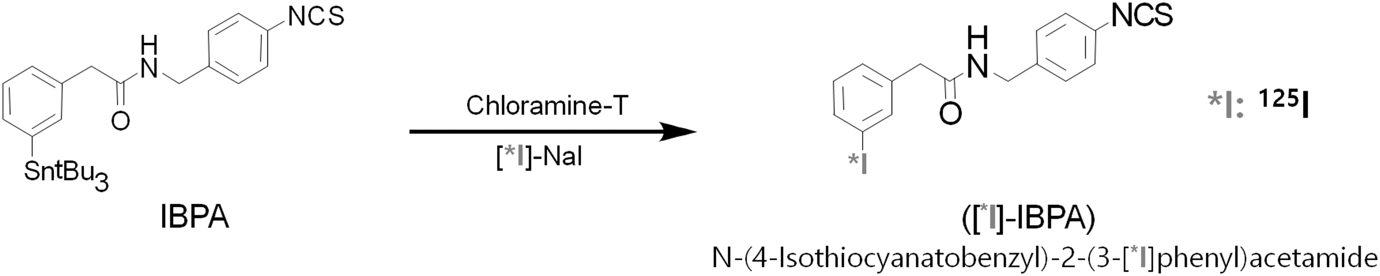
Chemical structures of IBPA and [125I]-IBPA. IBPA, N-(4-isothiocyanatobenzyl)-2-(3-(tributylstannyl)phenyl) acetamide.
Conjugation of [125I]-IBPA to rituximab
[125I]-IBPA was combined with rituximab. Rituximab is a mAb against the protein CD20 (Roche). Rituximab (10 mg/mL) in carbonate buffer (pH 9.5) was added to [125I]-IBPA. The mixture was incubated for 2 hours at 37°C. Labeled mAb was refined with PD-10 desalting columns (GE Healthcare) and eluted BupH phosphate buffer (pH 7.2). Radiochemical purity was evaluated using the AR 2000 radio-TLC scanner (Eckert & Ziegler). 25
Radioiodination of rituximab
[125I]-Rituximab was produced by directly labeling rituximab with 125I. Rituximab (10 mg/mL) in BupH phosphate buffer (pH 7.2) was added to Na125I, followed by 10 μL of chloramine-T (1 mg/mL). The reaction was quenched with sodium metabisulfite (2.5 mg/mL) after 10–20 seconds at room temperature. The labeled mAbs were eluted using a Zeba™ Spin desalting column (Thermo Scientific) with BupH phosphate buffer. Radiochemical purity was estimated by radio-TLC. All radioiodination studies were performed as described previously. 25
Thermal stability of [125I]-rituximab and [125I]-IBPA-rituximab
Samples of [125I]-rituximab and [125I]-IBPA-rituximab were stored at 4°C and 25°C. Radiochemical purity was measured by radio-TLC for 28 days.
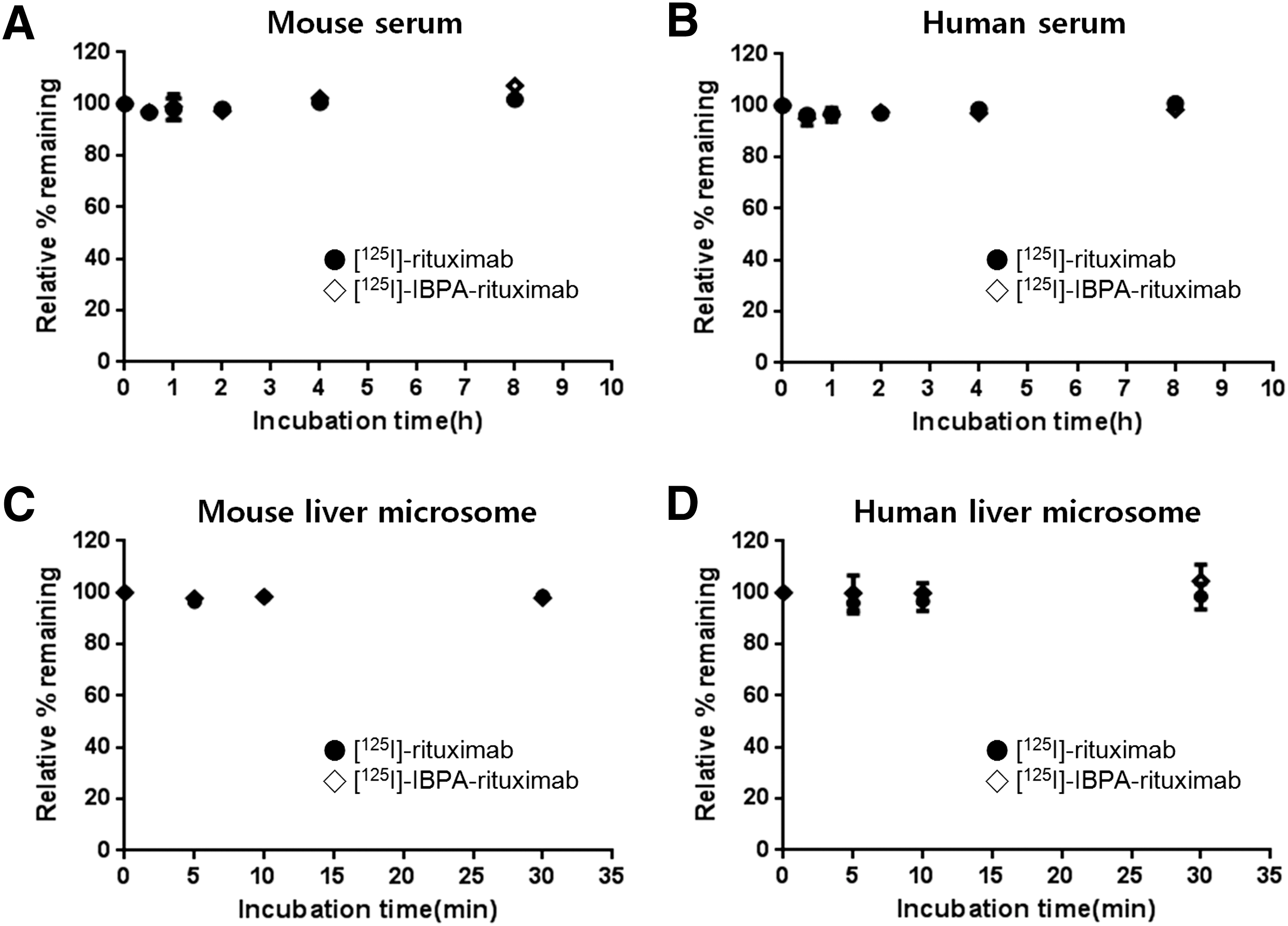
Stability of [125I]-rituximab and [125I]-IBPA-rituximab in human and mouse serum
To estimate the stabilization of [125I]-rituximab and [125I]-IBPA-rituximab in vitro, [125I]-rituximab or [125I]-IBPA-rituximab (0.37 MBq) was added to control human or mouse serum (Innovative Research, Inc.). The mixtures were incubated in a shaking water bath (Personal-11SD TAITEC) at 37°C. Acetonitrile was added to the mixture at 0.5, 1, 2, 4, and 8 hours (n = 3) after incubation. The mixtures were centrifuged for 10 minutes at 13,200 rpm, 4°C in a model 5810R apparatus (Eppendorf). The precipitants and supernatants were collected. Radioactivity was counted using a gamma counter (Perkin Elmer, Inc.). 25
Stability of [125I]-rituximab and [125I]-IBPA-rituximab in human and mouse liver microsomes
[125I]-Rituximab or [125I]-IBPA-rituximab (0.37 MBq) was added to the mixture, which consists of 0.1 M KH2PO4 (pH 7.4), 0.1 M MgCl2, and human or mouse liver microsomes (5 mg/mL; BD Gentest). The mixture was preincubated in a shaking water bath at 37°C with 40 rpm, and the reaction was started by addition of 10 mM NADPH. The reaction was quenched by addition of acetonitrile at 5, 10, and 30 minutes (n = 3) of incubation. The mixtures were centrifuged for 10 minutes at 13,200 rpm, 4°C. The precipitants and supernatants were collected. Radioactivity was measured using a gamma counter.
Cell lines and cultures
The Farage human B cell lymphoma and Daudi human B lymphoblast cell lines, which are CD20 positive, were obtained from the American Type Culture Collection (ATCC). The cells were grown in Hyclone™ RPMI (Thermo Scientific), including 10% fetal bovine serum (JR Scientific) and 1% antibiotics (Thermo Scientific). The medium was exchanged twice per week. The cells were cultured at 37°C, 5% CO2.
Cell binding assay
Serial dilutions of Farage and Daudi (1.6 × 10⁷–0.5 × 10⁶ cells) cells in 1% bovine serum albumin in phosphate-buffered saline (PBS) were incubated with [125I]-rituximab or [125I]-IBPA-rituximab (<0.004 MBq, specific activity 1 mCi/mg, final conc. 30–40 ng) at 4°C for 1 hour (n = 3). Each dilution step was added nonradiolabeled rituximab (1 mg/mL, final conc. 25 μg) for estimating nonspecific binding. The cells were washed three times with casein blocking buffer (1% casein in PBS). The radioactivity of the cells was counted using a gamma counter. The immunoreactivity of radiolabeled mAb was evaluated as described previously. 26
Planar images of radiolabeled monoclonal antibodies in nude mice
The animal experiments were used in 6-week-old female BALB/c nude mice (Central Lab. Animal). For imaging research, mice were given 2% isoflurane/1% O2 inhalation anesthesia. After tail vein injection of [125I]-rituximab or [125I]-IBPA-rituximab (3.7 MBq) (n = 2), static scans of each mouse were acquired at 24 and 48 hours using an Inveon SPECT scanner (Siemens Preclinical Solutions) equipped with a low-energy all-purpose collimator. The images were acquired until 100,000 counts (about 14 minutes) per total body image. Quantification of [125I] uptake in the region of interest of the body and thyroid was performed using the AMIDE image analysis software. 25 All animal studies were approved by the Institutional Animal Care and Use Committee of the KIRAMS and did not use thyroid blocking to evaluate the deiodination of [125I]-rituximab or [125I]-IBPA-rituximab in vivo. Student's t-test was used to determine statistical significance in thyroid radioactivity between [125I]-rituximab/[125I]-IBPA-rituximab at the 95% confidence level, with p < 0.05 considered significantly different.
Biodistribution of radiolabeled monoclonal antibodies in nude mice
Biodistribution studies of [125I]-rituximab or [125I]-IBPA-rituximab were performed in nude mice. After tail vein injection with [125I]-rituximab or [125I]-IBPA-rituximab (0.1 MBq), animals were sacrificed at 48 hours postinjection (n = 5). Blood and organs were extracted and weighed, and their radioactivity was counted using a gamma counter.
Whole-body autoradiography of [125I]-rituximab and [125I]-IBPA-rituximab in nude mice
After intravenous injection of [125I]-rituximab or [125I]-IBPA-rituximab at a dose of 0.1 MBq/head, mice were sacrificed at 48 hours postinjection (n = 2). The carcasses were frozen in a deep freezer and embedded in carboxymethylcellulose. Sections were obtained with a thickness of 40 μm. The whole-body sections were placed on an imaging plate (BAS-SR2040; Fuji Film Co.) for 24 hours. Exposed imaging plates were scanned using a BAS-5000 device (Fuji Film Co.).
Pharmacokinetics of [125I]-rituximab and [125I]-IBPA-rituximab in nude mice
After intravenous injection of [125I]-rituximab (0.83 MBq) or [125I]-IBPA-rituximab (0.77 MBq) in nude mice, blood samples were collected at 2, 4, 8, 24, 48, 72, 168, 336, and 504 hours (n = 5). Plasma was divided by centrifugation for 5 minutes at 13,200 rpm. Plasma samples were immediately treated with acetonitrile. The samples were centrifuged for 10 minutes at 13,200 rpm, 4°C. The radioactivity of the supernatants and precipitants was counted using a gamma counter. The pharmacokinetic parameters were evaluated with a noncompartmental method using the WinNonlin software (ver 2.0; Pharsight Corp.). 25
Results
Chemical synthesis
The iodo-IBPA standard and its precursor, IBPA, were synthesized. Iodo-IBPA and IBPA were prepared at comparable overall yields (iodo-IBPA, 81%; IBPA, 54%). The chemical purity of iodo-IBPA and IBPA was confirmed to be more than 98.2% by HPLC.
Radioiodination
The radioiodination yield of IBPA was 76.20% ± 4.37% (n = 5). The radiolabeled yield of [125I]-rituximab and [125I]-IBPA-rituximab was 55.25% ± 9.40% and 52.00% ± 9.90% (n = 3), respectively. The radiochemical purity of [125I]-rituximab and [125I]-IBPA-rituximab exceeded 99%.
Thermal stability of [125I]-rituximab and [125I]-IBPA-rituximab
The percentage of [125I]-rituximab and [125I]-IBPA-rituximab remaining up to 28 days after incubation at 4°C and 25°C exceeded 99% and 93%, respectively (Fig. 2).
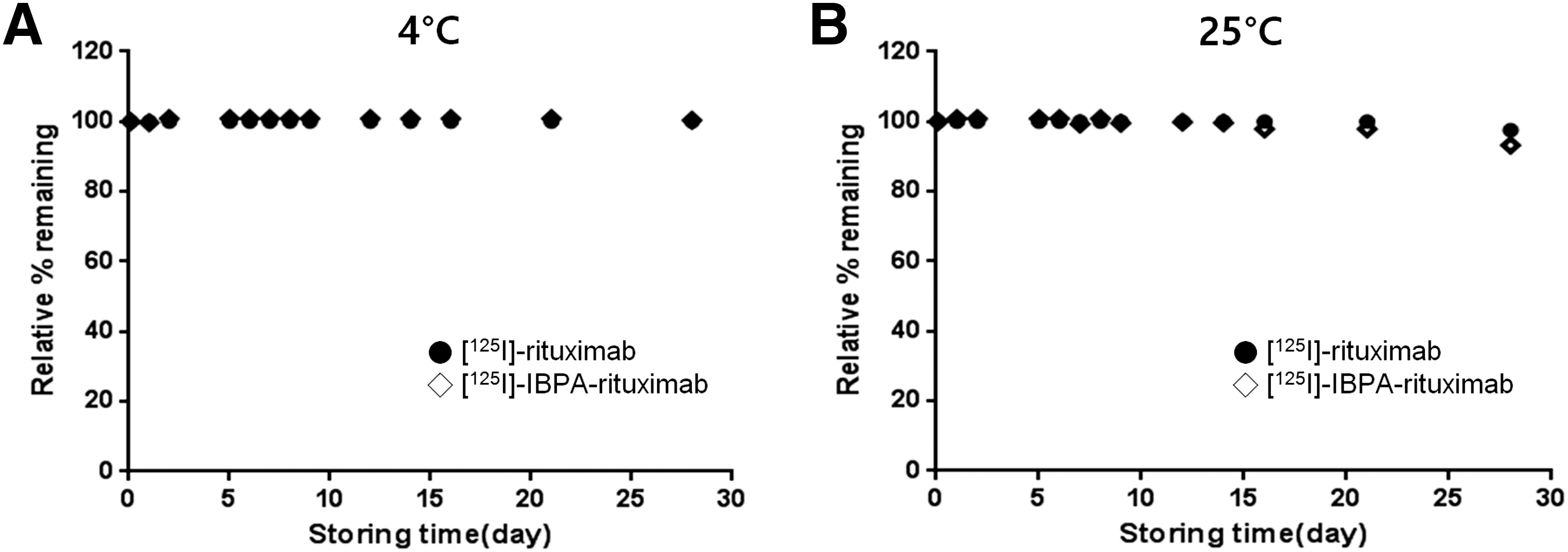
Average percentage of [125I]-rituximab (●) and [125I]-IBPA-rituximab (◊) remaining as a function of time during storage at
Stability of [125I]-rituximab and [125I]-IBPA-rituximab in human and mouse serum
In human and mouse serum, [125I]-rituximab and [125I]-IBPA-rituximab were stable. The percentage of [125I]-rituximab and [125I]-IBPA-rituximab remaining exceeded 96% up to 8 hours after incubation in mouse serum. The percentage of [125I]-rituximab and [125I]-IBPA-rituximab remaining exceeded 95% at up to 8 hours after incubation in human serum (Fig. 3).
Average percentage of [125I]-rituximab (●) and [125I]-IBPA-rituximab (◊) remaining as a function of time during incubation in
Stability of [125I]-rituximab and [125I]-IBPA-rituximab in human and mouse liver microsomes
In human and mouse liver microsomes, [125I]-rituximab and [125I]-IBPA-rituximab were stable. The percentage of [125I]-rituximab and [125I]-IBPA-rituximab remaining exceeded 96% up to 30 minutes after incubation in human and mouse liver microsomes (Fig. 3).
Cell binding assay
A Lindmo assay 26 was carried out to identify the immunoreactive fraction of [125I]-rituximab and [125I]-IBPA-rituximab. Nonspecific binding was calculated by using unlabeled rituximab to block the binding sites on the cell surface; this was subtracted from the total binding (TB) to calculate the specific binding (SB) of [125I]-rituximab and [125I]-IBPA-rituximab to cells of both Farage and Daudi lymphoma cell lines. Figure 4 shows the percentage of specifically bound radiolabeled antibody to the total applied radioactivity as a function of cell concentration. In this plot, the plateau SB/TB percentage values of [125I]-IBPA-rituximab was more than 30% and that of [125I]-rituximab was more than 10% for the highest concentration of Farage cells (Fig. 4A and Supplementary Table S1). In this plot, the plateau SB/TB percentage value of [125I]-IBPA-rituximab was more than 60% and that of [125I]-rituximab was more than 40% for the highest concentration of Daudi cells (Fig. 4B and Supplementary Table S2). However, the precise value of the plateau cannot be extracted from this plot. A double inverse plot of TB/SB as a function of inverse cell concentration (1/cells) was designed to calculate the immunoreactive fraction. Fitting a straight line through the data of the 2–4 higher cell concentrations allowed determination of the intercept value at the ordinate, which equals 1/r, where r represents the immunoreactive fraction of the total amount of antibody. These data indicated a linear relationship between TB/SB and 1/cells. The immunoreactive fraction of [125I]-rituximab and [125I]-IBPA-rituximab was found to be r = 1/2.136 = 0.468 and r = 1/1.515 = 0.660 with Daudi cells, respectively. The immunoreactive fraction of [125I]-rituximab and [125I]-IBPA-rituximab was found to be r = 1/7.379 = 0.136 and r = 1/2.995 = 0.334 with Farage cells, respectively.
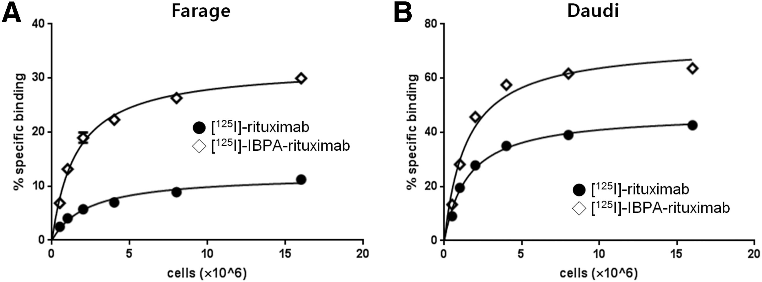
Cell binding assay for the determination of the immunoreactive fraction of [125I]-rituximab (●) and [125I]-IBPA-rituximab (◊) in
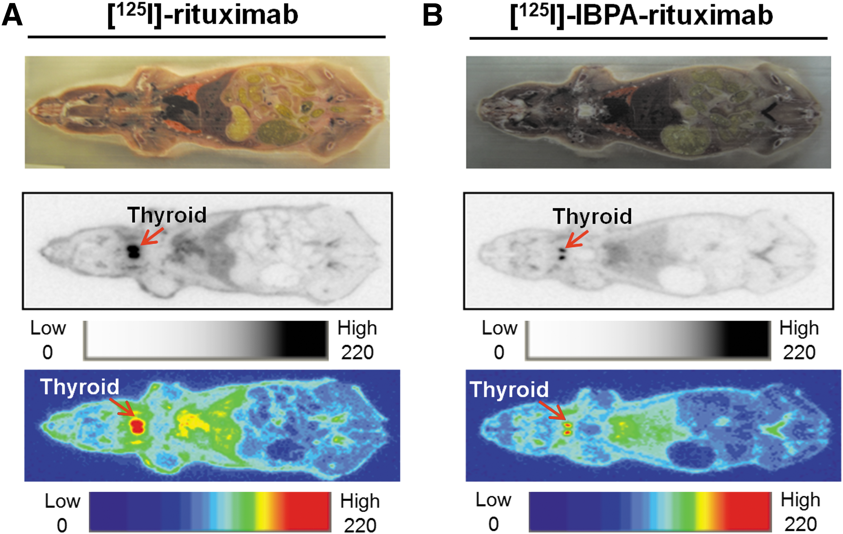
Planar images of [125I]-rituximab and [125I]-IBPA-rituximab in nude mice
Low radioactivity was detected in the thyroid following [125I]-IBPA-rituximab injection, while high radioactivity was detected in the thyroid following [125I]-rituximab administration at 24 and 48 hours postinfusion (Fig. 5). The integrated radioactivity in the thyroid region of [125I]-rituximab-treated mice (25,674.46 ± 799.34 cpm) was about 2.4-fold higher than that of [125I]-IBPA-rituximab-treated mice (10,819.62 ± 714.55 cpm) at 48 hours postinfusion (p < 0.005).

In planar images of
Biodistribution of [125I]-rituximab and [125I]-IBPA-rituximab in nude mice
Biodistribution profiles of [125I]-rituximab or [125I]-IBPA-rituximab in nude mice are shown in Figure 6. Uptake of radioactivity in the thyroid was lower when [125I]-IBPA-rituximab was used instead of [125I]-rituximab. The thyroidal uptake value after injection of [125I]-rituximab (0.79% ± 0.23%ID) was approximately sevenfold higher than that after infusion of [125I]-IBPA-rituximab (0.12% ± 0.01%ID), and the difference was statistically significant (p < 0.01).

Biodistribution of [125I]-rituximab (■, dose, 54.2 μg/0.1 MBq/head) and [125I]-IBPA-rituximab (□, dose, 55 μg/0.1 MBq/head) obtained 48 hours after a single i.v. injection in nude mice.
Whole-body autoradiography of [125I]-rituximab and [125I]-IBPA-rituximab in nude mice
Whole-body autoradiography showed that accumulation of [125I]-rituximab and [125I]-IBPA-rituximab in thyroid tissue was higher than that in other tissues (Fig. 7). In the thyroid region, a higher level of radioactivity accumulated in [125I]-rituximab than in [125I]-IBPA-rituximab-treated mice.
Whole-body autoradiograms of
Pharmacokinetics of [125I]-rituximab and [125I]-IBPA-rituximab in nude mice
The clearance of [125I]-IBPA-rituximab from plasma was similar to that of [125I]-rituximab (Fig. 8). In the plasma of nude mice, the mean terminal half-life of [125I]-rituximab and [125I]-IBPA-rituximab was 140.3 ± 28.5 and 195.9 ± 31.7 hours, respectively (Table 1). The half-life of [125I]-rituximab was shorter than that of [125I]-IBPA-rituximab (p < 0.05). The volume of distribution (V z) for [125I]-rituximab and [125I]-IBPA-rituximab was 2.6 ± 0.6 and 3.6 ± 0.5 mL, respectively (p < 0.05). Other pharmacokinetic parameters were not significantly different.
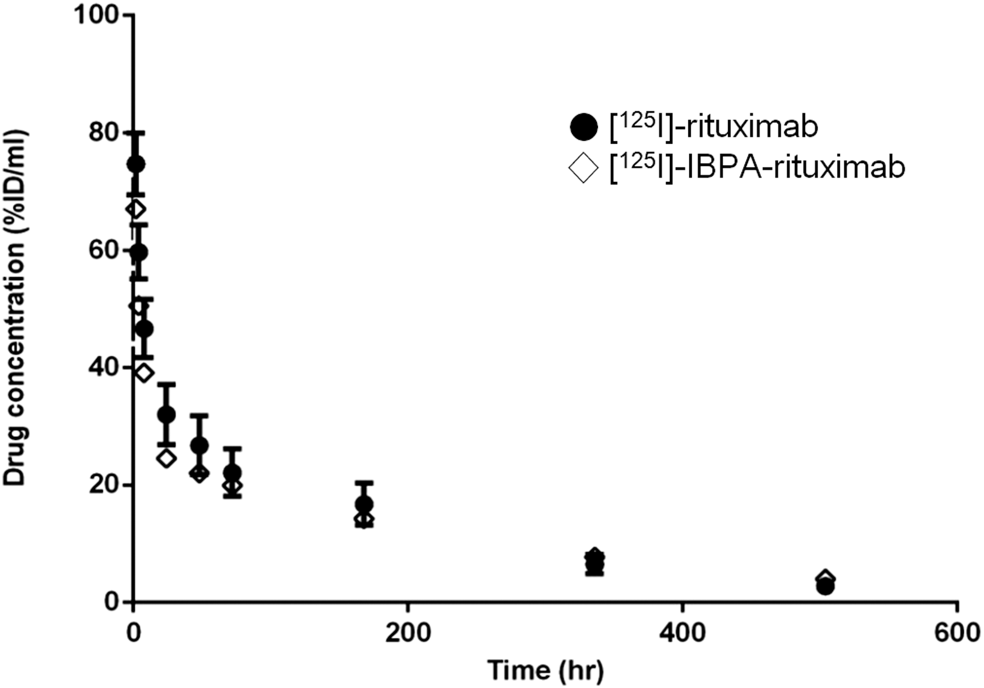
Average plasma radioactivity concentration versus time in nude mice after an i.v. injection of [125I]-rituximab (•, dose, 54.8 μg/0.83 MBq/head; n = 5) and [125I]-IBPA-rituximab (⋄, dose, 53.8 μg/0.77 MBq/head; n = 5). Blood samples were collected from each group (n = 5) at 2, 4, 8, 24, 48, 72, 168, 336, and 504 hours postinjection. Values are presented as mean %ID/mL ± SD. %ID/mL, percent injected dose per one milliliter of plasma.
Dose, 54.3 μg/0.8 MBq/head; n = 5.
C 0, concentration extrapolated to time zero; t1/2, λz, terminal half-life; AUCinf, area under plasma concentration–time curve to infinite time; V z, volume of distribution;
Cl, clearance.
IBPA, N-(4-isothiocyanatobenzyl)-2-(3-(tributylstannyl)phenyl) acetamide; %ID/mL, percent injected dose per one milliliter of plasma.
Discussion
Radioiodinated mAbs are of interest for diagnosis and radiotherapy of cancer. 27,28 However, after administration in vivo, radioiodinated mAbs frequently undergo deiodination, resulting in stomach and thyroid uptake of radioiodine and decreasing the concentration of radioiodinated mAb available for tumor binding. 29 Therefore, previous studies have focused on the development of a new radioiodination linker that is metabolically more stable in vivo. 25
Organic solvents, used for purification by HPLC, could be toxic to humans. 30,31 However, [125I]-IBPA can be purified using an aqueous solvent HPLC condition 32 due to its different chemical structure compared to compounds used previously (which included an isothiocyanate functional group for protein labeling). 30,31 This enhances the safety of administration and reduces the cost by decreasing the number of quality inspection items.
In this study, the potential of radioiodinated rituximab created using a linker, N-(4-isothiocyanatobenzyl)-2-(3-(tributylstannyl)phenyl) acetamide (IBPA; Republic of Korea, 10-1550399, August 31, 2015) was evaluated in vitro and in vivo.
The radiolabeled yield of [125I]-IBPA (70%–80%) was improved compared to previous reports (23%–55%). 30,31 [125I]-IBPA-rituximab was stable at 4°C and 25°C for up to 28 days (Fig. 2). Therefore, this method produces a stable conjugate that is suitable for long-term storage and routine use. Also, [125I]-IBPA-rituximab was stable in vitro (human serum, mouse serum, human liver microsomes, and mouse liver microsomes) (Fig. 3). Since the active site of the deiodinase enzyme is not complementary to [125I]-IBPA, 125I cannot be released from [125I]-IBPA-rituximab by the action of that enzyme. 33 The immunoreactive fraction of [125I]-IBPA-rituximab was approximately twofold that of [125I]-rituximab (Fig. 4).
Catabolism of radioiodinated antibodies is indicated by uptake of radioiodine into the thyroid and stomach. 16 –18 In biodistribution studies, the thyroid uptake of radioiodine decreased about sevenfold (p < 0.01) and radioactivity in the stomach ([125I]-rituximab; 1.79% ± 0.17%ID/g, [125I]-IBPA-rituximab; 0.96% ± 0.05%ID/g) decreased about twofold (p < 0.005) at 48 hours using the IBPA (Fig. 6). Furthermore, the thyroid uptake of [125I]-rituximab was about 2.4-fold higher than that of [125I]-IBPA-rituximab at 48 hours (p < 0.005), as revealed by planar imaging (Fig. 5). Clearly, the in vivo stabilization of radioiodinated mAb using IBPA was considerably enhanced compared to that of directly radioiodinated mAb.
To evaluate the effectiveness of IBPA in the systemic circulation, plasma radioactivity concentration and time for [125I]-IBPA-rituximab and [125I]-rituximab were determined. The biological half-life of [125I]-rituximab was considerably shorter than that of [125I]-IBPA-rituximab in nude mice (p < 0.05) (Table 1). These results indicate that [125I]-rituximab is eliminated from the body more rapidly than [125I]-IBPA-rituximab. Catabolites of [125I]-rituximab were discharged and metabolized more rapidly than those of [125I]-IBPA-rituximab. Therefore, [125I]-IBPA-rituximab lasted longer in the blood without being deiodinated, suggesting a prolonged duration of efficacy.
Conclusion
These results demonstrate that IBPA can improve the stability of radioiodinated rituximab in vivo. The considerable improvement in stability can lower the dose necessary for RIT. The reduced dose can decrease the risk of treatment-related toxicity to normal tissues while maintaining therapeutic efficacy.
In conclusion, IBPA is a potential bifunctional linker for stable radioiodinated proteins for use in RIT in patients with CD20-expressing B cell NHL.
Footnotes
Acknowledgment
This research was supported by the National R&D Program through the National Research Foundation of Korea (NRF) funded by the Ministry of Science, ICT & Future Planning (No. 1711026888).
Disclosure Statement
No competing financial interests exist.
References
Supplementary Material
Please find the following supplemental material available below.
For Open Access articles published under a Creative Commons License, all supplemental material carries the same license as the article it is associated with.
For non-Open Access articles published, all supplemental material carries a non-exclusive license, and permission requests for re-use of supplemental material or any part of supplemental material shall be sent directly to the copyright owner as specified in the copyright notice associated with the article.