
Abstract
Objective:
To study the effects of Numb gene expression on radiation sensitivity of nonsmall cell lung cancer (NSCLC) stem cells.
Materials and Methods:
The side population (SP) cells A549-SP were transfected with pcDNA3.1 (pcDNA3.1 group), pcDNA-Numb (pcDNA-Numb group) and shRNA-Numb (shRNA-Numb group). Real-time quantitative polymerase chain reaction and Western blot were performed to determine Numb expression; MTT method was used to measure the proliferation activity change of the NSCLC stem cells both before and after irradiation with different doses of 60Coγ ray; Hoechst staining and Annexin V-FITC/PI were used to detect the apoptosis of the NSCLC stem cells; and colony-forming assay was used to determine the effect of Numb expression on radiation sensitivity of the NSCLC stem cells.
Results:
Increased mRNA and protein expressions of the A549-SP cells were found in the pcDNA-Numb group, and decreased mRNA and protein expressions were found in the shRNA-Numb group. The optical density value of the cells decreased in the pcDNA-Numb group but increased in the shRNA-Numb group. The cells with over-expressed Numb showed obvious nuclear condensation and fragmentation; the apoptosis rate increased significantly. The cells with knockdown Numb showed less nuclear damage; the apoptosis rate significantly decreased. After irradiation, the cells in the pcDNA-Numb group showed decreased survival rate, clonality, and the values of D0, Dq, N, and SF2; whereas the cells in the shRNA-Numb group showed the opposite trend.
Conclusions:
Radiation sensitivity of NSCLC stem cells was enhanced with the increase of Numb expression. Determination of Numb expression helped to evaluate the response of lung cancer to radiotherapy, which was important for guiding tumor treatment clinically.
Introduction
Nonsmall cell lung cancer (NSCLC), accounting for 85% of all lung cancer, is defined as any malignant epithelial lung tumor that is lacking in a small-cell component, and it is composed histologically of squamous cell carcinoma, adenocarcinoma, and large-cell carcinoma. 1 –3 It is estimated that ∼20% of patients with NSCLC have locally advanced (stage III) disease when initially diagnosed and are confronted with a lower than 14% 5-year survival rate. 4,5 Radiotherapy combined with chemotherapy has been popularly recommended for the patients with locally advanced NSCLC. 6,7 It could contribute to controlling local disease and improving 5-year survival rate by about 4.5%. 8,9 Once persistent tumor or locoregional recurrence is diagnosed after concurrent chemoradiotherapy, curative treatments are limited and often palliative. 10 One explanation of recurrence lies in that cancer therapy focuses often on tumor cells but not on cancer stem cells (CSCs), which will repopulate the tumor and thus cause recurrence when the tumor cells are reduced or depleted by treatment. 11 CSCs are reported to be the unique source of any tumor cells at early stages of tumorigenesis, lead to tumor propagation, and relapse at advanced stages. 12 Accumulating studies suggest that targeting lung CSCs may be a novel strategy for NSCLC patients. 13,14
Numb family proteins (NFPs), including Numb and Numb-like (Numbl), are involved in the specification and differentiation of CSCs. 15,16 NFPs play multiple roles in normal and disease conditions in a cell context-dependent manner. 17 Numb is one of the clathrin-associated sorting proteins and combines with several other endocytic proteins. 18 The gene is located on chromosome 14q24.3 that encodes the phosphotyrosine-binding domain and the proline-rich region domain. 19 Numb is reported to function as a component of the adherens junction to regulate cell adhesion and migration, and to get involved in the ubiquitylation of p53 (Trp53) and Gli1 to regulate cancer initiation. 15 A critical factor of the high mortality in patients with NSCLC is the development of radioresistance, and, thus, overcoming radioresistance in NSCLC is currently an important challenge. 20 CSCs have been recognized as tumorigenic cells that have the ability of differentiating and self-renewing and that are resistant to conventional therapy, including chemotherapy and radiation therapy. 21 Although some cancer cells represent radioresistance as a result of the function of specific target proteins and accumulating studies suggest the potential of radioresistance-related factors as novel therapeutic targets, the protein profiles and factors associated with radioresistance in NSCLC cells and the molecular mechanisms behind them are poorly understood. 20 Our study, therefore, tried to explore whether and how the Numb gene affected the radiation sensitivity of NSCLC stem cells, in the hope of improving the effect of radiotherapy for NSCLC patients.
Materials and Methods
Cell culture and stem cell screening
Human lung adenocarcinoma A549 cell line (purchased from the cell bank of Peking Union Medical College) was cultured in Dulbecco's minimum essential medium (DMEM) containing 10% fetal bovine serum (FBS) at 37°C in 5% CO2 under saturated humidity, and the cells were passaged by general procedure. Side population (SP) cells (subpopulation of stem cells) and non-SP cells were sorted through staining and flow cytometry. Second-generation A549 cells in the logarithmic growth phase were digested by trypsase, neutralized, centrifuged for isolation of supernatant, washed twice with phosphate-buffered saline (PBS), and finally re-suspended using DMEM containing 2% FBS for a single-cell suspension. The cells were added with Hoechst 33342 until the concentration reached 5 μg/mL, bathed in water at 37°C for 90 minutes, centrifuged for isolation of supernatant, washed once by using PBS containing 2% FBS, and finally re-suspended in icy PBS containing 2% FBS. Proinsulin (PI) was then added until the concentration reached 2 μg/mL. The cells were then detected by a flow cytometer and divided into non-SP cells and SP cells (subpopulation of stem cells).
Construction of Numb gene vector and transfection of SP cells
Eukaryotic expression plasmid pcDNA3.1 (bought from Shanghai Institutes for Biological Sciences) was used in construction of recombinant plasmid pcDNA-Numb and shRNA-Numb, which were then transformed into intestinal competent cells and screened. The bacterial fluid (10 mL) containing recombinant plasmid was sequenced by Shanghai BioSune Co. Ltd. (Shanghai, China). The sequencing results were correct. Vacant pcDNA3.1, over-expressed pcDNA-Numb, and gene knockout shRNA-Numb were transfected with A549-SP cells in the logarithmic growth phase, respectively; then, the cells were grouped into pcDNA3.1 group, pcDNA-Numb group and shRNA-Numb group.
The SP cells were seeded into a 60 mm-sized humidified incubator and cultured overnight at 37°C in 5% CO2 until they were 80%–90% fused. A mixture of plasmid/liposome lipofectamin 2000 was prepared in a sterile EP tube, including 12 μL lipofectamin 2000 and 500 μL serum-free medium, which was rested for 5 minutes at room temperature, and 6 mg empty plasmid and 500 μL serum-free medium, which were mixed evenly and rested for 20 minutes for a complex of DNA with liposome. The SP cells in the three groups were washed by serum-free medium. The mixture was added with serum-free medium (without antibiotics), which after mild mixing was added into three incubators (60 mm) for transfection. The cells were then placed in an incubator at 37°C in 5% CO2, and 6–8 hours later, they were cultured in complete medium for another 24–48 hours. The SP cells were collected from each group for further use. 22
Real-time quantitative polymerase chain reaction for Numb mRNA expression
NSCLCs in the logarithmic growth phase were chosen for extracting the total RNA of stem cells according to RNA isolation Kit (Ambion, Inc., Austin, TX). The RNA sample (5 μL) taken independently from pcDNA3.1 group, pcDNA-Numb group and shRNA-Numb group was diluted 20 times via RNA enzyme-free ultrapure water. The optical density (OD) at 260 and 280 nm wavelengths was determined using an ultraviolet spectrophotometer, and concentration and purity of RNA were detected. The ratio of OD260/OD280 was between 1.8 and 2.0, which guaranteed the RNA purity. Reverse-transcription reaction: cDNA was synthetized by reverse transcription that served as the template for PCR according to the First-Strand cDNA Synthesis Kit (Thermo Fisher Scientific, Inc., GmbH, Germany). Reaction conditions were as follows: 37°C for 15 minutes to 98°C for 5 minutes. The real-time quantitative polymerase chain reaction (RT-qPCR) was performed using the ABI 7500 PCR system (Beckman Coulter, Inc., Brea, CA), and the reaction conditions were as follows: predenaturation for 2 minutes at 95°C, denaturation for 30 seconds at 95°C, annealing for 30 seconds at 55°C, and extension for 20 seconds at 72°C; the procedures were repeated 40 times. Primers of internal reference GAPDH and Numb were designed using primer design software Primer Premier 5.0. After being compared with the genes in GenBank, the primers were synthetized by Beijing Genomics Institute (Beijing, China). Forward primer of GAPDH: 5′-TGGTCACCAGGGCTGCTT-3′, and reverse primer: 5′-AGCTTCCCGTTCTCAGCCTT-3′. Forward primer of Numb: 5′-CAATCTCCTACCTTCCAAGGG-3′, and reverse primer: 5′-CGGACGCTCTTAGACACCTC-3′. Relative quantitative analysis: At the end of the reaction, the cycle threshold (Ct) of each reaction tube was obtained. Ct meant the number of amplification cycles when the fluorescence intensity reached a given threshold value. 2−ΔΔCt referred to the odds ratio of target gene expression between the experimental group (pcDNA-Numb group and shRNA-Numb group) and the control group (pcDNA3.1 group), in which ΔΔCt = ΔCt the experimental group − ΔCt the control group, and ΔCt = CtmRNA − CtGAPDH. 23
Western blot for Numb protein expression
The stem cells taken out from the primary culture medium were washed twice by the precooling PBS, digested by pancreatin, and finally centrifuged for 5 minutes at 1200 rpm. After isolation from supernatant, the cells were re-suspended by PBS and centrifuged again. The cells were split by protein extraction lysate buffer (100 μL/50 mL) and then rested on ice for 30 minutes. Centrifugation was carried out at 4°C and at 12,000 rpm for 10 minutes. The supernatant was isolated and put into a 0.5 mL-sized centrifuge tube, which was kept at −20°C for later use. BCA fluid (BCA reagent A:BCA reagent B = 50:1) was used for dissolution of protein. The final concentration of the PBS was adjusted to 0.5 mg/mL. The protein samples (0, 1, 2, 4, 8, 16, 20 μL, independently) of each group were seeded into wells of a 96-well plate, ensuring each well contained 20 μL with the shortage made up with PBS. After test solution (200 μL) was added, diluted standard samples were added to each well (10 μL), followed by light shaking, incubation at 37°C for 30 minutes, and cooling to room temperature. The OD value of each sample was measured using a microplate reader. The standard curve was drawn, and the protein concentration of each sample was calculated. Concentrated gel (5%) and separation gel (10%) were prepared. The supernatant of each group was calculated. The solution containing 50 μg proteins was taken for electrophoresis for 1–2 hours, with a voltage of 60–120 V. After the electrophoresis, wet electrotransfer was applied to transfer the proteins to polyvinylidene fluoride (PVDF) membrane; the PVDF membrane was rinsed with deionized water and soaked in formaldehyde for a few seconds; and the gel was then transferred to Coomassie brilliant blue dye for staining. Sealing fluid was used to adjust the concentration of the first antibody of Numb, which was then incubated overnight with the sealed PVDF membrane in a cold chamber at 4°C. The PVDF membrane combined with first antibody of Numb was washed three × 10 minutes with Tris-buffered saline-Tween 20 (TBST) solution. The PVDF membrane was then incubated for 1–2 hours with diluted second antibody of Numb at room temperature. The PVDF membrane combined with second antibody of Numb was washed three × 10 minutes with TBST. 24 Scotographic imaging was performed in accordance with the instructions of the ECL Kit; the gel image was used to analyze the net OD value and molecular weight of the target zone. The molecular weight of Numb protein was 70 kDa and that of the internal reference GAPDH was 37 kDa.
3-(4,5-Dimethylthiazol-2-yl)-2,5-diphenyltetrazolium bromide assay for the proliferation activity change of Numb over-expressed and Numb knockdown NSCLCs
After transfection, the three groups of stem cells were re-seeded to a new plate for a 24–72 hour incubation at 37°C with 5% CO2. Some of the cells underwent 3-(4,5-dimethylthiazol-2-yl)-2,5-diphenyltetrazolium bromide (MTT) detection after 48 and 72 hours of culture; whereas some underwent MTT detection after irradiation with different doses (0, 2, 4, 6, 8, and 10 Gy) of 60Coγ ray. When the fusion degree reached about 80%, the cells were washed twice with PBS, followed by conventional digestion with trypsin and percussion with straw for a single-cell suspension. To each well of cells, 20 μL MTT solution was added (5 mg/mL; Sigma Company, St. Louis, MO) for color generation, followed by continuous incubation for 4 hours at 37°C with 5% CO2. At the termination of the culture, the culture medium was discarded. Dimethyl sulfoxide (DMSO, 150 μL) was added to each well, followed by light shaking for 10 minutes to promote dissolution of the crystal. The OD value of each well was detected and recorded at 490 nm wavelength with the enzyme-linked immunosorbent assay instrument after 48 and 72 hours of incubation. A graph with the OD value as ordinate and with the interval as abscissa was drawn to demonstrate the changes in the OD value of pcDNA3.1 group, pcDNA-Numb group and shRNA-Numb group at a set time point with different doses of 60Coγ rays, to determine the proliferation of the three groups of stem cells. 25
Apoptosis detection
Morphological changes in apoptosis of the SP cells were determined using the Hoechst 33258 staining method. After transfection and 72 hours of culture, the culture solution was discarded and the cells were fixed with 0.5 mL fixation fluid, followed by rest at room temperature for 10 minutes. After that, the fixation fluid was discarded, followed by PBS washing for three times. Hoechst 33258 staining solution (0.5 mL) was added after the PBS solution was discarded. Five minutes later, the staining solution was discarded, followed by PBS washing for three times. The stem cells in each group were placed on the coverslips containing antifluorescence quenching mounting medium for fluorescence microscopy observation at 350 nm ultraviolet light. 26
Annexin V-fluorescein isothiocyanate (FITC) was performed to detect the apoptosis rate of the SP cells. The three groups of NSCLCs were independently treated with dialkyldithiophosphate (DDP) (2.5 g/L). The procedures performed 24 hours later included washing with PBS, a reaction for 10 minutes in a six-well plate with Annexin V-FITC (5 μL) and its binding liquid (195 μL) in the dark, immersion in an ice bath for 10 minutes with the binding liquid of Annexin V-FITC (190 μL) and PI (10 μL), and mounting. To the flow tube, PBS was added (400 μL), followed by analysis with flow cytometry.
Cell colony-forming assay
Each group of transfected NSCLC stem cells was radiated with different doses (0, 2, 4, 6, 8, and 10 Gy) of 60Coγ rays, with the source target distance set at 90 cm and an absorbed dose rate of 0.5 Gy/min. According to the irradiation dose and transfection carrier, the NSCLC stem cells were divided into 3 teams and 18 groups and incubated in six-well plates. The specific grouping was as follows: A1 (0 Gy), A2 (2 Gy), A3 (4 Gy), A4 (6 Gy), A5 (8 Gy), and A6 (10 Gy); B1 (0 Gy), B2 (2 Gy), B3 (4 Gy), B4 (6 Gy), B5 (8 Gy), and B6 (10 Gy); and C1 (0 Gy), C2 (2 Gy), C3 (4 Gy), C4 (6 Gy), C5 (8 Gy), and C6 (10 Gy). The incubation was terminated until the clone could be observed with the naked eye, and the culture medium was discarded. The NSCLC stem cells were then washed twice with PBS and fixed with methanol for 30 minutes, followed by removal of the methanol, staining with Giemsa, washing with water, and drying. The six-well plates were then put upside down and superimposed with gridded transparent films, followed by observation under a low-power lens and counting of the number of visible cell clones. 27
The mathematical model “multi-target click” in SigmaPlot10.0 software was applied for curve fitting, from which the values were obtained, including extrapolation number (N), quasithreshold dose (Dq), survival fraction of the cells after 2 Gy irradiation (SF2), and mean lethal dose (D0), and the survival curve of the cells was drawn. The colony-forming efficiency (CFE) and survival fraction (SF) of the cells were calculated: CFE (%) = colony number/number of inoculated cells × 100%; SF = colony number in a group with a dose of gray/number of inoculated cells in the group × CFE.
Statistical analysis
All the data were analyzed using SPSS 21.0 (SPSS, Inc., Chicago, IL). Categorical data were represented by ratio or percentage, and measurement data were represented by mean ± standard deviation (
Results
SP cells sorting by flow cytometry
To investigate the effects of Numb gene on radiation sensitivity of NSCLC stem cells, we reviewed a previous study and found that the fluorescence detection on A549-SP cells and non-SP cells can be used to compare the expression levels of SP cells and non-SP cells. 28 In this study, after being marked by Hoechst 33342 and PI, the lung adenocarcinoma A549 cells were sorted by flow cytometry; the dead cells with positive PI were abandoned. The cells with negative PI were divided into positive Hoechst 33342 cell subsets and into negative or weak positive cell subsets according to mark differences of Hoechst 33342, in which the latter subsets accounted for 3.7% of the total cell count (Fig. 1A). After being sorted, the SP and non-SP cells were observed under a fluoroscope; the non-SP cells emitted a strong blue light, and the SP cells emitted a weak blue light (Fig. 1B, C).

Sorting of the lung adenocarcinoma A549 cells
The mRNA and protein expression of Numb of the three groups
It has been reported that the Numb gene was significantly up-regulated in patients with liver cancer, 24 and our study revealed that the mRNA and protein expression levels increased significantly in the NSCLC stem cells that were transfected with over-expressed Numb (pcDNA-Numb) compared with the ones in the control group that were transfected with empty plasmid (pcDNA3.1) (both p < 0.05). However, the mRNA and protein expression levels decreased significantly in the NSCLC stem cells that were transfected with knockdown Numb (shRNA-Numb) compared with the ones in the control group (both p < 0.05) (Fig. 2).
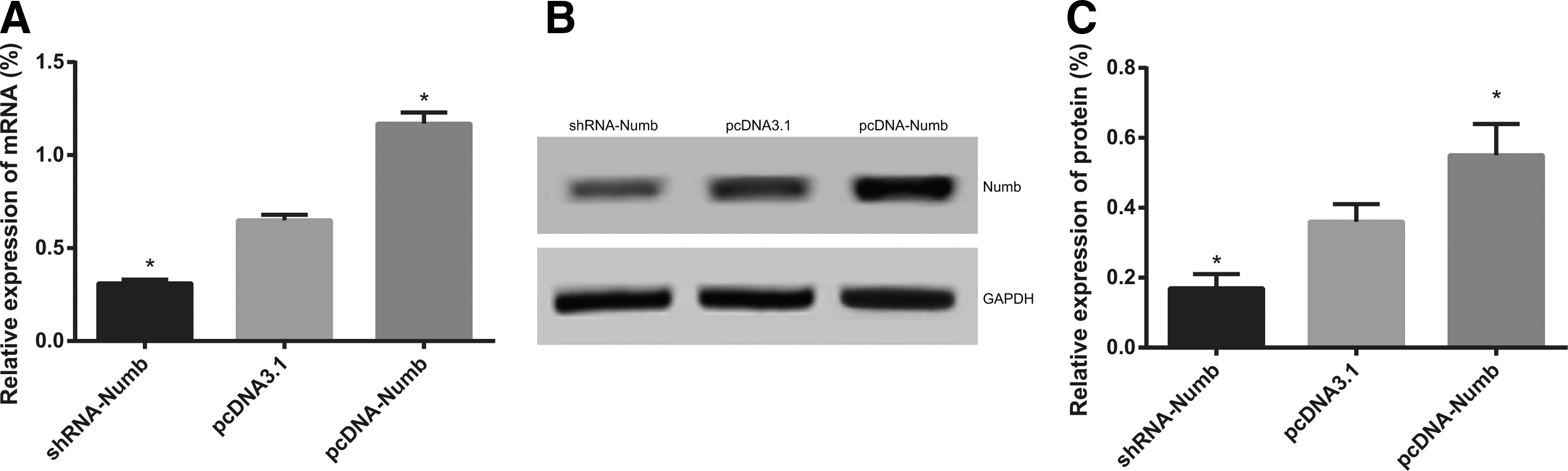
The mRNA and protein expression of Numb in the pcDNA3.1 group, pcDNA-Numb group and shRNA-Numb group
Proliferation of NSCLC stem cells with over-expressed and knockdown Numb
The MTT method was performed to detect the cell survival rate in each group, and the chart of the proliferation in each group after 48 and 72 hours of transfection was drawn (Fig. 3). A previous study has shown that knockdown of Numb gene may promote the proliferation of Ishikawa cells detected by cholecystokinin-8 (CCK8), 25 and by further study, we found that the OD value of the A549-SP cells after 48 and 72 hours of transfection decreased significantly in the pcDNA-Numb group compared with the pcDNA3.1 group (both p < 0.05), indicating that the proliferation of the cells in the pcDNA-Numb group was inhibited. The OD value of the A549-SP cells after 48 and 72 hours of transfection increased significantly in the shRNA-Numb group compared with the pcDNA3.1 group (both p < 0.05), indicating that the proliferation of the cells in the shRNA-Numb group was promoted. The results showed that over-expressed Numb might inhibit the proliferation of the NSCLC stem cells.
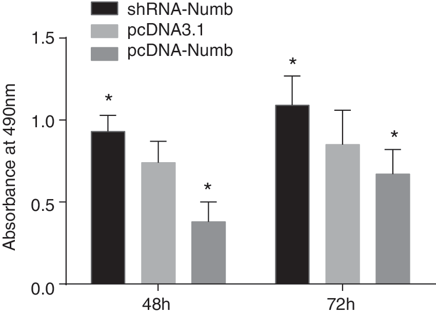
The proliferation results of the NSCLC stem cells after 48 and 72 hours of transfection. *Compared with the pcDNA3.1 group, p < 0.05. NSCLC, nonsmall cell lung cancer.
Apoptosis of NSCLC stem cells with over-expressed and knockdown Numb
Over-expression of Numb could promote apoptosis rate, and karyopyknosis and karyorrhexis were observed in NSCLC cells 29 ; in the present study, the apoptotic morphology and apoptosis rate were observed under a fluoroscope. Compared with the pcDNA3.1 group, the cells in the pcDNA-Numb group showed bright blue staining and dense fluorescence (Fig. 4A), obvious nuclear condensation and fragmentation, indicating a significant increase of apoptosis rate; the cells in the shRNA-Numb group showed less nuclear damage, and the apoptosis rate also decreased (Fig. 4B, C). The results showed that over-expressed Numb might promote the apoptosis of the NSCLC stem cells.
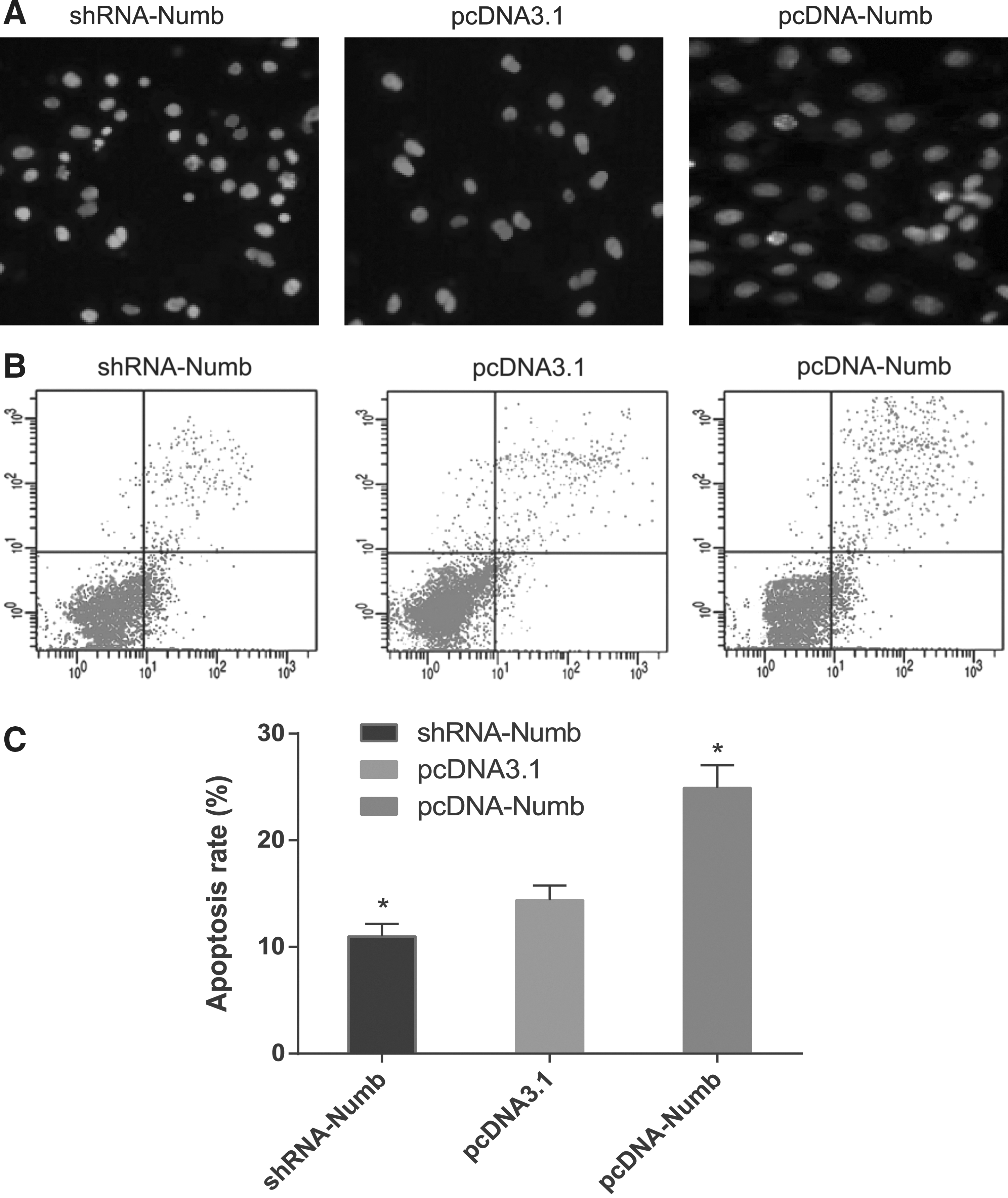
The apoptosis of the NSCLC stem cells in the pcDNA3.1 group, pcDNA-Numb group and shRNA-Numb group
Effects of Numb on radiation sensitivity of NSCLC stem cells
The OD value of each group of cells was detected by the MTT method. The cell survival rate of the cells irradiated with different doses of 60Coγ ray in the pcDNA3.1 group, pcDNA-Numb group and shRNA-Numb group was measured and compared (Fig. 5). A previous study showed that the survival rates of H292 and H1975 cells decreased along with the increased dosage. 29 In our study, the NSCLC stem cells after transfection in each group were irradiated with 60Coγ rays with different dosages of 0, 2, 4, 6, 8, and 10 Gy. The results showed that with the same radiation dosage, the survival rate of the cells transfected with over-expressed Numb decreased significantly compared with that of the cells transfected with vacant plasmid (p < 0.05), indicating that Numb over-expression might enhance the radiation sensitivity of NSCLC stem cells. The survival rate of the cells transfected with knockdown Numb increased significantly compared with that of the cells transfected with vacant plasmid (p < 0.05), indicating that Numb low expression might enhance the radiation resistance of NSCLC stem cells. In the three groups, the survival rate of the cells decreased with the increase of the doses of 60Coγ ray, suggesting that the lethality of radiotherapy to NSCLC stem cells is enhanced with the increase of the doses of 60Coγ ray.
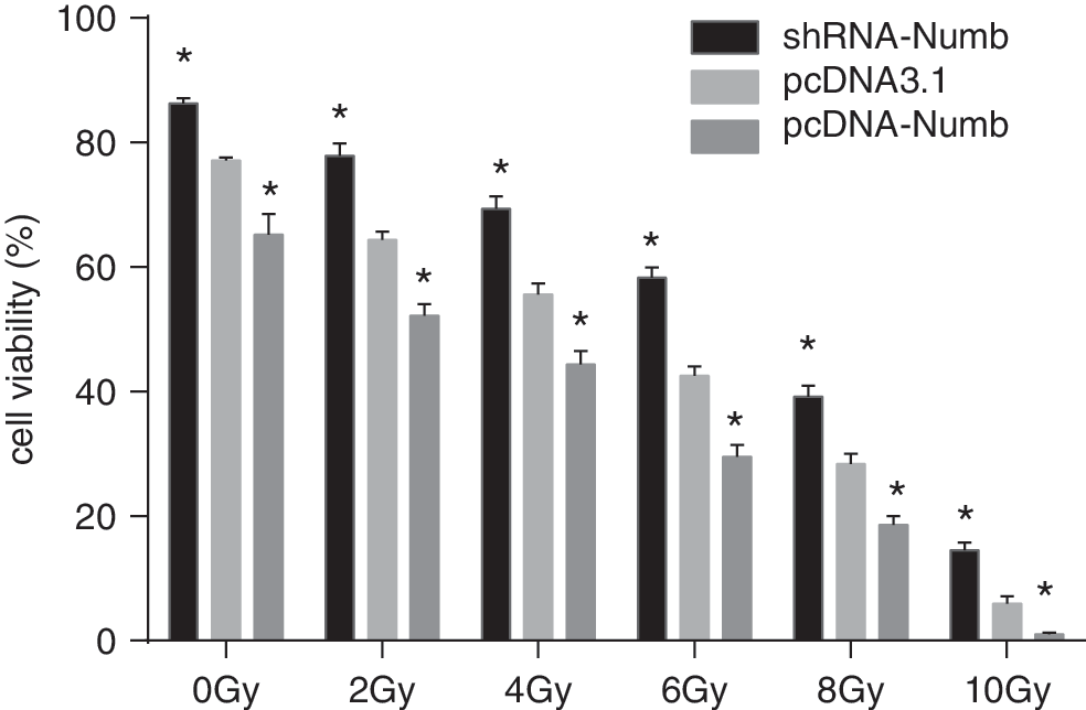
The survival rate of the NSCLC stem cells in each group with different doses of 60Coγ ray using the MTT method. *Compared with the pcDNA3.1 group, p < 0.05. MTT, 3-(4,5-dimethylthiazol-2-yl)-2,5-diphenyltetrazolium bromide.
The incubation ended with the appearance of cell clones that could be observed with the naked eye. The CFE was determined under a low-power lens; the selected doses were 2, 10 and 6 Gy (Fig. 6). The clonality of the cells in pcDNA-Numb group was significantly decreased compared with that of the cells in pcDNA3.1 group, indicating that Numb over-expression might enhance the radiation sensitivity of NSCLC stem cells. The clonality of the cells in the shRNA-Numb group was significantly enhanced compared with that of the cells in pcDNA3.1 group, indicating that Numb low expression might enhance the radiation resistance of NSCLC stem cells.

The colony-forming assay in the pcDNA3.1 group, pcDNA-Numb group and shRNA-Numb group (the selected doses were 2, 10, and 6 Gy).
According to the colony-forming assay (Table 1 and Fig. 7), compared with those of the cells in the pcDNA3.1 group, the values of D0, Dq, N, and SF2 of the cells in the pcDNA-Numb group decreased significantly (p < 0.05); however, the values increased significantly in the shRNA-Numb group, indicating that the radiation sensitivity of NSCLC stem cells was enhanced with the increase of Numb expression. Specifically, Numb over-expression might enhance the radiation sensitivity of NSCLC stem cells, and the radiation resistance of NSCLC stem cells might be related with the low expression of Numb.

The survival curves of the NSCLC stem cells for determination of the effects of Numb expression on radiation sensitivity of NSCLC stem cells.
Compared with the pcDNA3.1 group, p < 0.05.
Discussion
CSCs have exhibited radioresistant and chemoresistant properties. 30 To overcome radiation resistance in treatment of LSCLC, we shed light on the Numb gene. Numb gene expression was regulated in our study by vector construction and determined using RT-qPCR and Western blot. We explored the effects of the Numb gene on NSCLC stem cells, including proliferation, apoptosis, and radiation sensitivity, for the sake of substantiating our findings.
In the present study, we found that the NSCLC stem cells transfected with pcDNA-Numb showed inhibited proliferation and promoted apoptosis as observed from the results of MTT and staining. Over-expressed Numb was demonstrated to have the ability of inhibiting the proliferation of human clear cell renal cell carcinoma cells, and the suppressive effects were supposed to be attributed to down-regulation of cyclin D1 or MMP-9 expression. 31 Hong et al. reported that mRNA expression of Numb isoform 1 (Numb-1) was down-regulated in 66.7% of primary esophageal quamous cell carcinoma tissues, and they explained that ectopic expression of Numb-1 inhibited cell proliferation via inducing G2/M phase arrest. 32 Yingjie et al. showed that Numbl could suppress cell viability, inhibit cell proliferation, and promote cell apoptosis of lung cancer cells by abrogating TRAF6-induced activation of nuclear factor-kappa B (NF-κB). 33 Based on all that has been mentioned earlier, we speculate that the proliferation promotion and apoptosis inhibition of Numb gene might attribute to a similar mechanism, which needs to be verified in further research.
The most important finding in our study was the effect of Numb gene expression on the radiation sensitivity of NSCLC stem cells. We found that the survival rate of the NSCLC stem cells in the three groups declined with the increase of irradiation, and that the NSCLC stem cells with over-expressed Numb showed decreased survival rate, clonality, and the values D0, Dq, N, and SF2; whereas the cells with knockdown Numb showed the opposite effect. Radiotherapy could be used to control tumors in the early stage of tumor procession. 34 The optimal radiation dose for patients concurrently treated by chemotherapy and radiotherapy is typically 60–70 Gy, although 74 Gy has also been suggested. 8 Our study selected six different radiation doses for the tissues, ranging from 0 to 10 Gy, which indicated an obvious therapeutic effect with the survival rate changes in cells with different irradiation doses. Numb as a tumor suppressor has been reported to play a biological function via several pathways, including NOTCH, p53, and Hedhehog. 35 Studies have shown that tumor radioresistance might be associated with specific molecules, which sheds light on the mechanisms of cellular radioresistance: For example, p53 as apoptosis modulators regulate the radioresistance phenotype of lung and pancreatic cancers. 20 Yun et al. reported that depletion of hepatoma-derived growth factor-related protein 3 could induce apoptotic sensitization of radioresistant A549 cells via reactive oxygen species-dependent p53 activation. 36 Further, Numb enters into a tricomplex with p53 and the E3 ubiquitin ligase HDM2, thereby preventing ubiquitination and degradation of p53, and the loss of Numb expression may cause decreased p53 levels and increased chemoresistance. 37,38 We supposed that Numb has effects on the radiotherapy sensitivity of NSCLC cells via its involvement in the ubiquitylation of p53. In addition, P53 is a stress-responsive tumor suppressor encoded by the TP53 gene. 39 The p53 molecule can bind to DNA and regulate gene expression to prevent instability of the genome. 40 P53 was also reported to regulate the growth of malignant tumor cells, invasion, senescence, and apoptosis. 41 –43 Carcinomic DNA cells usually get damaged during radiation. 21 Then, we assumed that p53 might affect the radiation through its correlation with DNA.
To summarize, we explored the correlation between Numb gene expression and NSCLC stem cells and found that Numb gene inhibited proliferation, promoted apoptosis, and enhanced radiation sensitivity of NSCLS stem cells. However, due to limitations of the time period and the cost, we did not conduct the study on the effects of the expression of p53 and HDM2 on radiation sensitivity of NSCLC stem cells. Thus, the underlying mechanism put forward here needed verification in more studies in the future.
Footnotes
Acknowledgment
The authors express their sincere appreciation to the reviewers for their helpful comments on this article.
Disclosure Statement
No competing financial interests exist.