
Abstract
Deregulated expressions of mucins have been found in various malignancies and play a pivotal role in carcinogenesis. MUC5AC, as a secreted mucin, is reported to be aberrantly expressed during epithelial cancer progression, including colon cancer. However, the mechanisms of the oncoprotein MUC5AC in the initiation of colon cancer requires further investigation. Here, we collected colon cancer tissues (n = 20) and corresponding paracancerous tissues (n = 20) and found that the expression of MUC5AC was significantly elevated in colon cancer tissues when compared with the corresponding paracancerous tissues. Immunofluorescence indicated that all colon cancer cell lines, including HT29, SW620, and the normal human intestinal epithelial cells FHC, showed the positive expression of MUC5AC, and SW620 exhibited the highest expression. Moreover, knockdown of MUC5AC in SW620 cells remarkably suppressed cell vitality and promoted apoptosis and G1 cell cycle arrest, resulting in the impaired ability of colony formation. Furthermore, the inhibition of MUC5AC in SW620 cells dramatically repressed the cell migration and invasion. These results demonstrated that MUC5AC as an oncogene could be a promising target in the treatment of colon cancer.
Introduction
Even with the advancements in technology and health awareness (e.g., the adoption of widespread screening efforts), the prevalence of colorectal cancer (CRC) is still the third leading cause of cancer-related death in the United States and has been elevating in developing countries, including China. 1,2 Epidemiological statistics indicate that ∼1,360,000 new CRC cases will be diagnosed worldwide. Overall, the 5-year survival rates for patients with CRC are about 65%. 3,4 Many environmental factors, such as an unhealthy diet and exposure to tobacco and other occupational hazards, have been demonstrated to destroy the mucus barrier of the gastrointestinal system and to induce the dysfunctions of oncogenes and tumor suppressor genes that are involved in the initiation and development of colon cancer. 5 Therefore, further investigations on the cellular and molecular mechanisms of colon cancer are crucial for the discovery of more effective therapeutic targets.
Mucins belong to a heterogeneous group of high-molecular-weight, large O-glycoproteins that are composed of a long peptidic chain linked with oligosaccharidic chains and are predominantly expressed at the apical surface of the epithelial cells. Epithelial mucins could be structurally divided into two families: membrane-tethered mucins (e.g., MUC1, MUC3A, MUC3B, and MUC4) and secreted or gel-forming mucins (e.g., MUC2, MUC5AC, and MUC5B). 6,7 Under physiological conditions, tissue-specific expressions of MUC, particularly in the aerodigestive and genitourinary system, have been found to play an essential role in lubricating and protecting the epithelial and body lumens against harmful exogenous and endogenous environmental stresses (e.g., bacteria, drugs, and toxins) and also in cellular adhesion, differentiation, and immunity via transmitting growth and survival signals to the interior of the cell. 8 Mounting evidence, however, indicates that the levels of MUCs are significantly deregulated during carcinogenesis. 6,9
Membrane-bound mucins MUC1 and MUC4 have been extensively studied. In non-small cell lung cancer (NSCLC), high MUC1 expression was frequently detected in adenocarcinomas that indicated poorer overall survival and disease-free survival of patients with NSCLC. 10 Swartz et al. reported that the mRNA expression of MUC4 was remarkably enhanced in invasive pancreatic adenocarcinoma but not detectable in normal pancreas. 11 In pancreatic cancer cells, tumor-associated MUC5AC as a functional immunosuppressive agent could support the escape of pancreatic cancer cells from immunosurveillance in pancreatic cancer progression. 12 In addition, the upregulated level of serum MUC5AC was assessed in biliary tract cancer, 13 and MUC1 and MUC5AC expression was also found to be overexpressed during colon cancer progression. 14
Although the clinical significance of mucins was identified in various cancers, the mechanism of its tumorigenetic role was not well understood. MUC1 cytoplasmic tail (MUC1-CT) could suppress the p53-dependent apoptotic response to DNA damage, coactivate p21 at the transcriptional level, and attenuate activation of Bax transcription to inhibit the activation of intrinsic apoptotic pathways in the genotoxic stress response. 15 MUC5AC knockdown resulted in significantly decreased expression of integrins (α5, β1, β3–β5) and the phosphorylation of FAK, leading to the impaired ability of migration in the lung cancer cell line. 16 Of note, the mechanisms of oncoprotein MUC5AC in colon cancer need to be further identified.
In this study, the expression of MUC5AC was found to be significantly elevated in colon cancer tissues when compared with the corresponding paracancerous tissues in the clinical setting. Moreover, abrogation of MUC5AC in SW620 cells could impair cell vitality and induce the apoptosis and G1 cell cycle arrest, leading to the decreased ability of colony formation and cell migration and invasion. This suggests that inhibition of MUC5AC could be a potential tumor therapeutic strategy for the treatment of colon cancer.
Materials and Methods
Cell culture, tissue collection, and reagents
The human colon cancer cell line HT29, SW620 cells, and the normal human intestinal epithelial cells FHC were cultured in Dulbecco's modified Eagle's medium (DMEM) that was supplemented with 10% FBS (Life Technologies), ampicillin, and streptomycin at 37°C, under 5% CO2 conditions. Colon tissues (n = 20) and corresponding paracancerous tissues (n = 20) were collected from The Second Affiliated Hospital of Guilin Medical University. According to WHO classifications, all of the patients diagnosed with primary colon cancer were confirmed by hematoxylin and eosin staining by experienced pathologists. Patients who were diagnosed with autoimmune or other malignant diseases and pregnant or lactating individuals were excluded from our experimental group. No patients underwent preoperative chemotherapy and/or radiotherapy. This study was approved by the Research Ethics Committee of The Second Affiliated Hospital of Guilin Medical University. Written informed consent was obtained from all of the patients. SiRNA of MUC5AC or control was purchased from RiboBio siRNA-MUC5AC:5′-CCCUGCUCCUGGAAUAAAUTT-3′ (Guangzhou, China). Anti-MUC5AC and GAPDH antibodies were obtained from Cell Signaling Tech (Denver, MA).
Immunohistochemistry
The expression of MUC5AC was analyzed immunohistochemically on 2-μm-thick, formalin-fixed, and paraffin-embedded specimen sections. Slides were incubated in three washes of xylene for 5 minutes each and were followed by two washes of 100% ethanol for 10 minutes, 95% ethanol for 10 minutes, and ddH2O for 5 minutes each. Antigen unmasking was prepared by boiling in pH 9.0, 10 mM Tris/1 mM EDTA, blocked with 3% hydrogen peroxide for 10 minutes at room temperature, and washed. The primary antibody was then incubated with the FFPE specimen sections at 4°C overnight, and the EnVision Detection System kit (DAKO, Denmark) was used for the DAB chromogen followed by nuclear staining using hematoxylin. Neutral gum was used to cover the sliders, and drying occurred at room temperature for counting.
Immunofluorescence assays
The cells were fixed with 4% formaldehyde in phosphate-buffered saline (PBS) for 15 minutes and rinsed thrice with PBS. Then, cells were permeabilized with 100% methanol for 10 minutes at −20°C, blocked with 3% bovine serum albumin in PBS for 60 minutes, and incubated with primary antibodies overnight at 4°C. After rinsing three times in PBS, coverslips were incubated in fluorochrome-conjugated secondary antibody for 1–2 hours at room temperature in the dark and then stained the nucleus with DAPI (Bioword, China). The coverslips were mounted onto the glass slides with neutral gum and observed by an FV10i confocal microscope (OLYMPUS, Japan).
Cell transfection
According to the manufacturer's protocol, 2 × 10⁵/Well SW620 cells were cultured in a 12-well plate, transfected with siRNA-control/MUC5AC by Lipofectamine2000 (Invitrogen), and cultured for the indicated time.
RNA isolation and quantitative real-time PCR
According to the standard RNA isolation protocol, total RNA from tissues or cells was extracted using Trizol reagent (Invitrogen). The reactions were performed on the ABI7500 Real-Time PCR System (Applied Biosystems), and the expression levels of MUC5AC were normalized to GAPDH for gene expression. The primers were purchased from GenePharma (Shanghai, China) and are listed next:
Western blots
Cells for western blots were collected, and total protein was isolated from the cell samples according to the manufacturer's protocol. The total proteins were dissolved in SDS-PAGE loading buffer, heated at 100°C for 5 minutes, separated on 10% polyacrylamide gel, and transferred to nitrocellulose membranes (Amersham Biosciences). The membranes were blocked in 5% nonfat milk in TBST buffer (Tris Buffer Saline containing 0.1% Tween-20) for 1 hour at room temperature, and, subsequently, incubated overnight at 4°C by the appropriately diluted primary antibodies. After washing with TBST buffer, the blots were then incubated with HRP-conjugated secondary antibody for 1 hour at room temperature. After washing with TBST buffer, the signal of proteins was detected.
CCK-8 assay
After the transfection, cells were harvested and washed with PBS; the cell counting kit-8 (Kumamoto, Japan) mixed with DMEM was used for cell viability assay; and the absorbance was measured at 450 nm by a microplate reader.
Colony formation assay
After transfection for the indicated time, the cells were harvested and resuspended in complete medium containing 10% FBS and were seeded into 6-well plates for 10 days. Using 0.1% crystal violet, cells fixed with methanol for 15 minutes were visualized under a dissection microscope (Olympus, Japan) and colonies consisting of 50 cells or more were counted.
Flow cytometry assay
For the apoptosis analysis, the cells were fixed in cold 70% ethanol at −20°C for 2 hours. Then, cells were treated with 10 mg/mlRNase and stained with 2 μL of annexin V. These were mixed with 2 μL of Propidium iodide (eBioscience), used according to the manufacturer's instructions, and quantified by flow cytometry on an FACS Calibur instrument.
Cell migration and invasion assays
The migration and invasion ability of SW620 cells was measured by transwell chambers with an 8-μm pore size. For migration assays, 6 × 10⁴ cells suspended in DMEM were seeded in the upper chamber and the lower chamber contained 500 μL DMEM/10% FBS. For invasion assays, a 50 μL mixture of Matrigel (BD Biosciences) and DMEM (1:1) was precoated in the upper chamber overnight. SW620 cells were incubated for 24 hours, and cells that did not migrate or invade were removed by using a cotton swab. The cells were stained by using crystal violet staining and counted under an inverted microscope. Four random views were selected to count the cells, and the independent experiments were repeated three times.
Statistical analyses
Using the Statistical Package for Social Sciences version 16.0 (SPSS 16.0; SPSS, Inc., Chicago, IL) and the Prism statistical software package (Version 5.0; Graphpad Software, Inc.), the statistical analyses were performed. Unpaired t-tests or Mann–Whitney U tests were used to compare the two groups, and multiple-group comparisons were analyzed with one-way ANOVA. P < 0.05 was considered statistically significant. All experiments were performed at least three times.
Results
The expression of MUC5AC is elevated in colon cancer tissues
The tight control of mucins expression is essential to the normal tissue homeostasis, whereas the deregulation of mucins leads to excess of inflammation and even cancer. In this study, we collected colon cancer tissues (n = 20) and corresponding paracancerous tissues (n = 20). The mRNA expression of MUC5AC was analyzed by quantitative real-time PCR (Q-PCR), and the results showed that MUC5AC expression was significantly up-regulated in colon cancer tissues than that in the corresponding paracancerous tissues (Fig. 1A). To confirm the results, immunohistochemistry was performed and it was found that the protein level of MUC5AC was also enhanced in the tumor tissues (Fig. 1B), which implied that the increased mRNA and protein expression of MUC5AC in tumor tissues could be involved in the development of colon cancer.

The mRNA
Colon cancer cell lines harbor the positive expression of MUC5AC
To further investigate the function of MUC5AC in colon cancer, three cell lines were utilized, including two colon cancer cell lines HT29 and SW620 and one normal human intestinal epithelial cell line, FHC. The mRNA level of MUC5AC was determined and showed that the colon cancer cell lines HT29 and SW620 had higher mRNA levels of MUC5AC than that in normal human intestinal epithelial cells, FHC (Fig. 2A). In addition, using the immunofluorescence, we found that all three cell lines expressed the MUC5AC and the protein level of MUC5AC was the highest in SW620 (Fig. 2B), which provided the way to explore the tumorigenetic role of MUC5AC in colon cancer in vitro.
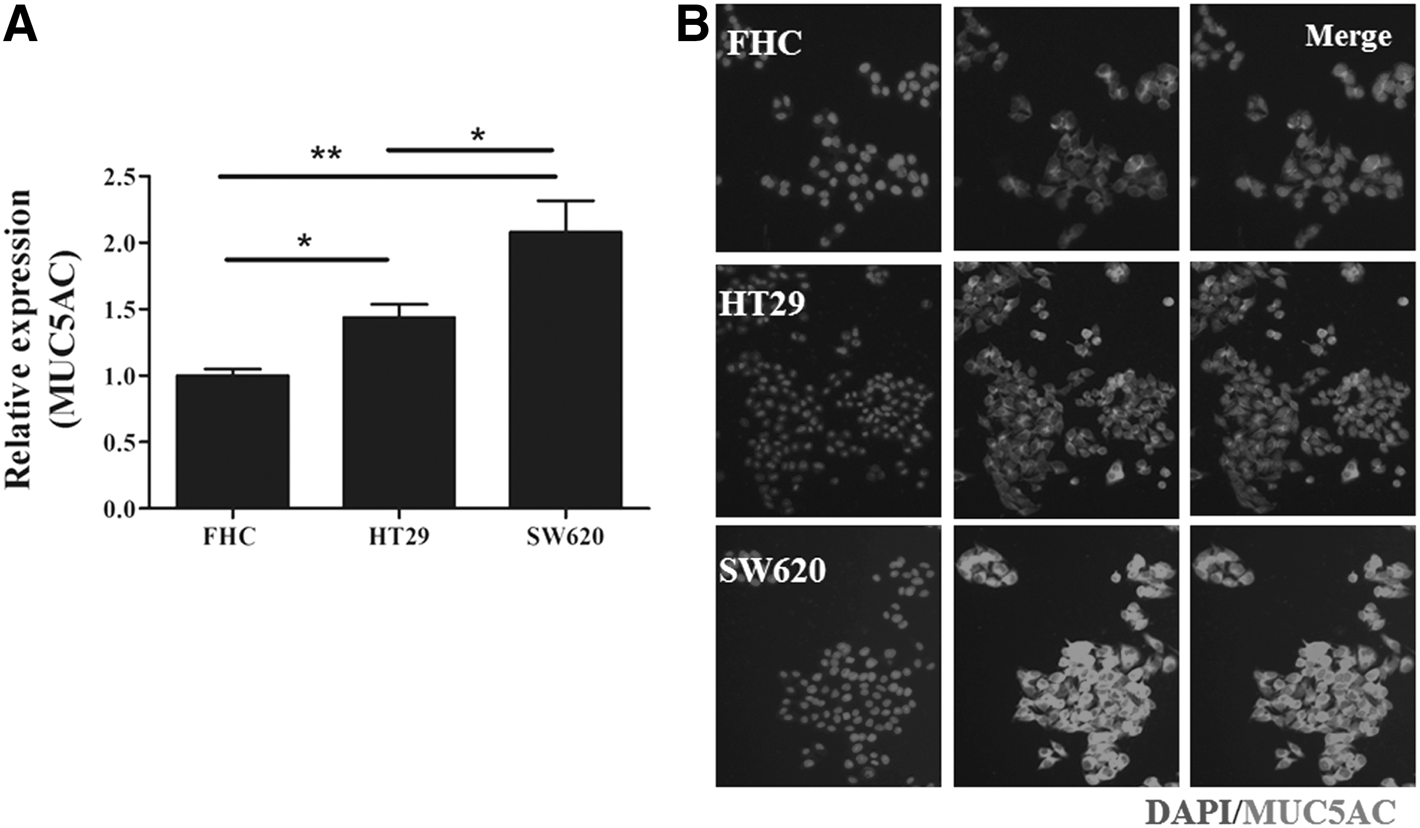
The mRNA
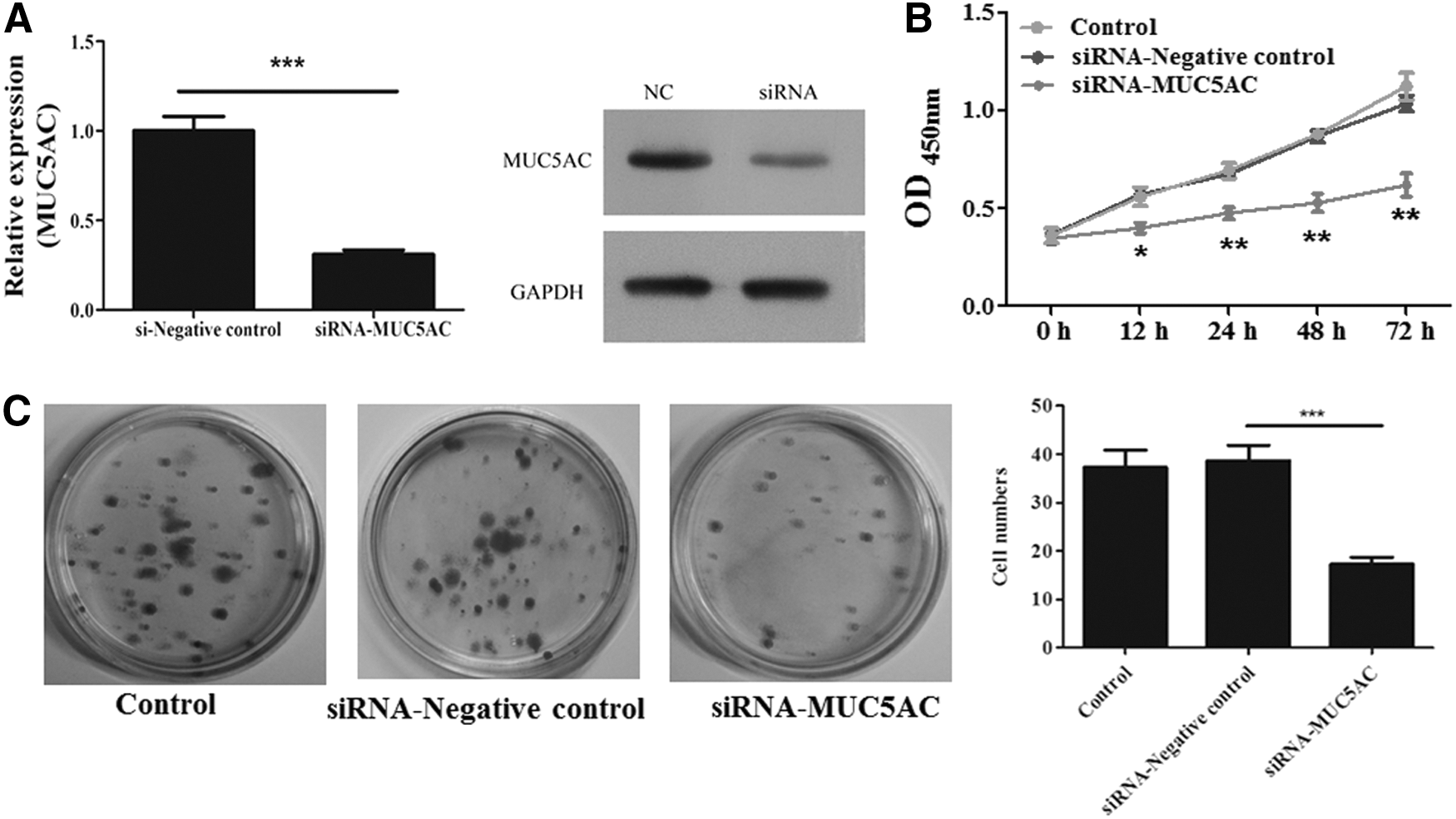
Inhibition of MUC5AC could negatively regulate the cell vitality and colony formation
Given that the mRNA and protein expression of MUC5AC was increased in colon cancer tissues and cell lines, MUC5AC might play a key role in tumor progression. According to the highest expression of MUC5AC in SW620, we overexpressed the siRNA of MUC5AC in the SW620 cell line for indicated times and determined the impacts of MUC5AC on the cell vitality. After the successful knockdown of MUC5AC (Fig. 3A), the CCK-8 assay was performed and indicated that the transfection of siRNA-MUC5AC could notably impair the cell vitality but the siRNA-control had no significant impacts on the cell vitality of SW620 (Fig. 3B). Moreover, colony formation assay also showed that the inhibition of MUC5AC by siRNA could remarkably abrogate the ability of colony formation of the SW620 cell line (Fig. 3C). These results indicated that inhibition of MUC5AC might decelerate the progression of colon cancer by regulating the cell vitality and colony formation.
Over-expressed siRNA-MUC5AC or negative control in SW620 cells
Inhibition of MUC5AC promotes apoptosis and cell cycle arrest
We hypothesized that the inhibition of MUC5AC might affect the apoptosis and cell cycle of SW620 cell line to decrease the cell vitality and colony formation. Therefore, we transfected the siRNA-control or siRNA-MUC5AC into the SW620 cell line, and the apoptosis and cell cycle were assessed. The results showed that the apoptosis rate was significantly enhanced when SW620 was overexpressed in siRNA-MUC5AC (Fig. 4A). The cell cycle was found to be arrested in G1, but siRNA-control had no effects on the apoptosis and cell cycle of SW620, which implied that the decreased cell vitality and colony formation were attributed to the MUC5AC-related apoptosis and G1 arrest (Fig. 4B).

The apoptosis
Inhibition of MUC5AC significantly abrogates the migration and invasion of SW620 cells
We had found the positive role of MUC5AC in the apoptosis and proliferation of colon cancer; further analysis was focused on its role in the metastasis in lung cancer. Transwell assay was performed, and the results showed that SW620 cells ectopically expressing siRNA-MUC5AC could significantly abrogate the ability of migration and invasion (Fig. 5A and B), indicating that the inhibition of MUC5AC could be a promising strategy to overcome the metastasis in colon cancer.

The migration
Discussion
As a major public health problem, CRC is the third most common cancer and the leading cause of mortality and morbidity throughout the world. 17 Environmental factors such as diet and dietary habits or physical inactivity and intrinsic factors such as mutations, activations, and deletions of oncogenes and tumor suppressor genes comprehensively interacted with each other to initiate and promote the progression of colon cancer. 5 The imbalance of the aerodigestive system has been demonstrated to increase the incidence of colon carcinogenesis. Mucins are found to be large O-glycoproteins expressed on the epithelia that provide a protective barrier against harsh insults from toxins, as well as pathogenic microbes, especially in the aerodigestive and genitourinary system, and their dysfunction are reported to be linked with inflammation and cancer. 18 In this study, we investigated the role of MUC5AC in colon cancer. The collected colon cancer tissues and cell lines indicated that the mRNA and protein level of MUC5AC was notably up-regulated in tumor sections and colon cell lines when compared with the normal control. In vitro, the inhibition of MUC5AC via siRNA-MUC5AC in the colon cancer cell line SW620 could induce the apoptosis and G1 arrest to repress the cell vitality and the colony formation. Of importance, the migration and invasion of SW620 cells could also be abrogated by the inhibition of MUC5AC and these results suggested that the MUC5C could be conducive to the targeted therapy of colon cancer.
Currently, mucins are broadly classified structurally into two main families: membrane-bound mucins (MUC1, MUC3A, MUC3B, MUC4, MUC12, MUC13, MUC15, MUC16, MUC17, and MUC20) and secreted or gel-forming mucins (MUC2, MUC5AC, MUC5B, MUC6, MUC7, MUC8, and MUC19). 19 Within the membrane-bound mucins, the gene encoding MUC1 is found to be located at 1q21, whereas MUC4 is located at chromosome locus 3q29. MUC3A, MUC3B, MUC11, MUC12, and MUC17 genes are located at 7q22.1. The secreted mucins MUC2, MUC5AC, MUC5B, and MUC6 are encoded by genes located in a cluster on chromosome 11p15. 20 It has been widely accepted that deregulation of mucin expression is not only a consequence but also a potential contributor to inflammation and cancer. As a mouse model of inflammatory bowel disease (IBD), IL10-/-mice overexpressing human MUC1 have higher IBD-like inflammation scores and higher incidence of the colitis-associated colon cancer initiation when compared with IL-10−/−mice. 21 In colon cancer, Krishn et al. had reported that MUC2 and MUC4 expression was found to be down-regulated whereas MUC1 and MUC5AC expression was up-regulated during adenoma-adenocarcinoma progression. 14 MUC5AC has also been shown to be an early oncofetal marker of colon carcinogenesis. 6 Here, we confirmed that the colon cancer tissues harbored higher expression of MUC5AC, as previously reported. Interestingly, decreased MUC5AC expression was associated with increased aggressiveness of gastric cancers, including tumor invasion and lymph node metastasis, which indicated that MUC5AC has a distinct function in different cancers. 22
However, the mechanism of mucins in the development of cancer has not been well clarified. Deregulation of transmembrane mucine (e.g., MUC1) is involved in constitutive activation of growth and survival pathways such as the Wnt/β-catenin and nuclear factor-κB pathways through their CT for the survival and proliferation of tumor. 8 Lau et al. found that epidermal growth factor stimulates Src-dependent MUC1 cleavage and nuclear localization to activate the invasion and metastasis-related gene integrin αvβ5, which emphasized the role of MUC1 in epidermal growth factor (EGFR)-dependent tumor cell metastasis. 23 EGFR-mediated activation was also found to up-regulate MUC5AC by means of ERK activation, which, in turn, leads to cancer cell proliferation in lung cancer. 24 Besides, silencing of MUC5AC participated in decreased proliferation and metastatic properties of lung cancer cells through the regulation of FAK signaling. 16 Our results indicated that, in colon cancer, the inhibition of MUC5AC could significantly induce the apoptosis and cell cycle arrest and also repress the metastatic properties of the colon cancer cell line SW620.
In conclusion, according to the increased expression of MUC5AC in colon cancer, the inhibition of MUC5AC was found to impair the proliferation of colon cancer by inducing cell apoptosis and cell cycle arrest. In addition, the strategies effectively targeting MUC5AC could be instrumental for preventing the metastasis of colon cancer.
Footnotes
Disclosure Statement
No competing financial interests exist.