




Abstract
Noninvasive in vivo tracking of adopted immune cells would help improve immunotherapy on glioblastoma. In this study, the authors tried to track adoptive CD8+ T lymphocytes in an in situ GL261 glioblastoma mouse model with magnetic resonance imaging (MRI). CD8+ T lymphocytes from spleen of preimmunized GL261 glioblastoma mice were labeled with superparamagnetic iron oxide, with polylysine as transfection agent. From Prussian blue staining, the labeling efficiency was 0.77% ± 0.06%, without altering cell viability and function. From anti-CD8, and anti-dextran staining, superparamagnetic iron oxide could be seen in the cytoplasm. In vitro imaging of agar gel mixtures with different concentrations of labeled CD8+ T lymphocytes was done with a 3.0T MR T2*WI sequence. Higher cell concentrations showed lower signal values. Twenty-four hours after tail vein injection of labeled and unlabeled CD8+ T lymphocytes, imaging of GL261 mice brain showed black spots at the periphery of the tumor in the labeled group only. Brain tumor pathology further verified infiltration of labeled CD8+ T lymphocytes in the tumor. Thus, preimmunized CD8+ T lymphocytes could be efficiently labeled with superparamagnetic iron oxide and tracked both in vitro and in vivo with 3.0T MRI.
Introduction
Glioblastoma (GBM) is the most common and aggressive primary brain tumor in adults. 1 Despite the current standard therapy of surgical resection, radiotherapy, and chemotherapy, GBM still grows rapidly afterward. Immunotherapy using cytotoxic (CD8+) T lymphocyte (CTL) is emerging to be an interesting area of research and potentially an important treatment option for GBM patients, 2,3 while improvements in clinical outcomes are modest. 4 –6 Noninvasive in vivo tracking of adopted T lymphocytes would be helpful in improving treatment outcomes for GBM patients.
Magnetic resonance imaging (MRI) provides excellent intrinsic contrast and high spatial resolution and is an indispensible tool for imaging in the central nervous system (CNS). It has been used in imaging different immunotherapies in brain tumor: imaging therapeutic nanoparticles 7,8 ; imaging intracellular oxygen dynamics by fluorine-19 MRI 9 ; imaging DCs with T1 contrast agent Gd(III)-HP-DO3A 10 ; tracking immunotherapy with changes with different MR sequences 11,12 ; tracking volume and signal changes of brain tumor by dynamic contrast-enhanced MRI, 13 and so on. Whereas there were very few studies about direct labeling and tracking of T lymphocytes to brain tumor.
Superparamagnetic iron oxide (SPIO) particles are a class of MRI contrast agent that is composed of nanosized iron oxide crystals coated with dextran or carboxydextran. It endows cells of interest with straightforward hypointense contrast enhancement after labeling. 14 Compared to T1 agents, SPIO-based T2 agents appear to be the preferred MRI contrast agents for monitoring cells due to the high sensitivity and excellent biocompatibility. Extensive efforts have been devoted to the development of SPIOs, 15 while ferumoxides (Feridex, Berlex Laboratories; and Endorem, Guerbet) and ferucarbotran (Resovist, Bayer HealthCare) are the two particles that are clinically available. Ferucarbotran was coated with carboxydextran, with a hydrodynamic diameter ranging between 45 and 60 nm. It is smaller than ferumoxides (120–180 nm) in size, which would be easier for endocytosis of T lymphocytes.
In this work, the authors labeled CTLs with a magnetic nanoparticle (MNP)—ferucarbotran and a transfection agent polylysine (PLL). They tracked the therapeutic T lymphocytes targeting GL261 glioblastoma in a murine mouse model with 3.0T MR.
Materials and Methods
CD8+ T lymphocytes
CD8+ T lymphocytes were isolated from the spleen of mice with GL261 tumor in the brain, using MACS separation system (CD8a+ T Cell Isolation Kit II; Miltenyi Biotec, Auburn, CA) and resuspended in RPMI1640 complete medium (10% fetal bovine serum, 100 U/mL penicillin, and 100 U/mL streptomycin). Cells were then activated and proliferated for 3–5 days in 24-well plates precoated with anti-CD3 (5 μL/mL, anti-mouse CD3 functional grade purified; eBioscience), supplemented with anti-CD28 (1 μg/mL, anti-mouse CD28 functional grade purified; eBioscience) and IL-2 (10 μL/mL, recombinant mouse IL-2).
Cell labeling
Different concentrations (100, 50, 25, 15, and 5 μg/mL) of SPIO (ferucarbotran, 0.5 mmol Fe/mL; Bayer HealthCare) and PLL (3 μg/mL; Sigma, St. Louis, MO) were gently shaken for 1 hour to get SPIO–PLL mixture. Stimulated CD8+ T lymphocytes were incubated with this mixture for 3 hours (37°C, 5% CO2). To quantify the labeling efficiency, Prussian blue staining of labeled cells was analyzed by counting the ratio of cells with blue spots (a minimum of 100 cells/FOV was analyzed, 10 FOVs from each slice). Stimulated CD8+ T lymphocytes without SPIO labeling were used as control. Viability of labeled (5 μg/mL SPIO, 3 μg/mL PLL) and control T cells was evaluated via Trypan blue staining following the formula: cell viability = (total number of cells − the number of blue stained cells)/total number of cells × 100% (a total of 30 groups [100 cells in each group] from each sample were counted and analyzed).
GL261 tumor cells were transfected with lentivirus vectors (lenti-Gluc) and cocultured with labeled (Labeled) and unlabeled CD8+ T lymphocytes (Unlabeled). GL261 tumor cells without CD8+ T lymphocytes were used as control (Sham). Twenty-four hours later, Gaussia luciferase (G-luciferase) level from medium of these groups was tested to assess GL261 tumor cell proliferation.
Cell staining
Slices of labeled (5 μg/mL SPIO, 3 μg/mL PLL) and control T lymphocytes were washed, dried, and fixed. Prussian blue staining: Slices were incubated with 2% potassium ferrocyanide solution and 2% hydrochloric acid mixture at 37°C for 30 minutes; washed and counterstained with 0.1% safranin solution.
Immunofluorescence staining: Slices were incubated with primary antibodies (CD8 alpha antibody, rat monoclonal; NOVUS Biologicals) overnight at 4°C; washed and incubated with secondary antibodies (goat anti-mouse IgG [H + L]/FITC, Jackson) for 1–1.5 hours at room temperature; and embedded with Hoechst 33258 as nuclear counterstain (no. B-2883; Sigma). Stained slices were analyzed with Olympus BX51 microscope equipped with an Olympus DP-70 digital acquisition system.
Electron Microscopy: Labeled T cells were pelleted, fixed with 2% glutaraldehyde in 0.1 M sodium cacodylate buffer (pH 7.2) for 1 hour at 4°C, treated with 0.5% OsO4 in cacodylate buffer for 1 hour, 0.25% uranyl acetate in acetate buffer (pH 5.0) overnight, and gradually dehydrated in ethanol (30%–100%). Samples were then immersed in 100% Epon/Araldite for 8 hours, placed in embedding molds, and polymerized in a 60°C oven for 48 hours. Samples were sectioned with a diamond and thin sections (100 nm) were picked up on copper grids and coated with carbon. Images were recorded on a Hitachi 7100 transmission electron microscope at 50kev, with an AMT Advantage 10 Image Acquisition System using a Kodak Megaplus 1.6i CCD camera system.
In vitro MRI
1 × 10⁶, 2 × 10⁶, 5 × 10⁶, 10 × 106 of SPIO-labeled CD8+ T lymphocytes were thoroughly mixed in 1% agar gel solution (corresponding amounts of unlabeled CD8+ T lymphocytes were also added to make the final cell count 10 × 106 in each sample). 10 × 106 unlabeled control CD8+ T lymphocytes were used as control. Samples were imaged with clinical 3.0T magnetic resonance (GE SIGNA VH/I 3.0T) and a mouse coil (inner diameter 3 cm). The axial images were acquired using a T2*WI sequence with the following parameters: gradient echo sequence, TR 900 mseconds, TE 20 mseconds, flip angle 15°, FOV 90 × 90 mm, matrix size 384 × 384, slice thickness 1.0 mm, NEX = 1, and resolution 0.23 × 0.23 × 1 mm. Signal value for each sample was calculated in the postprocessing working station (a total of 21 different FOVs from 3 different slices for each sample).
Animal models
Animal experiments were approved by the Institutional Animal Care and Use Committee of Fudan University. Mice (C57BL/6 to 8 weeks, female; Laboratory Animal Center, Medical College of Fudan University, China) were anesthetized with pentobarbital sodium and fixed in a stereotactic head frame (David Kopf Instruments, Tujunga, CA). GL261 tumor cells (1 × 10⁵) were injected into the deep frontal white matter (2.0 mm lateral and 1.2 mm anterior to the bregma) at a depth of 3.0 mm.
In vivo MRI
Three weeks after intracranial injection of tumor cells, control CD8+ T lymphocytes (2 × 10⁷, n = 4, without SPIO labeling) and labeled CD8+ T lymphocytes (2 × 10⁷, n = 6, SPIO labeling) were adoptively transferred via tail vein. Before and 24 hours after adoptive transfer, under general pentobarbital sodium anesthesia, mice brain was axially imaged (MR: GE SIGNA VH/I 3.0T; mouse coil: inner diameter 3 cm, US) using a T2*WI sequence (gradient echo sequence, TR 200 mseconds, TE 6 mseconds, flip angle 15°, FOV 50 × 50 mm, matrix size 256 × 256, slice thickness 1.0 mm, NEX = 2, resolution 0.20 × 0.20 × 1 mm). Right after imaging, mice brain were frozen and sectioned. Frozen brain sections were stained with Prussian blue, anti-CD8 (CD8 alpha antibody, rat monoclonal; NOVUS Biologicals; goat anti-mouse IgG [H + L]/FITC; Jackson), and anti-dextran (mouse monoclonal antibody, Clone DX-1, Stemcell Technologies; Cy3-conjugated AffiniPure mouse anti-goat IgG [H + L] Jackson) staining.
Statistical analysis
All data were processed with Prism version 5.0d. Cell viability was analyzed with unpaired t test. G-luciferase concentrations of different groups and in vitro MRI signal values of different cell samples were compared using one-way ANOVA. A level of p < 0.05 was considered statistically significant.
Results
SPIO labeling of CD8+ T lymphocytes
After 3–5 days of activation, CD8+ T lymphocytes became larger in size (Fig. 1A). From Prussian blue staining, iron nanoparticles showed as dispersed blue spots in the cytoplasm of labeled T lymphocytes, not in the control T cells (Fig. 1B, C), with a labeling efficiency of 0.77% ± 0.06% (5 μg Fe/mL, 3 μg/mL PLL). From the immunofluorescent staining, the green dextran coat of SPIO granules dispersed around the blue cell nucleus (Fig. 1D–F). Electromicroscopy images further verified the cytoplasm deposition of SPIO particles, which showed as several single large particles (Fig. 1G).

Incorporation of SPIO by CD8+ T lymphocytes.
The best labeling condition was figured out to be 5 μg Fe/mL with 3 μg PLL/mL. Three hours after labeling, the survival of labeled and unlabeled cells was 87.36% ± 1.07% and 89.57% ± 0.86%, respectively, with no significant difference (p = 0.5161, Fig. 1H). Cell structures were normal from electromicroscopy, showing no obvious toxic effect at cellular and subcellular levels. Twenty-four hours after coculture, the G-luciferase concentration from GL261 cells cocultured with labeled (141.7 ± 11.15, n = 6, p = 0.0039) and unlabeled CD8+ T lymphocytes (129.4 ± 14.09, n = 6, p = 0.0003) was lower than that from GL261 cells without CD8+ T lymphocytes (178.6 ± 22.08, n = 6, Fig. 1H). However, there was no significant difference between the labeled and unlabeled group (p = 0.4171). Thus, SPIO labeling did not alter the function of CD8+ T lymphocytes for inhibiting GL261 proliferation.
In vitro MRI of CD8+ T lymphocytes
Images of agar gel samples with different concentrations of labeled CD8+ T lymphocytes showed that samples with higher concentrations of labeled T lymphocytes gave lower signals. Except for the signal values between the 1 × 10⁶ cell concentration group and the control group, there was statistically significant difference in signal values between different concentration groups with the control group (p < 0.05, Fig. 2).
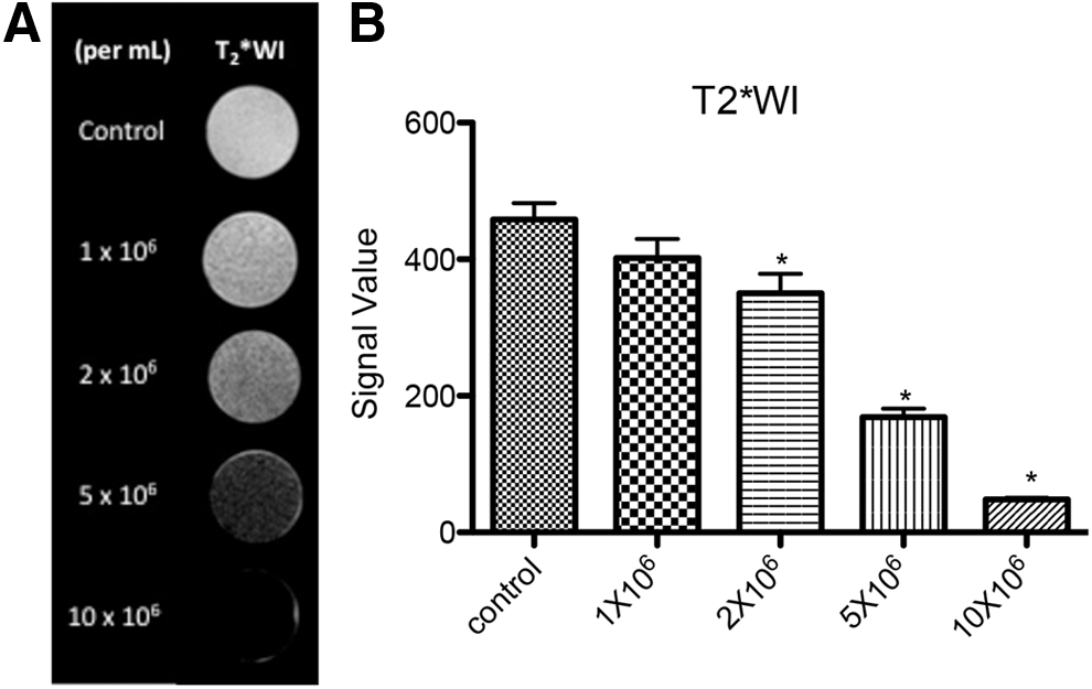
In vitro MR imaging of SPIO-labeled CD8+ T lymphocytes.
In vivo MR tracking of labeled CD8+ T lymphocytes
Serial mice brain images showed that 3 weeks after intracranial transplantation of GL261 tumor cells, all four mice in the control group and six mice in the cell tracking group developed brain tumor, which showed as high signal in the left hemisphere (some crossed the midline). In pretreatment images, there was no dark signal in or around brain tumor in these two groups (Fig. 3A–D). 24 hours after adoptive transfer, low signals (dark areas) were seen around the brain tumor in five of six mice in the cell tracking group (Fig. 3G, H), while none in the control group (Fig. 3E, F).
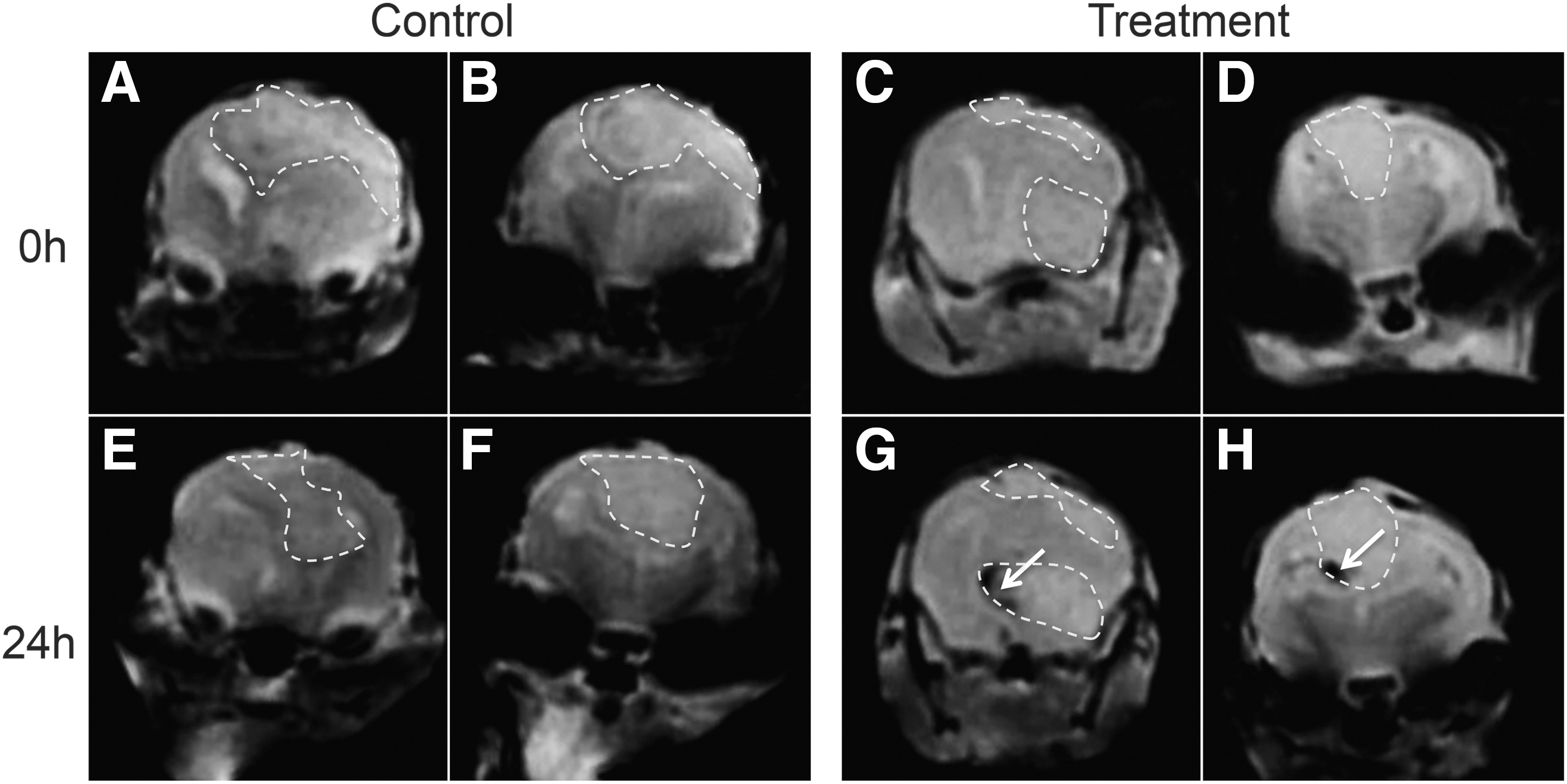
In vivo MR tracking of SPIO-labeled CD8+ T lymphocytes homing to GL261 brain tumor. Serial MR imaging was performed before (0 hour) and 24 hours after adoptive transfer of control and SPIO-labeled CD8+ T lymphocytes.
Histological results
Right after imaging, mice brains were sectioned and stained. From H&E staining, there was obvious nuclei heteromorphy at the tumor site (Fig. 4A). Prussian blue staining showed blue iron oxide deposition at the tumor site in the cell tracking group (Fig. 4B). Anti-CD8 immunofluorescence showed labeled CD8+ T lymphocytes (green fluorescence) at the tumor site in the cell tracking group (Fig. 4C–E) and this was further verified by colocalization of anti-dextran staining (Fig. 4F–H). These results showed SPIO-labeled CD8+ T lymphocytes recruiting to brain tumor.
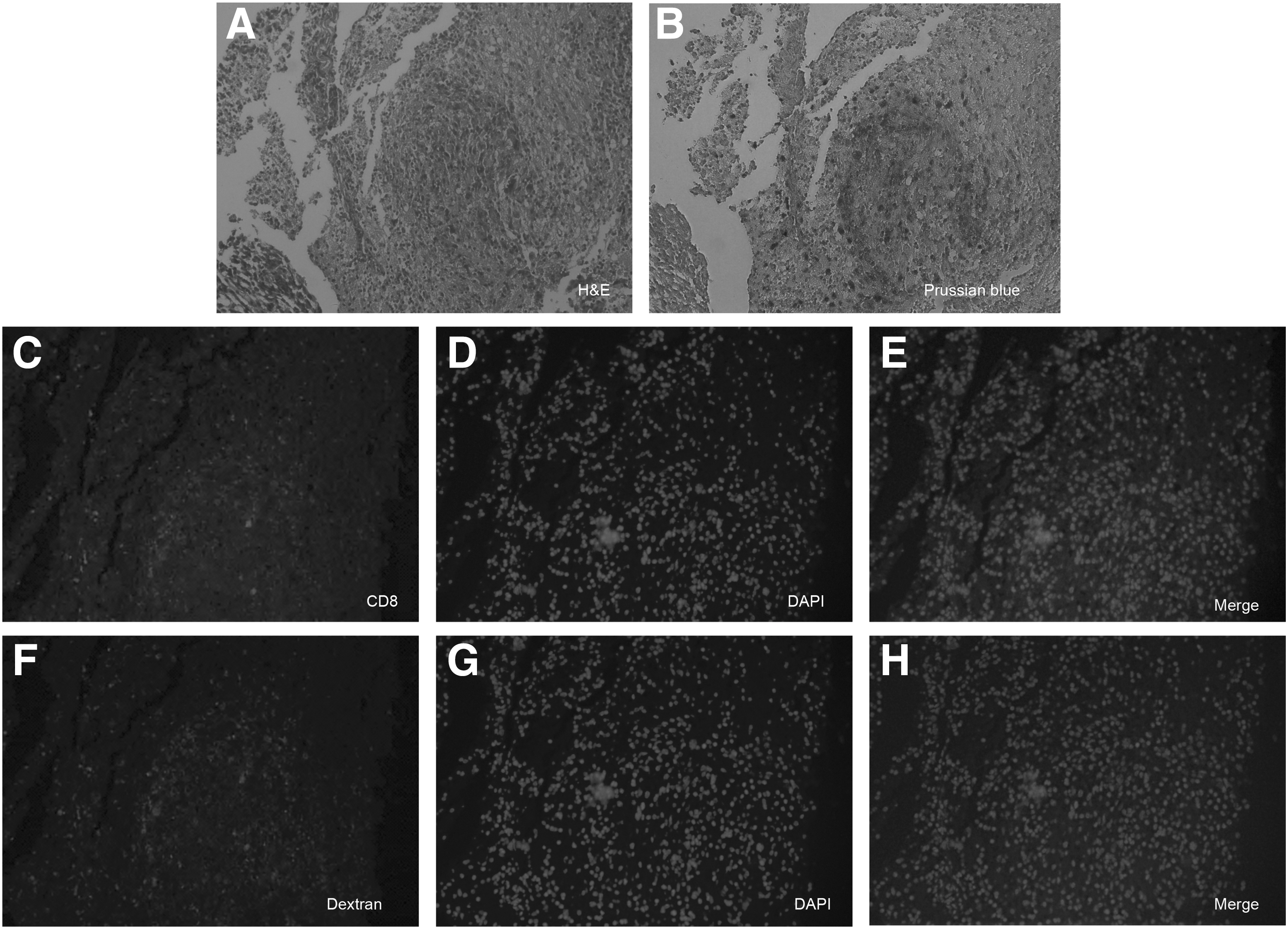
Histology of mouse brain after adoptive transfer of T lymphocytes.
Discussion
Noninvasive, reliable tracking of adoptive T lymphocytes recruiting to brain tumors would be helpful in quantifying the treatment efficacy and in development of more efficacious immunotherapy. In this study, the authors tracked SPIO-labeled CD8+ T lymphocytes targeting in situ GL261 glioblastoma mouse model with a clinical 3.0T MR.
There is significant interest in in vivo tracking of adoptive T lymphocytes. 16 –25 Regarding the animal models that have been used, most of the studies were done in immune-deficient mice. 11,19,23 –26 One advantage of immune-deficient mice is that tumor cell lines from human beings could be used, which is more clinically relevant. However, the immune environment plays a very important role in the capability of immunotherapy, which makes studies in immune-competent mice necessary. What is more, the blood–brain barrier and the immune privilege make brain tumors difficult and complex for adoptive immune cells to achieve a robust immune response 4 ; animal model in situ is also important in studying immunotherapy in CNS. In this study, the authors used the in situ GL261 murine model by direct intracranial injection of tumor cells to immune-competent mice, different from most of the previous studies of subcutaneous tumors in immune-deficient mice 16,17,25,27 ; thus, tracking adoptive transfer in this model is good for further studies in treatment outcome and improving immunotherapy.
SPIO is a class of intracellular MRI contrast agent that endows the cells of interest with straightforward hypointense contrast enhancement after proper labeling. 14 The poor phagocytic activity of activated T lymphocytes requires a fine labeling protocol without loss of cell viability. Physical properties of SPIO such as size, charge, and concentration are all important to efficient labeling of nonphagocytic cells. 28
Several methods may facilitate the labeling: surface modification of SPIO capsule, transfection agent assistance, cell surface receptor-mediated promotion of endocytosis, and so on. 29 –32 The most commonly accepted strategy is coating the anionic nanoparticles through electrostatic interactions with cationic transfection agents (TAs). 32 Based on the relative ratio of the transfection agent to nanoparticles, and at appropriate dilutions, macropinocytosis occurs. Different research groups have already compared different TAs in their ability to increase labeling efficiency and their toxicity. 33,34 Among these, the positively charged high-molecular-weight PLL significantly increases the efficiency of dextran-coated SPIO adherence to negatively charged cell surface.
To acquire the most efficient labeling of T cells, this study tried different SPIO-PLL ratios and the best condition was figured out to be 5 μg/mL SPIO with 3 μg/mL PLL. Compared to previous studies, the authors got a high labeling efficiency with a lower concentration of SPIO. 17,35,36 This could be because T lymphocytes were isolated from tumor preimmunized mice and further activated with necessary antibodies and cytokines, which could possibly enhance the phagocytosis of T lymphocytes. PLL is reported to be toxic with adverse effects occurring only above 2 μg/mL. 33,34 In this study, the authors used a PLL concentration of 3 μg/mL, which is slightly higher than the reported concentration. However, there is no toxic effect to lymphocytes, which could be because of the shorter incubation time (30 minutes vs. 24 hours) with T cells.
SPIO exhibits superparamagnetic properties and has been considered a novel intracellular contrast agent sensitive enough for in vivo real-time and long-term single-cell tracking by MRI. 30,37,38 Such labels are called “contrast agents” because they are not detected directly, but instead through their effect on local contrast of mobile water in tissues. Each SPIO nanoparticle gives an uneven weak magnetic field, altering the proton transverse magnetization phase, showing low signal in T2*WI sequence. In this case, intracellular localization and distribution of nanoparticles are important.
In this study, there is dense packing of SPIO particles inside cell vacuoles from the electron microscopy. This should lead to a larger local magnetic field and higher contrast in MR images, comparing to evenly distributed nanoparticles in the cell cytoplasm. It is an advantage of T lymphocytes with accumulated particles to act as several large particles with greater paramagnetic property. An in vitro study of agar gel samples with different concentrations of labeled T lymphocytes showed that with the increase of labeled cell concentration, signal decreased correspondingly. Since T cells are homogenously distributed in the agar gel and each cell contains nearly the same amount of SPIO nanoparticles, a higher labeled cell concentration gives higher SPIO concentration and greater signal loss. From this study, MRI signal intensity reduction induced by SPIO-labeled T cells can be detected with as few as 1 × 10⁶ T cells/mL, with an ∼80% cell labeling efficiency.
Different types of in vivo imaging technologies now allow for more careful investigation of adoptive immune cells. Bioluminescence imaging gives good susceptibility in CNS imaging, while the antitumor effect can only be described as the volume change of tumors, which is far from cellular level assessment 26,39,40 ; the limited depth permeability and poor spatial resolution also prohibited it from being used in the clinic. Nuclear imaging techniques exhibit superb tissue penetration through the skull and are highly quantitative. One clinical case report described T cell trafficking to human glioma with 18F-FHBG positron emission tomography imaging. 41 However, the low spatial resolution and the short half-life of radioisotope chemical material limited its application in long-term tracking of T cells in the CNS.
MRI provides excellent intrinsic contrast and high spatial resolution, and is an indispensable tool for longitudinal cell tracking and assessment in GBM at the cellular level. 18 Both Jacob's group 11 and Vrabec's group 12 have assessed immune response in brain tumor by indirectly describing signal changes of brain tumor in different sequences. Rygh's 13 group described immunotherapy in GBM by volume or signal changes of tumor with dynamic contrast-enhanced MRI. These studies were not direct evaluation of T cell accumulation in brain tumor. In this study, the accumulation of SPIO-labeled T lymphocytes directly showed as low signal around the brain tumor, corresponding to histology. To differentiate the therapeutic CD8+ T lymphocytes from the existing host CD8+ T lymphocytes, the presence of adoptively transferred CD8+ T cells was further confirmed by colocalization with SPIO via anti-dextran analysis.
One limitation of this study was that the control group was CD8+ T lymphocytes isolated from the spleen of GL261 brain tumor growing in mice without SPIO labeling. Since the aim of this study was to verify the possibility of tracking SPIO-labeled CD8+ T lymphocytes by MRI, the authors chose to use CD8+ T lymphocytes without SPIO labeling as control. Considering assessing the immune response, CD8+ T lymphocytes isolated from healthy mice and labeled with SPIO would be the ideal control group. In the future experiment for determination of the validity of this method, the authors would be sure to use that as the control group.
Conclusions
SPIO combined with PLL can simply and efficiently label CD8+ T lymphocytes while keeping cell viability and function. Results from this study provide important findings concerning the in vivo tracking of tumor-specific CD8+ T cells to brain tumor by MRI. This documentation of T-cell tracking provided critical parameters necessary to elicit elective antitumor immunity against CNS tumors, which would be beneficial for further evaluation of the biological activity of T cells and improve therapy efficiency.
Footnotes
Acknowledgments
This work was supported by the National Natural Science Foundation of China (81271633); Science Foundation of Qilu Hospital of Shandong University (2016QLQN26).
Disclosure Statement
There are no existing financial conflicts.