
Abstract
Objective:
Rhenium-188-HEDP is an effective radiopharmaceutical for the treatment of painful bone metastases from prostate cancer. The effectiveness of the β-radiation emitted by 188Re might be enhanced by combination with chemotherapy, using the radiosensitization concept. Therefore, the authors investigated the combined treatment of the taxanes, docetaxel and cabazitaxel, with 188Re in prostate carcinoma cell lines.
Materials and Methods:
The cytotoxic effects of single and combined treatment with taxanes and 188Re were investigated in three human prostate carcinoma cell lines (PC-3, DU 145, and LNCaP), using the colony-forming assay. The half maximal effective concentration (EC50) of all individual agents was determined. The combined treatment was studied at 0.25, 0.5, 1, 2, and 4 times the EC50 of each agent. The interaction was investigated with a regression model.
Results:
The survival curves showed dose-dependent cell growth inhibition for both the taxanes and 188Re. The regression model showed a good capability of explaining the data. It proved additivity in all combination experiments and confirmed a general trend to a slight subadditive effect.
Conclusions:
This proof-of-mechanism study exploring radiosensitization by combining 188Re and taxanes showed no synergism, but significant additivity. This encourages the design of in vivo studies. Future research should explore the potential added value of concomitant treatment of bone metastases with chemotherapy and 188Re-HEDP.
Introduction
In up to 90% of patients with prostate carcinoma, bone metastases will develop in the end stage (castration-resistant prostate carcinoma) of their malignant disease. 1,2 This complication often results in severe pain, impaired quality of life, risk of fractures, neurological symptoms, and hypercalcemia and it is a major cause of death. 1,3,4 The effective treatment of these complications is an unmet medical need in many patients. Current therapy of painful bone metastases usually consists of treatment with strong analgesics (e.g., opioids), external beam radiotherapy, or radionuclide therapy. Opioids are not always effective and may lead to serious side-effects. 5 External beam radiotherapy is suitable to treat a few localized metastases. For the treatment of diffuse osteoblastic skeletal metastases, bone-targeting radiopharmaceuticals, based on β- or α-emitting radionuclides, may be an effective and safe alternative. 6 –8 However, because effective treatment is not accomplished in all patients, there is an urgent need to improve the therapeutic modalities.
One of the approaches to augment the effectiveness of radiation-based therapy is combination with chemotherapy. The goal of this combined treatment, called chemoradiation or radiosensitization, is to enhance the eradication of malignant cells, for example, by overcoming radioresistance, while sparing healthy cells. 9 –11 In prostate cancer, targets of radioresistance include the apoptotic pathway, the DNA damage repair response, cell cycle regulation, and inflammatory response. 12,13 Combined treatment could be more efficacious using the same dosages or equally effective while using lower doses, thereby decreasing the toxicity. Different strategies used to reach this aim have been highlighted in recent reviews. 14,15
In preclinical research, additivity (i.e., enhanced cytotoxicity by combining chemotherapy and radiation) is a favorable outcome for clinical translation. When the combined effect is larger than the summation of the individual effects, the interaction is called supra-additivity or synergism. 16 The interaction is termed subadditivity or antagonism when the combined cytotoxicity is smaller than the summated individual effects. Although combining chemotherapy with external beam radiation has proceeded to a standard modality in some malignancies, 17,18 combination of chemotherapy with therapeutic radiopharmaceuticals has not been implemented in routine clinical care yet.
Rhenium-188-hydroxyethylidenediphosphonate (188Re-HEDP) is a bone-targeting therapeutic radiopharmaceutical based on the high-energy β-emitting radionuclide 188Re and the bisphosphonate etidronate. Clinical studies with this compound demonstrated its effectiveness and safety in metastatic prostate cancer patients. 19 –21 This radiopharmaceutical is, therefore, a good candidate for exploring the radiosensitization concept. However, no data are available of studies investigating the combined activity of 188Re and chemotherapy used in prostate carcinoma in a concurrent design. Docetaxel (DTX) is a first-line chemotherapeutic drug in advanced prostate cancer, while cabazitaxel (CTX) has been introduced for second-line treatment. 22,23 These agents act by stabilizing the microtubules, thus inhibiting cell progression through the G2/M phase of the cell cycle, resulting in cell arrest. Taxanes are known radiosensitizers. 24,25 During the G2/M phase, the cell is most radiosensitive because endogenic radioprotecting molecules are nearly absent in that phase. 26 Hence, the perturbed microtubule stability due to taxane interference compromises the DNA damage repair response following exposure to radiation and is thus an optimal target for enhancement of cytotoxic treatment by combination with 188Re. We, therefore, assessed the cytotoxic effects on prostate cancer cell lines of combined treatment of 188Re with taxanes.
Materials and Methods
Prostate Carcinoma Cell Lines and Cell Culture
The prostate carcinoma cell lines used in this study are PC-3, DU 145, and LNCaP. These well-known human cell lines have been derived from metastases from prostate cancer and exhibit different properties. 27 The cell lines were obtained from the American Type Culture Collection (CRL-1435, HTB-81, and CRL-1740; Rockville, MD). All cells were grown as monolayers in RPMI 1640 culture medium (Gibco, Life Technologies Europe, Bleiswijk, The Netherlands) supplemented with 10% fetal bovine serum (Gibco), 100 U/mL penicillin/streptomycin (Gibco), 1 mM sodium pyruvate (Sigma-Aldrich, St. Louis, MO), and insulin/transferrin/selenite medium supplement (Sigma-Aldrich). The cells were routinely cultured in 25-cm2 cell culture flasks (Greiner Bio-One, Frickenhausen, Germany) at 37°C in a humidified atmosphere of 5% CO2 and 95% air in a CO2 incubator (CB 150; Binder, Tuttlingen, Germany).
Colony-Forming Assay
For assessment of cytotoxic effects of the chemotherapeutic agents, 188Re and the combination of both, the colony-forming assay (CFA) was employed, a reliable cell survival assay determining the reproductive capacity of cells, which is considered the gold standard for research on radiosensitization. 28,29 In this method, also called clonogenic assay, the surviving fraction of viable cell colonies after exposure to cytotoxic agents is a measure of their cytotoxic efficacy. After incubation with one or two agents, the medium of the cultured cells was removed and the cells were washed twice with phosphate-buffered saline (PBS). After trypsinization, the remaining cells were collected and suspended in fresh medium and counted using a cell counter (CASY TT; Roche Innovatis, Bielefeld, Germany). A calculated volume corresponding to 200 or 400 cells was transferred in triplicate to 6-well plates (Greiner Bio-One). Fresh culture medium or conditioned medium for the LNCaP cell line (described before by Oprea-Lager et al. 30 ) was added and the well was swirled gently to obtain a homogeneous distribution of cells. The 6-well plates were incubated for 10 days at 37°C in the incubator. After incubation, the medium was removed and the colonies were fixed with 4% PBS-buffered formalin (JT Baker, Avantor, Deventer, The Netherlands) during a period of 1 hour, washed twice with demi-water, and stained with Giemsa solution (Merck Millipore, Darmstadt, Germany). After drying, the colonies were counted using a stereomicroscope (MS5; Leica AG, Heerbrugg, Switzerland). To be able to determine the plating efficiency, a blank sample consisting of medium, including cells, was added to each series of experiments.
Single-Agent Experiments—EC50 Determination
The EC50, being the concentration at which 50% of the colonies survive, was determined in triplicate for each cytotoxic agent in each cell line, using the time schedule that is shown in Figure 1A. A total of 50,000 cells for PC-3 and DU 145 or 100,000 cells for LNCaP were plated in cell culture flasks. To allow for cell attachment to the bottom of the flask, each agent was added 1 day later (Day 1) in different concentrations. The incubation time for the cytostatic agents was 3 to 4 days and, for 188Re, 1 to 3 days.
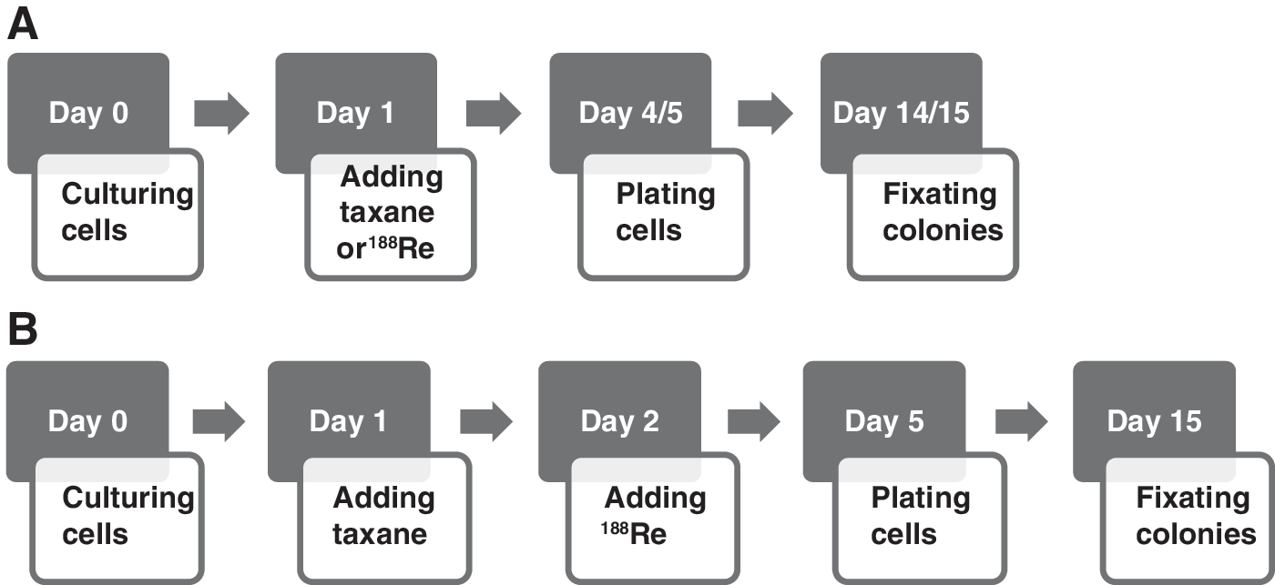
Time schedule of single-agent
Cytotoxic Agents
DTX concentrate for solution for infusion (Actavis, Hafnarfjordur, Island) and CTX (LC Laboratories, Woborn) were diluted in dimethyl sulfoxide (DMSO; Sigma-Aldrich) to obtain a stock solution of 2 mM, which was further diluted in PBS to reach final concentrations in the range of 0.5 to 5 nM for DTX and 0.5 to 25 nM for CTX. These ranges included the anticipated EC50 of both drugs.
188Re-perrhenate as eluate in sterile normal saline from a 188W/188Re generator (ITG, Garching, Germany) was diluted with sodium chloride 0.9% (B Braun Medical, Oss, The Netherlands) to a radioactivity concentration of 40 MBq/mL. This solution was further diluted to reach final radioactivity concentrations in the range of 0.5 to 5.0 MBq/mL. The authors deliberately chose to apply 188Re-perrhenate in a physiological solution instead of the clinically used 188Re-HEDP because 188Re-HEDP is an acidic solution that will negatively impact cell growth. Moreover, the ligand HEDP is not required for the proposed cytotoxic action of 188Re in cell lines.
Combination Experiments
For optimal cell growth inhibition, different incubation schemes of cytotoxic agents and 188Re were tested. The most efficacious treatment method (incubation for 1 day with DTX, followed by 3 days of incubation with 188Re, without washing step, data not shown) was implemented to analyze the combined effect of the taxanes and 188Re in all cell lines. Figure 1B represents the time schedule of the experimental design for the combination analysis.
In the combination experiments, culture flasks were seeded with 50,000 cells (LNCaP: 100,000 cells) and were incubated for 1 day at 37°C to achieve proliferating monolayers of cells. The next day, the culture flasks were allocated to four groups. The first group was treated with five different concentrations of the two taxanes for 1 day, followed by incubation with medium (including PBS) for 3 days. The second group was incubated for 1 day with medium (including PBS), followed by incubation with five different concentrations of 188Re. The third group was treated for 1 day with five different concentrations of taxanes, followed by incubation for 3 days with five different concentrations of 188Re. The fourth group (control) consisted of medium (with PBS), which was incubated for 4 days. The abovementioned five concentrations of the two taxanes and 188Re consisted of 0.25·EC50, 0.5·EC50, EC50, 2·EC50, and 4·EC50. A constant ratio design was applied for the combination experiments, resulting in the following data points: blank, 0.25·EC50 (taxane) + 0.25·EC50 (188Re), 0.5·EC50 (taxane) + 0.5·EC50 (188Re), EC50 (taxane) + EC50 (188Re), 2·EC50 (taxane) + 2·EC50 (188Re), and 4·EC50 (taxane) + 4·EC50 (188Re). All combination experiments were performed in triplicate.
Data Calculations
The surviving fraction was defined as the fraction (%) of colonies treated with a drug relative to the number of colonies without drug and was plotted in a survival curve. For each data point, the mean surviving fraction and the standard error of the mean (SEM) were calculated. The EC50 values were determined from the resulting survival curves and the mean EC50 and SEM were calculated.
Regression and Interaction Analysis
For the data analysis of the combination experiments, initially the software package CompuSyn (ComboSyn, Inc., version 1.0, 2004) was used. This software makes use of the median-effect principle, which is based on the mass action law. 31,32 Because CompuSyn could not handle some experimental data, information contained in these experimental points was lost.
Therefore, a regression and interaction analysis was performed. This analysis included additional experiments combining DTX and 188Re in the PC-3 cell line. The additional experimental runs were performed mostly in the interior of the original design space, with a balanced data point distribution across the area of interest. To be able to gather additional information, nonconstant ratio data points were chosen. For reference, some previously used data points, including the origin, were added.
To analyze the data from all combination experiments, a linear model with batch (accommodating potential differences between the experiments), single agent, taxane-188Re interaction (representing potential supra-additivity or subadditivity), and quadratic effects was fitted to the (transformed) data. Before performing this analysis, design points with obviously high leverage (being far from all other points and thereby exerting too much influence) were removed from the datasets. Different transformations were applied to the data and zero values were offset by 0.1%. The transformation (logistic, arcsin, or none) best satisfying the residual normality assumption accompanying the model (as evaluated by means of the normal probability plot) was selected.
The outcome of the regression and interaction analysis was expressed numerically as effect estimates and p-values. A negative sign of the effect estimate of the single agents indicates a negative effect: a larger dose results in smaller cell survival. Regarding the effect estimation of combined agents, a negative sign indicates supra-additivity, a positive sign subadditivity. R-squared (R 2) statistics (also called the coefficient of determination) was added as a measure of the capability of the model in explaining the data. This statistics consists of the correlation (squared) between the data and the prediction derived from the model and varies between 0 (indicating absence of explanatory power of the model) and 1 (indicating a perfect fit). Intermediate values represent various degrees of explanatory model performance. This statistics was mainly used to illustrate the added explanatory power of the interaction term in the fitted model.
To visualize the data generated by the model, heat maps were constructed. A heat map is a two-dimensional visualization of a data matrix. The rows and columns of the data matrix correspond to the settings of the studied agents, while each value of the data matrix represents the fraction of surviving cells at this combination of settings. These values are represented by colors. The shape of the color pattern of the heat maps indicates the type of interaction, comparable with the frequently used isobolograms. 33 A concave shape indicates supra-additivity, a convex shape subadditivity, and a more or less linear shape (mere) additivity.
Results
Single-Agent Experiments—EC50 Determination
The survival fractions of all single-agent experiments are provided in Supplementary Table S1 (Supplementary Data are available online at
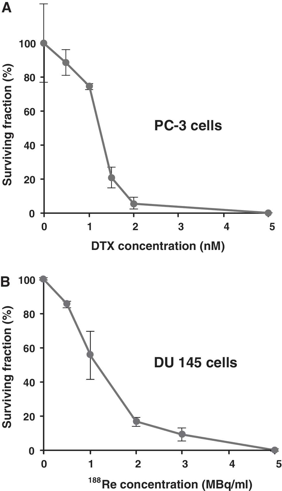
Dose–survival curve of PC-3 cells in the presence of DTX
The mean EC50 values calculated from the single-agent experiments for all cell lines and for all three agents used are displayed in Table 1.
The mean EC50 and SEM are displayed. The number of independent experiments is given in parentheses.
CFA, colony-forming assay; CTX, cabazitaxel; DTX, docetaxel; 188Re, rhenium-188; SEM = standard error of the mean.
Combination Experiments
The survival fractions of all combination experiments are provided in Supplementary Table S2. To visualize the combined effect of the cytostatic agent and 188Re, three-dimensional (3D) graphs were constructed. A representative 3D cell survival graph of a combination experiment is shown in Figure 3.
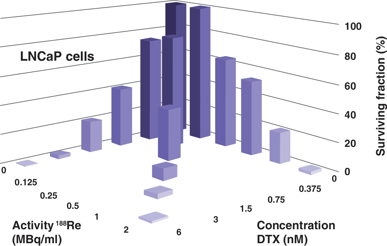
3D survival graph of LNCaP cells in the presence of DTX and 188Re in a combined CFA experiment. Data points: surviving fraction (%). The bars at the diagonal represent the cell survival in the presence of both agents. 3D, three-dimensional; CFA, colony-forming assay.
Regression and Interaction Analysis
The results generated by the regression model are summarized in Table 2. Supplementary Table S3 provides the raw data generated by the regression model for each dataset. Nearly all effect estimates concerning the single agents were significant and mostly large negative effects were observed. The regression model demonstrated a strong negative influence on cell growth for all agents and proved significant additivity. The effect estimates of the combined agents were positive and mostly small, indicating (slight) subadditivity. Furthermore, in most cases, the batch effect was found to be insignificant, indicating little environmental variation between the experiments (data not shown).
Effect estimation single agents: “−” small, negative; “−−” large, negative; “−−−” very large, negative; effect estimation combination: “0” no interaction, (mere) additivity; “+” small, subadditive; “++” large, subadditive. Detailed data are provided in the Supplementary Data.
The R-squared analysis derived from the model, with and without interaction term, is shown in Table 3.
R-squared values of the regression model fitted with and without the interaction effect.
Except for the experiments with CTX in LNCaP cells, the model showed a good capability of explaining the data. Moreover, the small difference between the R-squared calculations with and without interaction term demonstrates that this term adds little information.
For each cytotoxic drug-188Re-cell line combination, three heat maps were constructed based on the model fitted to the transformed data. A representative set of three heat maps is shown as an example in Figure 4.
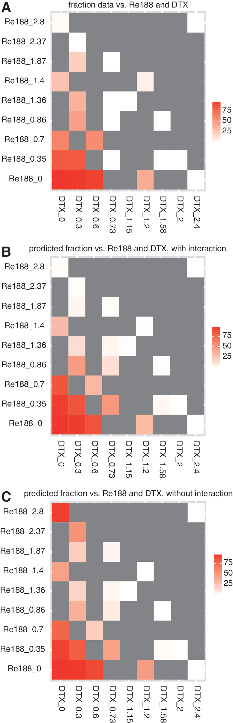
Heat maps of DTX and 188Re in PC-3 cells, including additional nonconstant ratio data points. For each grid point of the experimental design, the (averaged) surviving fraction is represented by color (value color coding in the legend to the right). The color gray represents unexamined grid points.
The heat maps demonstrated that the experimental data could be described well with the model (small difference between experimental and predicted data). Based on the shape of the color pattern, the heat maps showed mere additivity (color pattern linear shaped) or slight subadditivity (pattern somewhat convex shaped). Because the heat maps of the predicted fraction with and without interaction were nearly identical, the authors conclude that the interaction effect adds little to the prediction.
Discussion
To the best of the authors' knowledge, this is the first in vitro study to investigate the radiosensitization concept concurrently combining the therapeutic radionuclide rhenium-188 (188Re) and the frequently used taxanes, docetaxel (DTX) and cabazitaxel (CTX), in three human prostate carcinoma cell lines (PC-3, DU 145, and LNCaP). Using the CFA, all individual agents were shown to induce significant dose-dependent cell death in all cell lines. The regression model was well capable to explain the experimental data and confirmed the significant and mostly large negative effect on cell growth upon incubation with the individual drugs. The model demonstrated that combination of either of the two taxanes with 188Re resulted in significant additive effects. Interaction analysis indicated a trend toward slight subadditivity. However, this interaction effect is very small. Although one may argue that drawing conclusions based on one growth response assay is limited, CFA is the gold standard to determine cell reproductive death after treatment with ionizing radiation. 28,29 Moreover, the authors have replicated the experiments in different representative human prostate cancer cell lines to improve the robustness of their results.
In general, the EC50 values determined by carrying out the single-agent experiments corresponded well with the literature. 30,34 In PC-3 cells, the authors found the EC50 to be 1.0 nM for DTX and 1.6 nM for CTX. In a similar experimental design, Oprea-Lager determined these values to be around 1 nM for both drugs. 30,34 In DU 145 cells, the authors established an EC50 of 1.1 nM for DTX and 1.6 nM for CTX. These figures were 1 nM and 2 nM in a comparable study. 30 The EC50 in LNCaP cells was determined by to be 1.0 nM for DTX and 1.3 nM for CTX. In the study of Oprea-Lager, these values were 2 and 5 nM, respectively. This discrepancy might be the consequence of the behavior of LNCaP cells, which results in larger experimental variation than in other cell lines. The authors are the first to report EC50 values for 188Re in prostate carcinoma cell lines (1.0 MBq/mL in PC-3, 1.0 MBq/mL in DU 145, and 0.6 MBq/mL in LNCaP).
In the study, synergistic action between the two taxanes and 188Re was not demonstrated. As taxanes are proven radiosensitizers, 24,25 a synergistic effect when combined with the powerful β-emitting radionuclide 188Re was anticipated. In an earlier study, a supra-additive effect of 186Re and cisplatin was found in the MATLyLu rat prostate cancer cell line. 35 In this study, 186Re-HEDP and cisplatin were coincubated for 4 days and the interaction between both cytotoxic agents was evaluated using three different methods, including a predecessor of CompuSyn. There are several possible explanations for the different outcome of the present study. First of all, the authors used three human cell lines instead of one animal cell line. Second, the authors carried out all CFA experiments with five concentrations of each cytotoxic agent. Geldof et al. used three data points for cisplatin and two for 186Re-HEDP. This design yielded less information and might have resulted in less optimal data calculation. Third, cisplatin could be a more suitable agent for radiosensitization than taxanes. 28
The authors did several interesting findings. In the experiments using CTX, more variation was observed between the individual experiments. This may be a result of the poor solubility of this drug, which might lead to some variation in the concentration region of the experimental design. However, a quantitative assay of experimental samples did not reveal relevant deviations (data not shown). Furthermore, the LNCaP cell line exhibited slow growing performance, low plating efficiency values, and sensibility to environmental factors. This caused more variation than in the other cell lines.
Studying the radiosensitization phenomenon has yielded a number of methodic approaches. To calculate the effect of combined treatments, many data analysis methods have been developed, using different approaches and statistical techniques. 16,31 –33,36,37 There is still much debate on designating the most appropriate method for a particular study. Because of its widespread application, the authors initially chose to use the CompuSyn software and the constant ratio setup recommended. This program was originally developed for testing combinations of drugs and not for radiation-induced effects. Because data points resulting in fa values of 0 (100% survival) and 1 (no survival) cannot be handled by CompuSyn, information contained in these experimental points is lost. Furthermore, nonconstant ratio data points can add valuable information to strengthen the conclusions. 38 Moreover, it is difficult to draw conclusions from the CompuSyn output when the experiments yield divergent data. In addition, the values calculated by CompuSyn are not subject to established statistical analysis. These disadvantages have been recognized by others as well. 28,37
The regression model does not have these drawbacks. The information of all data points is processed and a balanced general conclusion can be drawn from a set of experiments. It provides insight in the influence of several variables (e.g., the interaction and batch effects) instead of generating a single figure, which is difficult to interpret.
Rhenium-188-HEDP is an effective radiopharmaceutical for the targeted treatment of painful skeletal metastases in prostate cancer patients. 19 –21 Being a powerful β emitter (E max 2.1 MeV), 188Re might be a suitable radionuclide to exert a synergistic effect when combined with other cytotoxic agents used in advanced prostate carcinoma, such as taxanes. Sequential application of DTX and 186Re-HEDP has been studied in humans. 39 Studies using sequential administration of DTX or CTX and 188Re-HEDP are underway. Although synergism between 188Re and DTX and CTX cannot be concluded from the study, significant additivity is clearly demonstrated and is a useful outcome as well. This conclusion encourages the design of in vivo studies. Of course, when this concept will be translated to in vivo models, other aspects have to be explored, such as pharmacokinetics and toxicity. The ultimate goal of in vivo studies in animals and humans is improvement of the therapeutic strategy. However, the type and extent of interaction may be less important for obtaining this goal due to other factors influencing the final treatment effectiveness. Of course, 188Re has to be administered as 188Re-HEDP when an animal bone metastasis model is used and in studies in patients with bone metastases.
Conclusions
This is the first systematic exploration of the radiosensitization principle using the therapeutic radionuclide 188Re and the taxanes, docetaxel and cabazitaxel, performed in three human prostate carcinoma cell lines. The authors proved strong negative effects on cell growth of the individual agents and a significant additive effect—but no synergism—when taxanes are combined with 188Re. The findings establish a proof of mechanism and constitute an encouragement for further research on the in vivo interaction between chemotherapy and 188Re to optimize treatment of advanced prostate carcinoma.
Footnotes
Acknowledgment
The research was partly funded by the Assistance Fund of Meander Medical Center.
Disclosure Statement
No competing financial interests exist.
References
Supplementary Material
Please find the following supplemental material available below.
For Open Access articles published under a Creative Commons License, all supplemental material carries the same license as the article it is associated with.
For non-Open Access articles published, all supplemental material carries a non-exclusive license, and permission requests for re-use of supplemental material or any part of supplemental material shall be sent directly to the copyright owner as specified in the copyright notice associated with the article.